. 2014 Oct 27;9(10):e110047.
doi: 10.1371/journal.pone.0110047. eCollection 2014.

Role of ARF6, Rab11 and external Hsp90 in the trafficking and recycling of recombinant-soluble Neisseria meningitidis adhesin A (rNadA) in human epithelial cells

Affiliations

Role of ARF6, Rab11 and external Hsp90 in the trafficking and recycling of recombinant-soluble Neisseria meningitidis adhesin A (rNadA) in human epithelial cells

Giuseppe Bozza et al. PLoS One. .

Abstract

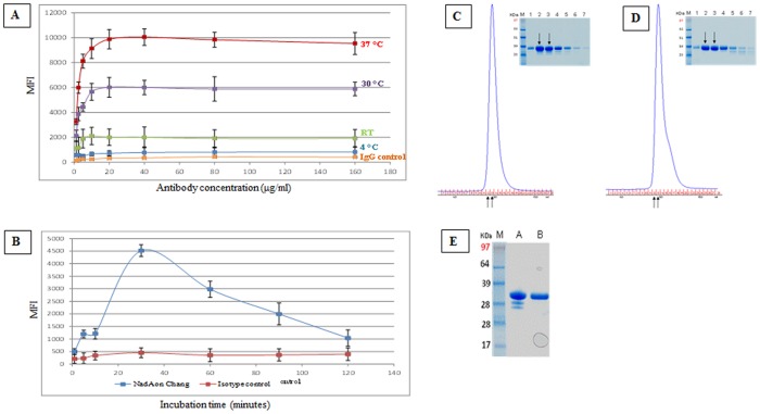
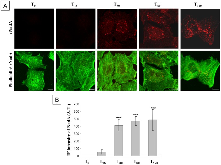
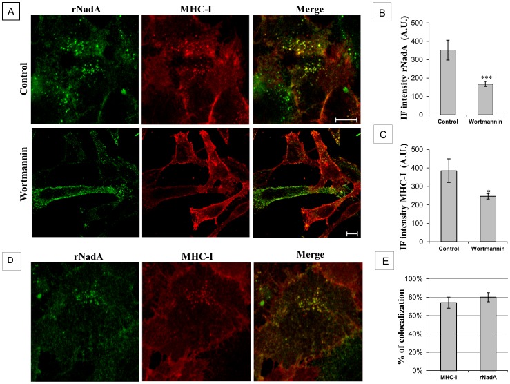
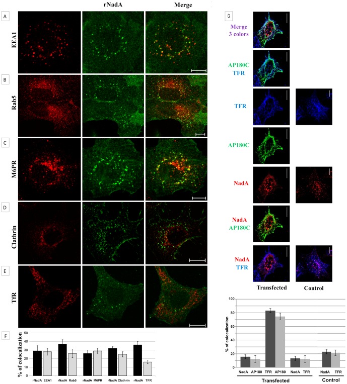
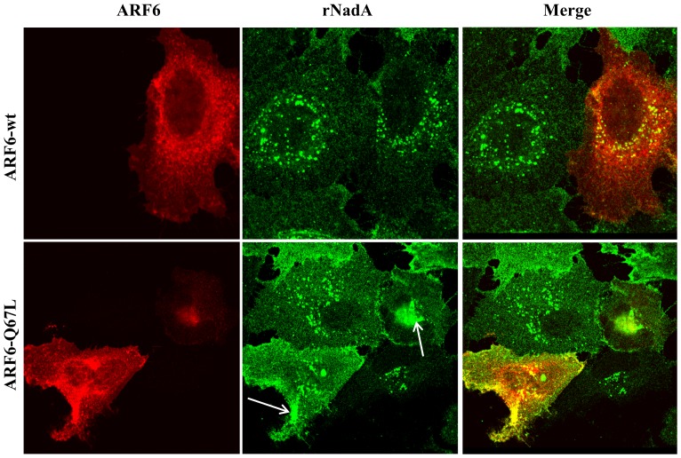
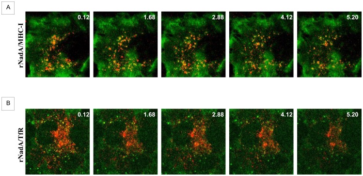
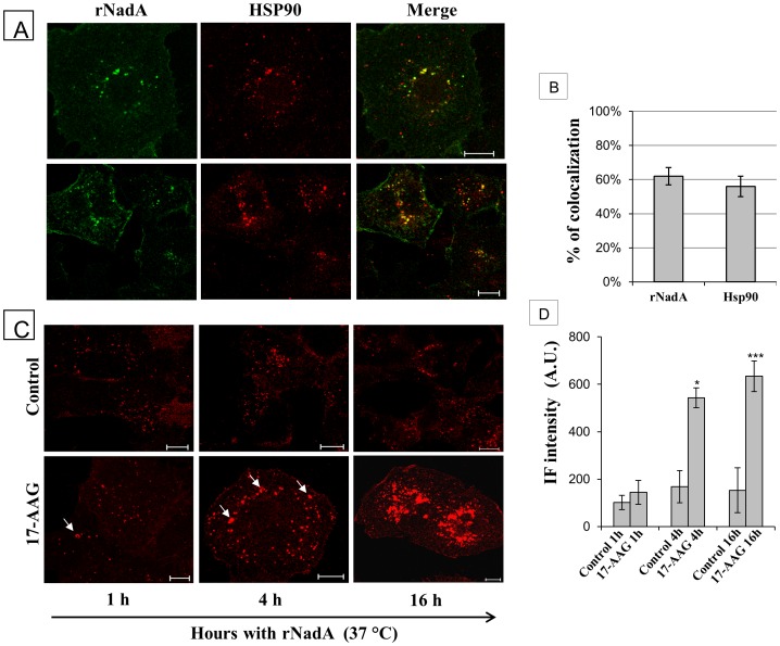
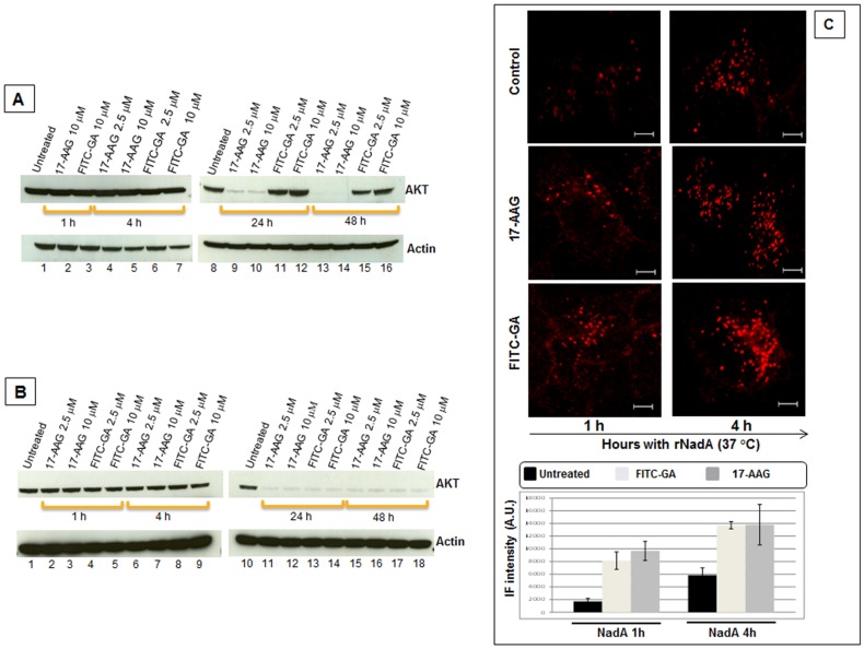
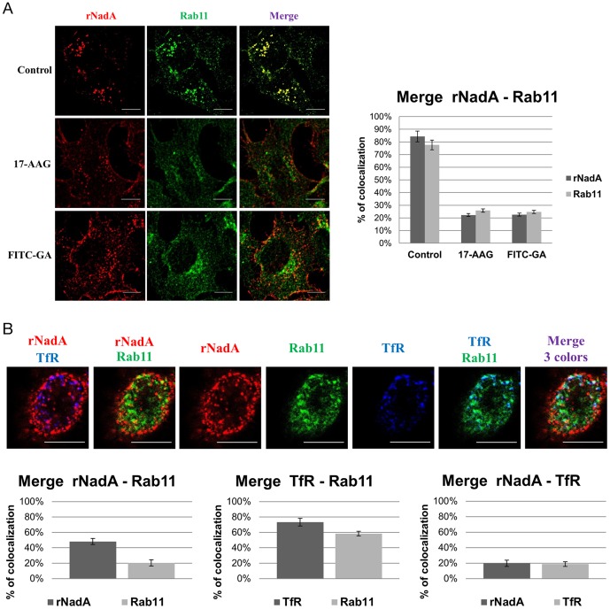
Neisseria meningitidis adhesin A (NadA) is a meningococcus surface protein thought to assist in the adhesion of the bacterium to host cells. We have previously shown that NadA also promotes bacterial internalization in a heterologous expression system. Here we have used the soluble recombinant NadA (rNadA) lacking the membrane anchor region to characterize its internalization route in Chang epithelial cells. Added to the culture medium, rNadA internalizes through a PI3K-dependent endocytosis process not mediated by the canonical clathrin or caveolin scaffolds, but instead follows an ARF6-regulated recycling pathway previously described for MHC-I. The intracellular pool of rNadA reaches a steady state level within one hour of incubation and colocalizes in endocytic vesicles with MHC-I and with the extracellularly labeled chaperone Hsp90. Treatment with membrane permeated and impermeable Hsp90 inhibitors 17-AAG and FITC-GA respectively, lead to intracellular accumulation of rNadA, strongly suggesting that the extracellular secreted pool of the chaperone is involved in rNadA intracellular trafficking. A significant number of intracellular vesicles containing rNadA recruit Rab11, a small GTPase associated to recycling endosomes, but do not contain transferrin receptor (TfR). Interestingly, cell treatment with Hsp90 inhibitors, including the membrane-impermeable FITC-GA, abolished Rab11-rNadA colocalization but do not interfere with Rab11-TfR colocalization. Collectively, these results are consistent with a model whereby rNadA internalizes into human epithelial cells hijacking the recycling endosome pathway and recycle back to the surface of the cell via an ARF6-dependent, Rab11 associated and Hsp90-regulated mechanism. The present study addresses for the first time a meningoccoccal adhesin mechanism of endocytosis and suggests a possible entry pathway engaged by N. meningitidis in primary infection of human epithelial cells.


Conflict of interest statement

Competing Interests: PM, VND, SS, BA, RR, and MP are employees of Novartis Vaccines, whose company funded this study. GB, RB, and MB were students working at Novartis and BB is a student still working at Novartis. EC was a post-doctoral fellow working at Novartis. MM is a Novartis Scientific collaborator and was responsible for this project. The antigen of this study is one of the components of Bexsero, a commercially available vaccine against meningococcus B. There are no further patents, products in development, or marketed products to declare. This does not alter the authors' adherence to all the PLOS ONE policies on sharing data and materials.

Figures

Figure 1
Figure 1. Purified rNadA binds Chang cells in a time- and temperature dependent manner.
A and B. Chang cells were incubated with 200 µg/ml rNadA and then washed with PBS – 1% FBS. Binding was detected using anti-NadA 9F11 mouse mAb and Allophycocyanin-conjugated goat anti-mouse secondary antibody. Analysis was performed with Canto II instrument, and mean fluorescence intensity is reported (MFI). A: Binding of rNadA to cells for 30 minutes at the indicated temperatures, and using varying concentrations of the primary antibody B: Binding of rNadA was performed at 37°C for a period of time ranging from 1 minute to 2 hours as indicated. C: SE-HPLC profile of rNadA at room temperature. D: SE-HPLC profile of rNadA after heating period of 4 hrs at 37°C. E: SDS-Page of collected fractions. Black arrows in D indicate the fractions pooled.
Figure 2
Figure 2. Time course of rNadA internalization.
A: Chang cells were incubated with 200 µg/ml rNadA at 37°C for 0–120 min and then fixed, permeabilized and double stained for rNadA (upper panels; red) and Alexa488-conjugated phalloidin (green). Merged images of rNadA and phalloidin are shown in the lower panel. Scale bar is 10 µm. B: Quantification of IF intensity of rNadA in the experiment illustrated in panel A. Data are mean ± s.e.m representative of two independent experiments, each assessing 10–15 cells, and expressed as Arbitrary Units (A.U.). ***p<0.001, compared to T0 treated cells (t-test).
Figure 3
Figure 3. PI3K inhibition impairs rNadA internalization and colocalization of rNadA with MHC-I.
A: Chang cells were treated with either vehicle (upper panel) or 100 nM Wortmannin (lower panel) for 1 h at 37°C, and then incubated with 200 µg/ml rNadA for an additional hour at 37°C in presence of the inhibitor or vehicle. Cells were then fixed, permeabilized and double stained for rNadA (green) and MHC-1 (red). Merged images are also shown. Scale bar 10 µm. B: Quantification of IF intensity of rNadA in the experiment illustrated in panel A. Data are mean ± s.e.m representative of two independent experiments, each assessing 20–25 cells, and expressed as Arbitrary Units (A.U.). ***p<0.001, compared to control cells (t-test). C: Quantification of IF intensity of MHC-I in the experiment illustrated in panel A. Data are mean ± s.e.m representative of two independent experiments, each assessing 20–25 cells, and expressed as Arbitrary Units (A.U.). *p<0.05, compared to control cells (t-test). D: Chang cells were treated with anti-MHC-I antibody for 1 h at 4°C and then incubated at 37°C for 1 h with 200 µg/ml rNadA. Cells were fixed and double stained for rNadA (green) and MHC-1 (red). Scale bar is 10 µm. E: Quantification of MHC-I and rNadA colocalization. MHC-I column indicate the percentage of MHC-I immunofluorescent pixels colocalizing with rNadA immunofluorescent pixels. Conversely, rNadA column indicate the percentage of rNadA immunofluorescent pixels colocalizing with MHC-I immunofluorescent pixels. Data are mean ± s.e.m representative of two independent experiments, each assessing 20–25 cells.
Figure 4
Figure 4. Colocalization of rNadA with endosome markers.
A: Chang cells were incubated with 200 µg/ml rNadA for 1 h at 37°C, then fixed, permeabilized and double stained for rNadA (green) and EEA1 ( A ), Rab5 ( B ), M6PR ( C), Clathrin ( D ) and TfR ( E ) (red). Merged images of the red and green signals are also shown. Images are representative of two independent experiments. Scale bar is 10 µm. F: Quantification of rNadA colocalization with endosome markers. The black columns indicate the percentage of rNadA immunofluorescent pixels colocalizing with the endosomal marker (EEA1, RAb5, M6PR, Clathrin, TfR as indicated) immunofluorescent pixels. Viceversa the grey columns indicate the percentage of the endosomal marker immunofluorescent pixels colocalizing with rNadA immunofluorescent pixels. Data are mean ± s.e.m representative of two independent experiments, each assessing 20–25 cells. G: Chang cells were transfected with an EGFR-AP180C expressing plasmid or with empty vector and incubated 24 hours at 37°C to recover. Cells were then incubated with rNadA, fixed and stained as indicated in point A. EGFR-AP190C is shown in green, rNadA in red and TfR in blu. Quantification was performed as indicated in point F.
Figure 5
Figure 5. rNadA intracellular distribution is affected by ARF6.
Chang cells were transfected in order to overexpress wild type ARF6 (ARF6-wt) or dominant-negative ARF6 (ARF6-Q67L) and then incubated overnight with 200 µg/ml rNadA at 37°C. Afterwards, cells were fixed, permeabilized and double stained for rNadA (green) and ARF6 (red). Merged images are also shown. Scale bar 10 µm. Images are representative of two independent experiments.
Figure 6
Figure 6. Time lapse rNadA internalization.
A: Chang cells were incubated for 1 h with Alexa633-conjugated rNadA (shown in red) and Alexa488-conjugated anti-MHC-I antibody (green). Cells were then washed and live images were recorded every 2 seconds by confocal microscopy. Five frames from the Movie S1 are shown. Time (seconds) is indicated in the top right of each panel. B: Chang cells were incubated for 1 h with Alexa488-conjugated rNadA (green) and Cy3-conjugated transferrin (red). Cells were then washed and live images were recorded every 4 seconds by confocal microscope. Five frames from the Supplementary Movie S2 are shown. Time (seconds) is indicated in the top right of each panel.
Figure 7
Figure 7. Colocalization of rNadA with HSP90.
A: Chang cells, stained in vivo with a rabbit polyclonal anti-HSP90, were incubated with rNadA for 1 h at 37°C, then fixed, permeabilized and double stained for rNadA (green) and HSP90 (red). Panels are taken from two different experiments. Merged images are also shown. Scale bar 10 µm. B: Quantification of rNadA and Hsp90 colocalization. rNadA column indicate the percentage of rNadA immunofluorescent pixels colocalizing with HSP90 immunofluorescent pixels. Conversely, HSP90 columns indicate the percentage of HSP90 immunofluorescent pixels colocalizing with rNadA immunofluorescent pixels. Data are mean ± s.e.m representative of two independent experiments, each assessing 20–25 cells. C: Chang cells pre-treated overnight with either vehicle (upper panel) or 0.5 µM 17-AAG (lower panel), were incubated with 200 µg/ml rNadA for 1 h, 4 h and 16 hrs as indicated, then fixed, permeabilized and stained for rNadA (red). Intracellular accumulation of rNadA clusters are indicated by arrows. Scale bar 10 µm. D: Quantification of IF intensity of rNadA in the experiment illustrated in panel C. Data are mean ± s.e.m representative of two independent experiments, each assessing 20–25 cells, and expressed as Arbitrary Units (A.U.). ***p<0.001, compared to 1 h of 17-AAG treated cells (t-test). *p<0.05, compared to 1 h of 17-AAG treated cells (t-test).
Figure 8
Figure 8. Inhibition of Hsp90 influences AKT degradation and intracellular accumulation of rNadA.
A: Chang cells were incubated for 1, 4, 24 and 48 hours with either 17-AAG or FITC-GA at two different concentrations. At the end of each period cells were washed, trypsinized and RIPA-buffer total lysate prepared. Proteins from each extract were separated on NuSieve gel, blotted, and AKT or Actin (loading control) were detected using the respective primary antibodies. B: Chang cells were pre-treated with 0.01% saponin in PBS for 30 seconds at room temperature, washed three times with PBS and then handled as described in A. C: Chang cells were pre-treated for 1 hour with either vehicle (upper panels), 10 µM 17-AAG (middle panel) or 10 µM FITC-GA (lower panel), and then incubated with 200 µg/ml rNadA at 37°C for 1 (left panels) or 4 hours (right panels). Cells were then fixed, permeabilized and stained for rNadA. The drugs were present during the entire incubation period. IF intensity was calculated as mean ± s.e.m in two independent experiments, each assessing 10–15 cells and expressed as Arbitrary Units (A.U.). Scale bar 10 µm.
Figure 9
Figure 9. rNada internalization and colocalization with Rab11 and TfR.
A: rNadA and Rab11 colocalization in presence of inhibitors. The control untreated Chang cells are shown in the upper panel. Pre-treatment of cells was performed for 1 hour with 10 µM 17-AAG (middle panel) or 10 µM FITC-GA (bottom panel). Cells were then incubated with 200 µg/ml rNadA at 37°C for 1 hour. Afterward, cells were fixed, permeabilized and stained for NadA. The drugs were present during the entire incubation period. Graph: rNadA columns indicate the percentage of rNadA immunofluorescent pixels colocalizing with Rab11 immunofluorescent pixels in untreated, 17-AAG and FITC-GA cells (from left to right) respectively. Conversely, Rab11 columns indicate the percentage of Rab11 immunofluorescent pixels colocalizing with rNadA immunofluorescent pixels in untreated, 17-AAG and FITC-GA cells (from left to right) respectively. B: Rab11, rNadA and TfR colocalization . A: Chang cells were transfected with EGFP-Rab11 plasmid, incubated 24 hours at 37°C. and then incubated with 200 µg/ml of rNadA at 37°C for 2 hours. Afterward, cells were fixed, permeabilized and stained for NadA (Red) and TfR (blu). Rab11 is colored in green. Graphs: rNadA columns indicate the percentage of rNadA immunofluorescent pixels colocalizing with Rab11 fluorescent pixels (left) or with TfR immunofluorescent pixels (right). Rab11column indicate the percentage of fluorescent pixels colocalizing with rNadA immunofluorescent pixels (left) or TfR immunofluorescent pixels (right). TfR columns indicate the percentage of TfR immunofluorescent pixels colocalizing with Rab11 fluorescent pixels (middle) or with rNadA immunofluorescent pixels (left). Data are mean ± s.e.m representative of two independent experiments, each assessing 20–25 cells.

Similar articles

Cited by

References

    1. Emonts M, Hazelzet JA, de Groot R, Hermans PW (2003) Host genetic determinants of Neisseria meningitidis infections. Lancet Infect Dis 3: 565–577. - PubMed
    1. Stephens DS (2009) Biology and pathogenesis of the evolutionarily successful, obligate human bacterium Neisseria meningitidis. Vaccine 27 Suppl 2: B71–77. - PMC - PubMed
    1. Hill DJ, Griffiths NJ, Borodina E, Virji M (2010) Cellular and molecular biology of Neisseria meningitidis colonization and invasive disease. Clin Sci (Lond) 118: 547–564. - PMC - PubMed
    1. Virji M (2009) Pathogenic neisseriae: surface modulation, pathogenesis and infection control. Nat Rev Microbiol 7: 274–286. - PubMed
    1. Carbonnelle E, Hill DJ, Morand P, Griffiths NJ, Bourdoulous S, et al. (2009) Meningococcal interactions with the host. Vaccine 27 Suppl 2: B78–89. - PubMed

Publication types

Grants and funding

This work is entirely funded by Novartis Vaccines; no external entities have contributed to cover project expenses. The funders had no role in study design, data collection and analysis, decision to publish, or preparation of the manuscript.