. 2014 Oct 22;55(10):6651-72.
doi: 10.1167/iovs.14-15502.

Chemistry and biology of the initial steps in vision: the Friedenwald lecture

Affiliations

Chemistry and biology of the initial steps in vision: the Friedenwald lecture

Krzysztof Palczewski. Invest Ophthalmol Vis Sci. .

Abstract

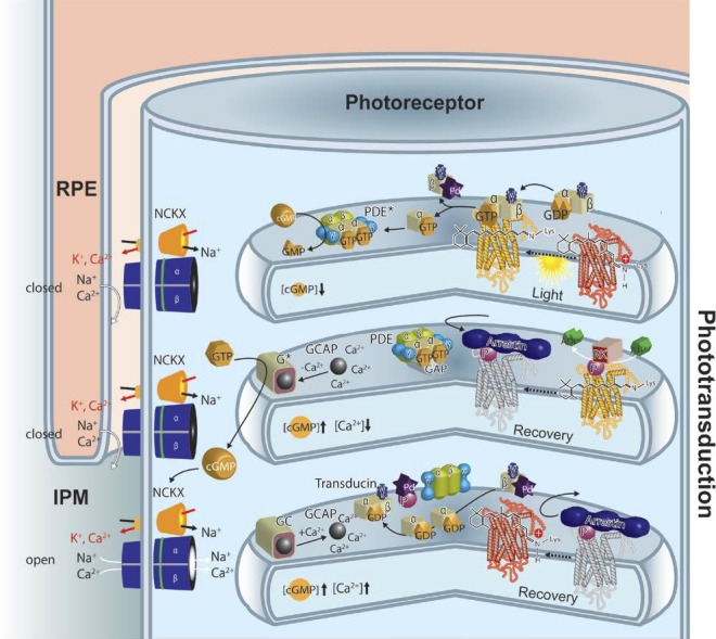
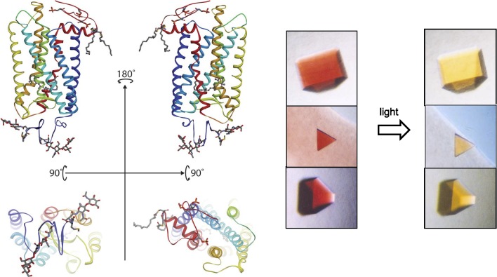
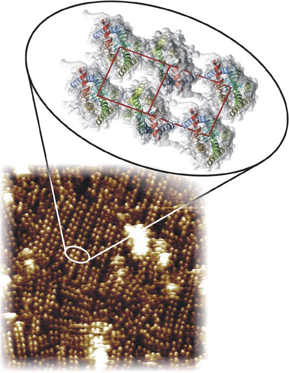
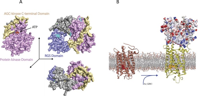
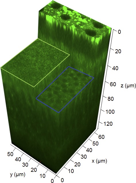
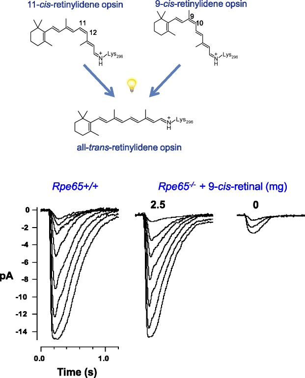
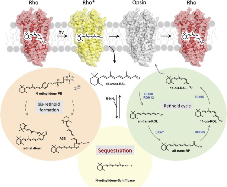
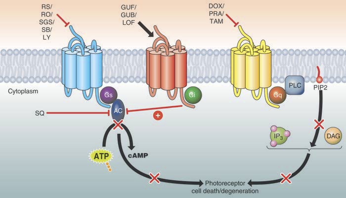
Visual transduction is the process in the eye whereby absorption of light in the retina is translated into electrical signals that ultimately reach the brain. The first challenge presented by visual transduction is to understand its molecular basis. We know that maintenance of vision is a continuous process requiring the activation and subsequent restoration of a vitamin A-derived chromophore through a series of chemical reactions catalyzed by enzymes in the retina and retinal pigment epithelium (RPE). Diverse biochemical approaches that identified key proteins and reactions were essential to achieve a mechanistic understanding of these visual processes. The three-dimensional arrangements of these enzymes' polypeptide chains provide invaluable insights into their mechanisms of action. A wealth of information has already been obtained by solving high-resolution crystal structures of both rhodopsin and the retinoid isomerase from pigment RPE (RPE65). Rhodopsin, which is activated by photoisomerization of its 11-cis-retinylidene chromophore, is a prototypical member of a large family of membrane-bound proteins called G protein-coupled receptors (GPCRs). RPE65 is a retinoid isomerase critical for regeneration of the chromophore. Electron microscopy (EM) and atomic force microscopy have provided insights into how certain proteins are assembled to form much larger structures such as rod photoreceptor cell outer segment membranes. A second challenge of visual transduction is to use this knowledge to devise therapeutic approaches that can prevent or reverse conditions leading to blindness. Imaging modalities like optical coherence tomography (OCT) and scanning laser ophthalmoscopy (SLO) applied to appropriate animal models as well as human retinal imaging have been employed to characterize blinding diseases, monitor their progression, and evaluate the success of therapeutic agents. Lately two-photon (2-PO) imaging, together with biochemical assays, are revealing functional aspects of vision at a new molecular level. These multidisciplinary approaches combined with suitable animal models and inbred mutant species can be especially helpful in translating provocative cell and tissue culture findings into therapeutic options for further development in animals and eventually in humans. A host of different approaches and techniques is required for substantial progress in understanding fundamental properties of the visual system.

Keywords: G protein–coupled receptor(s); membrane proteins; photoreceptors; phototransduction; protein structure; receptor phosphorylation; rhodopsin; signal transduction; vision.


Figures

Figure 1
Figure 1
Phototransduction in a rod outer segment. Phototransduction can be described in three stages shown from top to bottom in this cartoon. When light strikes rhodopsin (red), it causes isomerization of the 11-cis-retinylidene chromophore to an all-trans configuration and a conformational change in the opsin protein. This, in turn, leads to formation of a complex with the heterotrimeric G protein, transducin. Nucleotide exchange in the transducin α-subunit from guanosine diphosphate to GTP causes dissociation of transducin with formation of the transducin α-subunit. This subunit interacts with tetrameric cGMP–specific PDE6, whereas the transducin βγ-subunit complexes with phosducin. One activated rhodopsin molecule can activate dozens of transducin molecules in this first amplification stage of phototransduction. Displacement of the inhibitory γ-subunit activates PDE in the second amplification step of phototransduction. The resulting decrease in the concentration of cGMP is associated with a decrease in intradiscal Ca2+ concentration because cGMP is a ligand for cGMP-gated cation channels (shown in blue in the plasma membrane), nonselective channels that also allow passage of Ca2+ in their cGMP-bound state. The low Ca2+-level is maintained by the light-insensitive Na+/Ca2+-K+ exchanger, which extrudes Ca2+ ions out against a gradient in exchange for Na+ and K+ ions. Each of the above-activated molecules needs to return to its inactive state before absorption of the next photon. Thus, rhodopsin is phosphorylated at its C-terminus by GRK1 (or rhodopsin kinase [RK]), followed by binding of arrestin, a capping protein. Guanosine triphosphate is hydrolyzed by the α-subunit of transducin with the help of a GTPase-activating protein. Guanylate cyclase 1 and GC2 (GC, light/dark-brown box) are activated by Ca2+-binding proteins (GCAP1 and GCAP2, black ball) in their Ca2+-free forms to restore cGMP levels and open the cyclic nucleotide–gated cation channels in the plasma membrane. Guanylate cyclase-activating proteins are inactivated and GC activities return to their dark condition. Once GTP is hydrolyzed by the α-subunit of transducin along with phosphorylation of phosducin, the heterotrimeric G protein is restored. Opsin recombines with 11-cis-retinal and the rhodopsin thus formed is ready to be photoactivated. Note that all these processes take place on the cytoplasmic surfaces of disc and plasma membranes.
Figure 2
Figure 2
Rhodopsin structure and crystals. Left top: ribbon drawings of rhodopsin (PDB accession code: 1F88) in the plane of a disc membrane (two views rotated by 180°). Bottom: the intradiscal side (left) and cytoplasmic side (right) of this receptor. Right: a photo-stable crystal form of the dark inactive state (left) and light-exposed activated state (right) of the receptor. Crystal pictures are reproduced from Salom D, Le Trong I, Pohl E, et al. Improvements in G protein-coupled receptor purification yield light stable rhodopsin crystals. J Struct Biol. 2006;156:497–504. Copyright © 2006 Elsevier, Inc.; Salom D, Lodowski DT, Stenkamp RE, et al. Crystal structure of a photoactivated deprotonated intermediate of rhodopsin. Proc Natl Acad Sci U S A. 2006;103:16123–16128. Copyright © 2006 The National Academy of Sciences of the USA; and Salom D, Padayatti PS, Palczewski K. Crystallization of G protein–coupled receptors. Methods Cell Biol. 2013;117:451–468. Copyright © 2013 Elsevier, Inc., with permission from Elsevier, Inc. and the National Academy of Sciences.
Figure 3
Figure 3
Organization of rhodopsin in disc membranes. An AFM tomograph (tilted by 5°) shows the paracrystalline arrangement of rhodopsin dimers in a native disc membrane. The rhodopsin molecules protrude from the lipid bilayer by 1.4 ± 0.2 nm or one-fourth of their mass. Inset shows a model for the packing arrangement of rhodopsin molecules within the paracrystalline arrays in native disc membranes (PDB accession code: 1N3M). Helices of rhodopsin are colored as follows: helix I in blue, helix II in light blue, helix III in green, helix IV in light green, helix V in yellow, helix VI in orange, and helix VII and cytoplasmic helix 8 in red. The red box shows the close contacts between four molecules of rhodopsin. The dashed-line rectangle shows the longer contacts with neighboring dimers of rhodopsin. The native membrane structure and the molecular model of rhodopsin are reproduced from Liang Y, Fotiadis D, Filipek S, Saperstein DA, Palczewski K, Engel A. Organization of the G protein-coupled receptors rhodopsin and opsin in native membranes. J Biol Chem. 2003;278:21655–21662. Copyright © 2003 The American Society for Biochemistry and Molecular Biology, Inc.; and Fotiadis D, Liang Y, Filipek S, Saperstein DA, Engel A, Palczewski K. Atomic-force microscopy: rhodopsin dimers in native disc membranes. Nature. 2003;421:127–128, with permission from the American Society for Biochemistry and Molecular Biology and Nature Publishing Group.
Figure 4
Figure 4
Structure of rhodopsin kinase (GRK1) and phosphorylation of rhodopsin. (A) Overview of GRK1 with bound ATP (PDB accession code: 3C4W). The structural domain of regulators of G protein signaling is colored blue, the protein kinase domain violet, and the AGC-kinase C-terminal domain yellow. Atoms of the substrate ATP are shown as spheres. (B) Conceptual model of GRK1 docked to monomeric activated rhodopsin. Rhodopsin (PDB accession code: 1U19) is activated by light (PDB accession code: 3PQR) and only then does GRK1 bind to the cytoplasmic surface of the receptor. G protein–coupled receptor kinase 1 is rendered as molecular surface, rhodopsin as a bundle of transmembrane helices with connecting cytoplasmic and intradiscal loops. The active site of GRK1 could have access to the C-tail of a neighboring unactivated rhodopsin (not shown) in dimeric or oligomeric forms, allowing phosphorylation of an additional nonactivated rhodopsin in rod outer segments (high-gain phosphorylation).
Figure 5
Figure 5
Regulation of GC in photoreceptors. (A) Sensitivity of GC to physiological concentrations of free Ca2+ in rods and cones. (B) Structure of myristoylated GCAP1 in three orientations (PDB accession code: 2R2I). The N-terminal domain is colored blue (EF-hand 1 and EF-hand 2) and the C-terminal domain is red (EF-3 and EF-4). Ca2+ ions and the myristoyl groups are shown as dark-green spheres and green space–filling shapes, respectively. The surface is made semitransparent to reveal the buried myristoyl group. Red arrows point toward the myristoyl group. SAS, solvent accessibility surface.
Figure 6
Figure 6
The visual (retinoid) cycle. This metabolic renewal of 11-cis-retinal takes place in photoreceptor outer segments and the RPE. First, all-trans-retinylidene is hydrolyzed from opsin and all-trans-retinal diffuses to the cytoplasmic side where it is reduced to an alcohol by membrane associated all-trans-retinol RDH. Lack of adequate RDH activity can lead to LCA (or RP); all diseases are depicted in red letters. A fraction of all-trans-retinal is released into the intradiscal side. There all-trans-retinal and phosphatidylethanolamine form a Schiff base and together are transported into the cytoplasmic side via ABCA4. Lack of ABCA4 transport activity is associated with Stargardt disease, whereas polymorphisms in this gene are associated with AMD. Retinol diffuses from the cytoplasm to the RPE where it becomes esterified by LRAT to form fatty acid retinyl esters. Such esters have a propensity to coalescence, thermodynamically driving the transfer from photoreceptor to RPE cells. The esters then serve as substrates for the isomerization reaction catalyzed by the 65-kDa protein, RPE65. The resulting product, 11-cis-retinol, is oxidized to 11-cis-retinal by the 11-cis-retinol specific RDH5 and dual specificity (cis and trans-retinols) RDHs, including RDH10; 11-cis-retinal diffuses back into the photoreceptor outer segments, a process thermodynamically driven by the formation of stable visual pigments. Essential for transporting and protecting these retinoids are intracellular and extracellular retinoid-binding proteins such as CRALBP, CRBP1, IRBP and retinol-binding protein 4 (RBP4). Inactivating mutations in the LRAT and RPE65 genes are causes of childhood blindness because these genes are nonredundant, whereas mutations in RDHs and retinoid-binding proteins have less severe effects but can be associated with RP, cone-rod dystrophy, fundus albipunctatus, fundus albescens or Bothnia dystrophy. Retinoids are retained in the eye as a result of LRAT activity. In the bloodstream, retinoids are bound to RBP4 and then enter the eye by passive transport with the help of STRA6. Mutations in the STRA6 gene cause Matthew-Wood Syndrome, a severe disease that includes obesity and mental retardation, as well as faulty eye development and blindness, indicating the importance of STRA6 for retinol transport into the brain and eye.
Figure 7
Figure 7
Enzymatic mechanism of LRAT and the architecture of its active site. Transfer of an acyl moiety from phospholipid onto vitamin A occurs in two catalytic steps. The acyl moiety is first transferred onto a catalytic Cys residue forming a thioester. This thioester is then cleaved by a nucleophilic attack from retinol's activated hydroxyl group causing subsequent formation of the final retinyl ester product and restoration of the LRAT active site. A model of the human LRAT catalytic domain based on a LRAT/HRASLS3 chimeric protein reveals that the overall structure of the catalytic domain resembles the archetypical α/β fold of a NlpC/P60 protein. The architecture of the LRAT active site (blue) shows the positions of key residues involved in catalysis in relation to the native HRASLS3 protein depicted in gray (PDB accession code: 4DOT). The catalytic Cys161 residue is located at the N-terminus of a helix packed against a core of β-sheets containing the conserved His60 and His72 residues.
Figure 8
Figure 8
Crystal structure of RPE65. (A) Cartoon representation showing the beta-propeller fold of RPE65. The catalytic iron, located on the propeller axis, is coordinated by four conserved histidine (His) residues. Three second sphere Glu residues are also important active site components. Residues of Glu and His are shown in stick representations. (B) Dimeric structure of RPE65. The hydrophobic patches that mediate RPE65 membrane association (brown) are oriented in parallel. The entrance to the active site cavity (dashed black lines) is surrounded by residues comprising the hydrophobic patch (PDB accession code: 4F2Z I).
Figure 9
Figure 9
Three-dimensional 2PO microscopic image of a retina and RPE in the intact eye of a 5-month-old C57BL/6J-Tyrc–2J mouse. The retinal pigmented epithelium is at the top of the image at z = 0 μm. A section through the retinal inner segments outlined by the light-green rectangle is shown at a location 30 μm away from the RPE. Another section through the outer nuclear layer outlined in blue is shown at a location 40 μm away from the RPE. Images were obtained with a 730-nm excitation wavelength.
Figure 10
Figure 10
Rescue of vision in LRAT- and RPE65-deficient mice. Above: Endogenous 11-cis-retinal bound to opsin (rhodopsin) can be replaced by 9-cis-retinal to form a functional pigment (isorhodopsin) that differs by only a 60% reduction in quantum yield and approximately a ~15-nm hypsochromic shift in maximal light absorption. These differences minimally affect vision. However, 9-cis-retinoids are chemically more stable than 11-cis-retinoids and can be pharmacologically formulated for use in the treatment of LCA; 11-cis-retinoids formed in situ in the eye are photo- and thermally unstable without protective retinoid-binding proteins. Both rhodopsin and isorhodopsin form active meta II rhodopsin intermediates. Below: stimulus response families for Rpe65+/+ or Rpe65−/− mice supplemented with 0 and 2.5 mg of 9-cis-retinal. Light flash (10 ms) strengths were increased in 2-fold steps from the dimmest intensity. Reproduced with permission from Van Hooser JP, Liang Y, Maeda T, et al. Recovery of visual functions in a mouse model of Leber congenital amaurosis. J Biol Chem. 2002;277:19173–19182. Copyright © 2002 The American Society for Biochemistry and Molecular Biology, Inc.
Figure 11
Figure 11
Preservation of retinal structure and function by sequestering excess all-trans-retinal. Once released from rhodopsin, all-trans-retinal can enter the retinoid cycle, form bis-retinoids such as A2E, or persist as a free toxic aldehyde that impairs multiple processes and leads to cell death. High levels of A2E are associated with Stargardt disease and AMD; A2E also accumulates with age without any obvious disease as evidenced by elevated fluorescent material in the eyes of healthy individuals. Treatment with primary amines that form a Schiff base can lower the excessive amounts of all-trans-retinal that accumulate in transgenic mice. The same strategy could be beneficial as a treatment for AMD and/or Stargardt disease. Reproduced from Maeda A, Golczak M, Chen Y, et al. Primary amines protect against retinal degeneration in mouse models of retinopathies. Nat Chem Biol. 2012;8:170–178; with permission from Nature Publishing Group.
Figure 12
Figure 12
Systems pharmacological strategies prevent the development of light-induced photoreceptor degeneration. In appropriate mouse models of human retinopathies, antagonists of multiple Gs-coupled GPCRs prevented photoreceptor cell death (red bar, top left). Moreover, pharmacological activation of Gi-coupled GPCRs (black arrow, top middle) that suppress adenylate cyclase activity (red bar, middle) preserved retinal structure and function against an environmental insult (prolonged exposure to intense illumination). Direct inhibition of AC was also beneficial (red line). Additionally, Gq-coupled GPCRs can participate in photoreceptor degeneration, and inhibition of these GPCRs also proved effective in protecting photoreceptors from light-induced degeneration (red bar, top right). Reproduced with permission from Chen Y, Palczewska G, Mustafi D, et al. Systems pharmacology identifies drug targets for Stargardt disease-associated retinal degeneration. J Clin Invest. 2013;123:5119–5134. Copyright © 2013 American Society for Clinical Investigation.

Similar articles

Cited by

References

    1. Arshavsky VY, Lamb TD, Pugh EN Jr. G proteins and phototransduction. Annu Rev Physiol. 2002; 64: 153–187 - PubMed
    1. Orban T, Jastrzebska B, Palczewski K. Structural approaches to understanding retinal proteins needed for vision. Curr Opin Cell Biol. 2014; 27: 32–43 - PMC - PubMed
    1. Palczewski K, Orban T. From atomic structures to neuronal functions of G protein-coupled receptors. Annu Rev Neurosci. 2013; 36: 139–164 - PMC - PubMed
    1. Palczewski K. G protein-coupled receptor rhodopsin. Annu Rev Biochem. 2006; 75: 743–767 - PMC - PubMed
    1. Palczewski K. Chemistry and biology of vision. J Biol Chem. 2012; 287: 1612–1619 - PMC - PubMed

Publication types

MeSH terms