. 2014 Dec 12;289(50):34490-502.
doi: 10.1074/jbc.M114.604132. Epub 2014 Oct 15.

The E2 ubiquitin-conjugating enzyme UBE2J1 is required for spermiogenesis in mice

Affiliations

The E2 ubiquitin-conjugating enzyme UBE2J1 is required for spermiogenesis in mice

Paul-Albert Koenig et al. J Biol Chem. .

Abstract

ER-resident proteins destined for degradation are dislocated into the cytosol by components of the ER quality control machinery for proteasomal degradation. Dislocation substrates are ubiquitylated in the cytosol by E2 ubiquitin-conjugating/E3 ligase complexes. UBE2J1 is one of the well-characterized E2 enzymes that participate in this process. However, the physiological function of Ube2j1 is poorly defined. We find that Ube2j1(-/-) mice have reduced viability and fail to thrive early after birth. Male Ube2j1(-/-) mice are sterile due to a defect in late spermatogenesis. Ultrastructural analysis shows that removal of the cytoplasm is incomplete in Ube2j1(-/-) elongating spermatids, compromising the release of mature elongate spermatids into the lumen of the seminiferous tubule. Our findings identify an essential function for the ubiquitin-proteasome-system in spermiogenesis and define a novel, non-redundant physiological function for the dislocation step of ER quality control.

Keywords: ER Quality Control; ERAD Tuning; Endoplasmic Reticulum-associated Protein Degradation (ERAD); Gene Knockout; Male Infertility; Mouse Model; Spermatogenesis; Spermiogenesis; Ubiquitin-conjugating Enzyme (E2 enzyme).


Figures

FIGURE 1.
FIGURE 1.
Generation of Ube2j1fl/fl mice. Ube2j1−/− mice fail to thrive after birth. A, scheme of the targeting strategy. LoxP sites were introduced into introns 1 and 3 to allow excision of exons 2 and 3 by Cre recombinase. Circled A, B, and C indicate annealing sites for the genotyping primers. B, representation of the reads measured for Ube2j1 by RNA-Seq in representative Ube2j1+/+ (WT) or Ube2j1−/− (KO) MEF lines (passage 3). The bottom panel depicts the intron/exon structure of Ube2j1. C, immunoblot of MEF lysates from two individual Ube2j1+/+, Ube2j1+/−, or Ube2j1−/− embryos, each. Asterisk, potential splice variant. D, frequencies of sex and genotypes of pups from Ube2j1+/− parents at P0.5 (after birth) and P21 (weaning). E, weight of Ube2j1+/+, Ube2j1+/−, and Ube2j1−/− mice at age P0.5 (top) and P21 (bottom). *, p < 0.05; **, p < 0.01; ***, p < 0.001; not significant (n.s.); p > 0.05, as determined by one-way ANOVA with Tukey's Multiple Comparison Test.
FIGURE 2.
FIGURE 2.
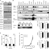
Ube2j1−/− MEFs and organs express higher levels of specific ER dislocation components, and Ube2j1−/− MEFs are more susceptible to SV40 infection. A, immunoblots of several components of the ER quality machinery in two independent Ube2j1+/+ (WT) or Ube2j1−/− (KO) MEF lines, respectively, show increased levels for specific ER quality control components in Ube2j1−/− MEFs. B and C, protein levels in organ lysates from a Ube2j1−/− mouse and a wild type littermate. SI, small intestine; LI, large intestine; SG, salivary gland; SM, skeletal muscle; LN, lymph nodes; Epi, epididymis. D, transcript levels of select genes involved in the ER stress response and ER dislocation are unaltered in Ube2j1−/− MEFs. Shown are average reads per kilobase per million mapped reads (RPKM) values as determined by RNA-Seq from two independent MEF cell lines (passage 3) per genotype. E, wild type or Ube2j1−/− MEFs were infected with different MOIs of SV40. 24 h after infection the expression of the large T antigen was measured by FACS. Shown are mean and standard deviation values of relative percentages of infected cells of three independent experiments. **, p < 0.01; ***, p < 0.001, as determined by Student's t tests. F, binding of cholera toxin B subunit (FITC-CTxB) to wild type or Ube2j1−/− MEFs. Shown are mean MFI values and standard deviation of two wild type or two Ube2j1−/− MEF lines, respectively, from one representative experiment.
FIGURE 3.
FIGURE 3.
Ube2j1-deficiency does not enhance induction of the UPR. A, representation of the reads measured for Xbp-1 by RNA-Seq. Shown are reads from unstimulated Ube2j1+/+ (WT) or Ube2j1−/− (KO) MEF lines (passage 3). The bottom panel depicts the intron/exon structure of Xbp-1. B, splicing of the Xbp1-mRNA after exposure of Ube2j1+/+ and Ube2j1−/− MEFs to tunicamycin as measured by RT-PCR. B, proteolytic processing of ATF6 after exposure of Ube2j1+/+ and Ube2j1−/− MEFs to tunicamycin measured by immunoblotting with an antibody directed to the N terminus of ATF6.
FIGURE 4.
FIGURE 4.
B cell development and antibody secretion are normal in absence of Ube2j1. A, FACS plots for markers of different B cell populations in the bone marrow, spleen and peritoneal cavity revealed no difference in B cell development in Ube2j1−/− mice. B and C, synthesis, maturation, and secretion of immunoglobulin does not differ significantly between Ube2j1+/+ and Ube2j1−/− B cells. Shown are autoradiographs of immunoprecipitates for μ chain of IgM and class I MHC heavy chain from B plasmablast lysates (left column) and supernatants (right column) from a pulse-chase experiment (B) without or (C) with pre-treatment (5 h) with tunicamycin.
FIGURE 5.
FIGURE 5.
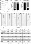
Ube2j1-deficiency causes male sterility in mice. A, total number of sperm cells in the epididymis of Ube2j1+/+ (WT) or Ube2j1−/− (KO) mice. *: p < 0.05, as determined by unpaired t test. B, progressive motility of Ube2j1−/− sperm cells is impaired. Shown are average percentages of (black) immotile, (white) non-progressively motile, and (gray) progressively motile Ube2j1+/+ (WT) or Ube2j1−/− (KO) sperm cells (n≈200 per mouse, 3 mice per genotype). C, assessment of sperm cell morphology. Average percentages of (black) morphologically normal, (white) acephalic and (gray) morphologically abnormal sperm cells as observed in an ethanol-fixed sperm smear are shown (n≈200 per mouse, 3 mice per genotype). D, photographs of ethanol-fixed Ube2j1+/+ (WT) or Ube2j1−/− (KO) spermatozoa. Note the abnormal head morphology in the knock-out sperm. E, percentages of embryo stages observed in an IVF experiment 1, 2, and 3.5 days after fertilization. Spermatozoa from Ube2j1+/+ (WT) or Ube2j1−/− (KO) mice (3 mice per genotype) were incubated with wild type oocytes (∼200 oocytes per sperm of one male) and embryos at the indicated stages were counted.
FIGURE 6.
FIGURE 6.
Male Ube2j1−/− mice have defects in spermatid differentiation. A, immunohistochemical staining for UBE2J1 (green), acrosomes (PNA lectin, red) and DNA (DAPI, blue) in testis cross-sections of a wild type mouse. Note the stage-specific expression of UBE2J1. No staining was detected in Ube2j1−/− testes (right panel). Scale bar, 10 μm. B, immunohistochemical staining for UBE2J1 (green) and the germ cell-specific DDX4 (red) in a wild type testis. C, PAS-stained cross-section of testes from Ube2j1+/+ (WT, top row) and Ube2j1−/− (bottom row) mice. Note the abnormal germ cell associations and spermiation defect at stage IX. Asterisks denote mislocated spermatids. Filled arrowheads, elongated spermatids to be released at the end of stage VIII. Empty arrowheads, next generation round spermatids. D, average testis weight in grams of Ube2j1+/+ (WT), Ube2j1+/− (HET), and Ube2j1−/− (KO) mice. Shown are mean testis weight of individual mice (data points), and overall average with standard deviation. Not significant (n.s.) indicates p > 0.05, as determined by one-way ANOVA with Tukey's Multiple Comparison Test.
FIGURE 7.
FIGURE 7.
Electron microscopy reveals structural abnormalities in Ube2j1−/− spermatozoa. A, cross sections of a Ube2j1−/− stage IX testis shows retained spermatids (asterisks) with cytoplasm (arrows) above a layer of elongate spermatids. ES, ectoplasmic specialization. B, wild type elongated spermatids in the testis. C and D, Ube2j1−/− and (E) wild type spermatids in cross sectioned epididymides. F and G, Ube2j1−/− and (H) wild type spermatids isolated from epididymides. Ube2j1−/− spermatozoa retain excess cytoplasm around the acrosome and neck region. Note vacuoles (wedges) surrounded by one or more membranes (F), and retained condensed ER (radial body, insets) in Ube2j1−/− spermatozoa (C, D, F, G). Scale bars, 2 μm (A and B); 500 nm (C–F); 100 nm (G); 500 nm (H).

Similar articles

Cited by

References

    1. Smith M. H., Ploegh H. L., Weissman J. S. (2011) Road to ruin: targeting proteins for degradation in the endoplasmic reticulum. Science 334, 1086–1090 - PMC - PubMed
    1. Claessen J. H., Kundrat L., Ploegh H. L. (2012) Protein quality control in the ER: balancing the ubiquitin checkbook. Trends Cell Biol. 22, 22–32 - PMC - PubMed
    1. Lester D., Farquharson C., Russell G., Houston B. (2000) Identification of a family of noncanonical ubiquitin-conjugating enzymes structurally related to yeast UBC6. Biochem. Biophys. Res. Commun. 269, 474–480 - PubMed
    1. Lenk U., Yu H., Walter J., Gelman M. S., Hartmann E., Kopito R. R., Sommer T. (2002) A role for mammalian Ubc6 homologues in ER-associated protein degradation. J. Cell Sci. 115, 3007–3014 - PubMed
    1. Mueller B., Klemm E. J., Spooner E., Claessen J. H., Ploegh H. L. (2008) SEL1L nucleates a protein complex required for dislocation of misfolded glycoproteins. Proc. Natl. Acad. Sci. U.S.A. 105, 12325–12330 - PMC - PubMed

Publication types