Review
. 2014 Nov;15(11):736-47.
doi: 10.1038/nrm3888. Epub 2014 Oct 10.

Signalling dynamics in the spindle checkpoint response

Affiliations
Review

Signalling dynamics in the spindle checkpoint response

Nitobe London et al. Nat Rev Mol Cell Biol. 2014 Nov.

Abstract

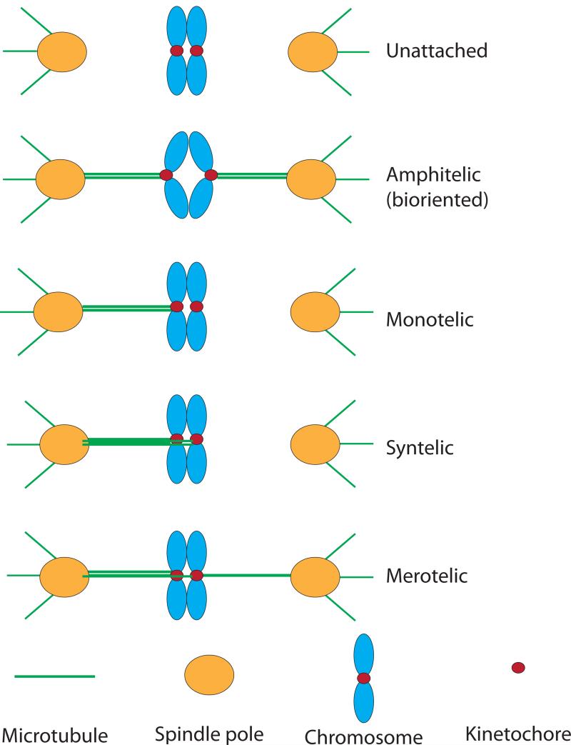
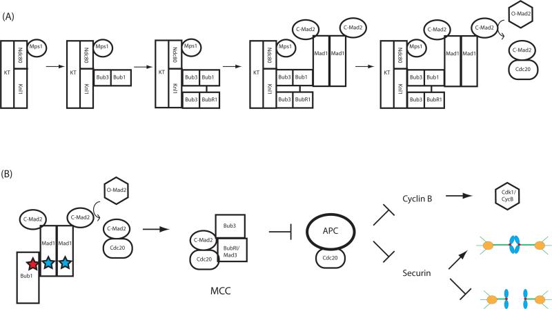
The spindle checkpoint ensures proper chromosome segregation during cell division. Unravelling checkpoint signalling has been a long-standing challenge owing to the complexity of the structures and forces that regulate chromosome segregation. New reports have now substantially advanced our understanding of checkpoint signalling mechanisms at the kinetochore, the structure that connects microtubules and chromatin. In contrast to the traditional view of a binary checkpoint response - either completely on or off - new findings indicate that the checkpoint response strength is variable. This revised perspective provides insight into how checkpoint bypass can lead to aneuploidy and informs strategies to exploit these errors for cancer treatments.


Figures

Figure 1
Figure 1. Kinetochore-microtubule attachment states on the mitotic spindle
Kinetochores can bind to microtubules attached to either spindle pole, making several different configurations possible. See text for description of each. Spindle poles nucleate spindle microtubules, shown attaching to kinetochores, as well as astral microtubules. Sister chromosomes are linked by the protein complex cohesin until anaphase onset. Microtubules tend to pull chromosomes towards spindle poles, resulting in tension across bioriented sister chromosomes. Syntelic attachments lead to spindle checkpoint activation whereas merotelic attachments do not necessarily do so because some tension is applied across sister kinetochores.
Figure 2
Figure 2. Kinetochore activation of the checkpoint through hierarchical checkpoint protein recruitment
(A) Assembly of a functional checkpoint-signaling complex on kinetochores is stepwise. Bub3 forms separate complexes with Bub1 and BubR1/Mad3. Mps1 activity recruits Bub1/3, and Bub1 permits localization of BubR1/Mad3. Next, a Mad1/2 heterodimer binds to the kinetochore through a Mad1-Bub1 interaction and possibly through an additional receptor. Kinetochore-bound Mad1/2 facilitates checkpoint activation through catalytic conversion of soluble O-Mad2 to C-Mad2. (B) Kinetochores catalyze production of the MCC. C-Mad2 generated through kinetochore Mad1/C-Mad2 binds to Cdc20, possibly through an intermediate conformation, I-Mad2 (not shown). Conserved motifs in Bub1 and Mad1 (stars) are necessary for checkpoint signaling (see text). Cdc20 and BubR1 localize to kinetochores in most organisms, although it is unresolved how this facilitates MCC formation. For instance, it remains to be determined whether kinetochore-bound or soluble BubR1 is incorporated into the MCC. The MCC inhibits APC/CCdc20 activity, and the critical mitotic targets of the APC/C (cyclin B and securin, see text) are thereby stabilized while the checkpoint is active.
Figure 3
Figure 3. Checkpoint protein regions and interactions
(A) Interacting regions of kinetochore and checkpoint proteins are depicted. Lines between regions indicate established interactions, and phosphoregulated interactions are shown in red. BubR1/Mad3 domains vary by organism with the pseudokinase domain absent from some lineages. Structures have been determined for interactions of Bub1-Bub3 and BubR1/Mad3-Bub3, Bub1/3-Knl1, BubR1/Mad3-Knl1, BubR1/Mad3-Mad2, and Mad1/2. MELT-like repeats exhibit poor sequence-level conservation and repeat numbers vary from 2 in some fungi and plants to 27 in Xenopus tropicalis,. Note that a single MELT-like motif interacts with three WD40 domains. C-helix, RLK, CD1, and Bub3-binding regions are all required for the checkpoint. RLK: A Mad1 motif required for Bub1 association. C-helix: the Mad1 C-terminal helix. CD1: “conserved domain 1” of Bub1,. TPR and WD40 are common protein domains. Diagrams are oriented with N-termini on the left. (B) Hypothetical BubR1 kinetochore binding mechanisms. (i) The KI2 motif of Knl1 interacts directly with BubR1, possibly facilitating recruitment of a single BubR1 molecule,,. (ii) Bub1 may also recruit BubR1, possibly through a Bub1-BubR1 interaction. Bub3 that is associated with BubR1 at the kinetochore may therefore be available for other interactions. Bub1:BubR1 stoichiometry at the kinetochore has not been systematically analyzed.
Figure 4
Figure 4. Possible spindle checkpoint silencing mechanisms at the kinetochore
Numerous mechanisms appear to be involved in silencing the checkpoint. Removal of Mad1/2 and inactivation of Mps1 may be the key silencing events at the kinetochore. (A) In human cells, the motor protein dynein transfers Mad1/2 and RZZ from kinetochores to microtubules, trafficking it towards the spindle pole. This is mediated by Spindly, which associates with RZZ and with dynein. (B) The phosphatase PP1 associates with kinetochores and is important for checkpoint silencing,-. PP1 activity promotes removal of Bub1/3, although it is not known whether it promotes dissociation of Bub1/3 from Mad1/2,. Other phosphatases may also be important for silencing. (C) Microtubules may silence the checkpoint by physically displacing Mad1 or inducing conformational change at the kinetochore that reduces Mad1 affinity. (D) Mps1 kinase activity removes itself from the kinetochore,. The substrates that mediate removal are unknown. It is also unknown what drives Mps1 (re)association with kinetochores, although Aurora B and Checkpoint kinase 2 (Chk2) may be major factors,-,,.
Figure 5
Figure 5. The graded checkpoint response
(A) Various treatments result in different degrees of checkpoint activity. Weak checkpoint activation results from minor defects, such as a lack of tension caused by Taxol treatment, which eliminates microtubule dynamics, or dimethyl anastrom (DMA) treatment, which collapses the mitotic spindle while preserving microtubule attachments. A weak checkpoint results in reduced mitotic duration (mitotic slippage), faster cyclin B degradation, and low kinetochore Mad2 levels. Complete disruption of microtubules results in a strong checkpoint response with an increased length of mitosis, slower cyclin B degradation, and increased Mad2 at kinetochores. The rate of securin degradation, a readout for APC/C activity and, inversely, checkpoint strength, correlates with the number of unattached kinetochores. (B) Hypothetical model for modulation of checkpoint strength through Mps1 activity. More severe perturbations may enhance or stabilize Mps1 phosphorylation on its targets, including Spc105 and Bub1. This in turn leads to greater kinetochore localization of Mad2 and a stronger checkpoint response indicated by greater MCC levels and less free Cdc20. Titration of Mps1 activity by inhibitor addition to checkpoint-arrested cells resulted in a proportionally attenuated checkpoint response, similarly to microtubule-directed drug treatments. Activity of the opposing phosphatase PP1 may also be responsive to microtubule attachment.
Figure 6
Figure 6. Timing of checkpoint signaling
Interphase: Prior to mitosis, nuclear pores promote production of the MCC through Mad1/2, similarly to kinetochores, although the upstream activation signal(s), if any, are unknown. Prometaphase/Metaphase: In most cell types, kinetochore-microtubule attachments are established following nuclear envelope breakdown and kinetochores, initially unattached, signal the checkpoint and thereby inhibit the APC/C. Metaphase: Upon biorientation at metaphase, kinetochore checkpoint signaling is silenced, leading to APC/C activity and the beginning of cyclin B degradation (arrow). Early Anaphase: At anaphase, attached kinetochores are no longer under tension. Rapid inactivation of Cdk1/cyclin B by the APC/C prevents these unattached kinetochores from signaling the checkpoint-,. Late Anaphase: Following segregation, APC/C activity destroys checkpoint proteins, including Bub1 and Mps1, fully silencing checkpoint control over the APC/C,,.

Similar articles

Cited by

References

    1. Gordon DJ, Resio B, Pellman D. Causes and consequences of aneuploidy in cancer. Nature Reviews Genetics. 2012;13(3):189–203. - PubMed
    1. Ricke RM, van Deursen JM. Aneuploidy in health, disease, and aging. The Journal of cell biology. 2013;201:11–21. - PMC - PubMed
    1. McIntosh JR. Motors or dynamics: What really moves chromosomes? Nature cell biology. 2012;14:1234–1234. - PMC - PubMed
    1. Maresca TJ, Salmon ED. Welcome to a new kind of tension: translating kinetochore mechanics into a wait-anaphase signal. Journal of Cell Science. 2010;123:825–835. - PMC - PubMed
    1. Hoyt MA, Totis L, Roberts BTS. cerevisiae genes required for cell cycle arrest in response to loss of microtubule function. Cell. 1991;66:507–517. - PubMed

Publication types

MeSH terms

Substances