. 2015 Feb;172(4):1005-19.
doi: 10.1111/bph.12964. Epub 2014 Dec 1.

Serelaxin-mediated signal transduction in human vascular cells: bell-shaped concentration-response curves reflect differential coupling to G proteins

Affiliations

Serelaxin-mediated signal transduction in human vascular cells: bell-shaped concentration-response curves reflect differential coupling to G proteins

M Sarwar et al. Br J Pharmacol. 2015 Feb.

Abstract

Background and purpose: In a recently conducted phase III clinical trial, RELAX-AHF, serelaxin infusion over 48 h improved short- and long-term clinical outcomes in patients with acute heart failure. In this study we used human primary cells from the umbilical vasculature to better understand the signalling mechanisms activated by serelaxin.

Experimental approach: We examined the acute effects of serelaxin on signal transduction mechanisms in primary human umbilical vascular cells and its chronic actions on markers of cardiovascular function and disease.

Key results: The RXFP1 receptor, the cognate serelaxin receptor, was expressed at the cell surface in HUVECs and human umbilical vein smooth muscle cells (HUVSMCs), human umbilical artery smooth muscle cells (HUASMCs) and human cardiac fibroblasts (HCFs), but not human umbilical artery endothelial cells. In HUVECs and HUVSMCs, serelaxin increased cAMP, cGMP accumulation and pERK1/2, and the concentration-response curves (CRCs) were bell-shaped. Similar bell-shaped CRCs for cGMP and pERK1/2 were observed in HCFs, whereas in HUASMCs, serelaxin increased cAMP, cGMP and pERK1/2 with sigmoidal CRCs. Gαi/o and lipid raft disruption, but not Gαs inhibition, altered the serelaxin CRC for cAMP and cGMP accumulation in HUVSMC but not HUASMC. Longer term serelaxin exposure increased the expression of neuronal NOS, VEGF, ETβ receptors and MMPs (gelatinases) in RXFP1 receptor-expressing cells.

Conclusions and implications: Serelaxin caused acute and chronic changes in human umbilical vascular cells that were cell background dependent. Bell-shaped CRCs that were observed only in venous cells and fibroblasts involved Gαi/o located within membrane lipid rafts.


Figures

Figure 1
Figure 1
The expression of RXFP1 and RXFP2 receptor mRNA and RXFP1 receptor protein in human primary umbilical vascular cells and human primary cardiac fibroblasts. qPCR (A) was utilized to show expression levels of RXFP1 and RXFP2 receptor mRNA in HUAECs, HUVECs, HUASMCs, HUVSMCs and HCFs relative to β-actin (n = 2). RXFP2 receptor mRNA was only measureable in the positive control. Cell surface RXFP1 receptor protein expression was determined by radioligand binding (B) utilizing [125I]-serelaxin and showed specific serelaxin binding in HEK-RXFP1 cells (n = 6), HUASMCs (n = 4), HUVECs (n = 4), HUVSMCs (n = 3) and HCFs (n = 3), but not in HUAECs (n = 2).
Figure 2
Figure 2
The effect of serelaxin on cAMP accumulation in human primary umbilical vascular cells and cardiac fibroblasts. Serelaxin treatment (30 min) increased cAMP accumulation in (A) HUVECs (n = 6), (B) HUVSMCs (n = 6) and (C) HUASMCs (n = 4), but not in (D) HCFs (n = 3). The serelaxin CRC was bell-shaped for HUVECs and HUVSMCs but sigmoidal for HUASMCs. For each cell type, the effect of PTX (50 ng·mL−1, 18 h) and wortmannin (100 nM, 30 min) pretreatment was determined after exposure to serelaxin (30 nM) for 30 min to determine the role of Gαi and PI3K. Statistical significance was assessed using a one-way anova with a Dunnett's post hoc test compared with serelaxin alone: *P < 0.05 and **P < 0.01.
Figure 3
Figure 3
The effect of serelaxin on cGMP accumulation in human primary umbilical vascular cells and cardiac fibroblasts. Serelaxin treatment (30 min) increased cGMP accumulation in (A) HUVECs (n = 7), (B) HUVSMCs (n = 5), (C) HUASMCs (n = 6) and (D) HCFs (n = 5). The serelaxin CRC was bell-shaped for HUVECs, HUVSMCs and HCFs but sigmoidal for HUASMCs. PTX (50 ng·mL−1, 18 h) and wortmannin (100 nM, 30 min) pretreatment significantly inhibited serelaxin (30 nM)-mediated cGMP accumulation in each cell type. Statistical significance was assessed using a one-way anova with a Dunnett's post hoc test compared with serelaxin alone: *P < 0.05 and **P < 0.01.
Figure 4
Figure 4
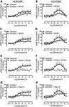
The role of G-protein coupling in serelaxin-mediated cAMP accumulation in human primary umbilical vascular cells. Pretreatment of HUASMC (n = 6) in (A) and of HUVSMC (n = 6) in (B) with the selective Gαs inhibitor NF449 (10 μM, 30 min) caused a rightward shift and reduced the E-max of the cAMP CRC to serelaxin without modifying the shape of the curve. Pretreatment of HUASMC (n = 7) in (C) and of HUVSMC (n = 6) in (D) with the selective Gαi/o inhibitor NF023 (10 μM, 30 min) reduced the E-max of the cAMP CRC to serelaxin and changed the shape of the curve observed with HUVSMC (D) from bell-shaped to sigmoidal. In both (E) HUASMC (n = 6) and (F) HUVSMC (n = 6), pretreatment with both NF449 and NF023 completely abolished serelaxin-mediated cAMP responses, showing that the responses result entirely from RXFP1 receptors interaction with G proteins. In (G) HUASMC (n = 6) and in (H) HUVSMC (n = 6), pretreatment with filipin III (1 μg·mL−1, 1 h), which disrupts lipid rafts, mimicked the effect of the Gαi/o inhibitor NF023 – reducing E-max and converting CRCs from bell-shaped to sigmoidal in HUVSMC.
Figure 5
Figure 5
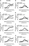
The role of G-protein coupling in serelaxin-mediated cGMP accumulation in human primary umbilical vascular cells. Pretreatment of HUASMC (n = 6) in (A) and of HUVSMC (n = 6) in (B) with the selective Gαs inhibitor NF449 (10 μM, 30 min) caused a rightward shift and reduced the E-max of the cGMP CRC to serelaxin without modifying the shape of the curve. Pretreatment of HUASMC (n = 7) in (C) and of HUVSMC (n = 6) in (D) with the selective Gαi/o inhibitor NF023 (10 μM, 30 min) reduced the E-max of the cGMP CRC to serelaxin and changed the shape of the curve observed with HUVSMC (D) from bell-shaped to sigmoidal. In both (E) HUASMC (n = 6) and (F) HUVSMC (n = 6), pretreatment with both NF449 and NF023 completely abolished serelaxin-mediated cGMP responses, showing that the responses result entirely from RXFP1 receptors interaction with G proteins. In (G) HUASMC (n = 6) and in (H) HUVSMC (n = 6), pretreatment with filipin III (1 μg·mL−1, 1 h), which disrupts lipid rafts, mimicked the effect of the Gαi/o inhibitor NF023 – reducing E-max and converting CRCs from bell-shaped to sigmoidal in HUVSMC.
Figure 6
Figure 6
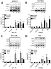
Changes in the expression of nNOS, VEGF and ETB receptors in human primary umbilical vascular cells and cardiac fibroblasts after serelaxin (1.68 nM) exposure for 24 and 48 h. In HUVECs (A), serelaxin treatment increased the expression of nNOS (n = 5), ETB receptors (n = 6) and VEGF (n = 7). In HUVSMCs (B), serelaxin treatment increased the expression of nNOS (n = 5) and ETB (n = 5) but not VEGF (n = 4); however, in HUASMC (C), serelaxin treatment increased the expression of nNOS (n = 6), ETB receptors (n = 7) and VEGF (n = 5). In HCFs (D), similar to HUVECs and HUASMCs, serelaxin treatment increased the expression of nNOS (n = 5), ETB receptors (n = 5) and VEGF (n = 5). A representative blot of each protein and β-actin, a loading control, is shown with the densitometry in each figure. Statistical significance was assessed using a one-way anova with a Dunnett's post hoc test compared with vehicle alone: *P < 0.05 and **P < 0.01.
Figure 7
Figure 7
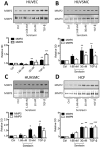
Changes in the activity of MMPs in human primary umbilical vascular cells and cardiac fibroblasts using zymography to assess changes in activity of MMP2 and MMP9 after long-term serelaxin exposure (1.68 and 30 nM) for 48 h. Serelaxin treatment increased the activity of MMP2 in (A) HUVECs (n = 5), (B) HUVSMCs (n = 6), (C) HUASMCs (n = 7) and (D) HCFs (n = 5). Serelaxin also significantly increased the activity of MMP9 but only in (B) HUVSMCs (n = 5) and (D) HCFs (n = 5) and not in (A) HUVECs (n = 5) and (C) HUASMCs (n = 5). Furthermore, and consistent with the lack of RXFP1 receptor expression (Figure 1), serelaxin had no effect on MMP activity in HUAECs (n = 2). A representative scan of each zymography is shown along with the densitometry in each figure. Statistical significance was assessed using a one-way anova with a Dunnett's post hoc test compared with control alone: *P < 0.05 and **P < 0.01.
Figure 8
Figure 8
Signal transduction mechanisms employed by serelaxin in human umbilical vascular cells and HCFs and their potential physiological effects. Short-term (<1 h) serelaxin stimulation produces cAMP/cGMP accumulation in vascular cells and pERK1/2 in all cells that is differentially regulated by Gαs, Gαi, GαOB and PI3K, and these pathways are likely to be involved in the vasodilator and anti-apoptotic effects of serelaxin respectively. In HUASMCs, the serelaxin-mediated cAMP or VEGF response did not involve GαOB and PI3K, whereas in HCFs, the RXFP1 receptor was not coupled to cAMP production. Longer term (24–48 h) serelaxin treatment increased VEGF expression involving both cAMP-dependent and cAMP-independent mechanisms, and these pathways are likely to be involved in angiogenesis. In addition, serelaxin treatment increased the activity of MMP2 and MMP9 to mediate its remodelling actions, but these enzymes are also secreted to convert big ET to ET1-32 that activates ETB receptors to further enhance vasodilatation (Conrad, 2010). Solid lines indicate mechanisms identified in the current study whereas dashed lines indicate previously established mechanisms.

Similar articles

Cited by

References

    1. Alexander SPH, Benson HE, Faccenda E, Pawson AJ, Sharman JL, Spedding M, et al. The Concise Guide to PHARMACOLOGY 2013/14: G protein-coupled receptors. Br J Pharmacol. 2013a;170:1459–1581. - PMC - PubMed
    1. Alexander SPH, Benson HE, Faccenda E, Pawson AJ, Sharman JL, Spedding M, et al. The Concise Guide to PHARMACOLOGY 2013/14: Ligand-gated ion channels. Br J Pharmacol. 2013b;170:1582–1606. - PMC - PubMed
    1. Alexander SPH, Benson HE, Faccenda E, Pawson AJ, Sharman JL, Spedding M, et al. The Concise Guide to PHARMACOLOGY 2013/14: Enzymes. Br J Pharmacol. 2013c;170:1797–1867. - PMC - PubMed
    1. Alexiou K, Wilbring M, Matschke K, Dschietzig T. Relaxin protects rat lungs from ischemia-reperfusion injury via inducible NO synthase: role of ERK-1/2, PI3K, and forkhead transcription factor FKHRL1. PLoS ONE. 2013;8:e75592. - PMC - PubMed
    1. Baker JG, Hill SJ. A comparison of the antagonist affinities for the Gi- and Gs-coupled states of the human adenosine A1-receptor. J Pharmacol Exp Ther. 2006;320:218–228. - PubMed

Publication types

MeSH terms

LinkOut - more resources