. 2014 Oct 23;514(7523):455-61.
doi: 10.1038/nature13808. Epub 2014 Oct 8.

Structure and immune recognition of trimeric pre-fusion HIV-1 Env

Affiliations

Structure and immune recognition of trimeric pre-fusion HIV-1 Env

Marie Pancera et al. Nature. .

Abstract

The human immunodeficiency virus type 1 (HIV-1) envelope (Env) spike, comprising three gp120 and three gp41 subunits, is a conformational machine that facilitates HIV-1 entry by rearranging from a mature unliganded state, through receptor-bound intermediates, to a post-fusion state. As the sole viral antigen on the HIV-1 virion surface, Env is both the target of neutralizing antibodies and a focus of vaccine efforts. Here we report the structure at 3.5 Å resolution for an HIV-1 Env trimer captured in a mature closed state by antibodies PGT122 and 35O22. This structure reveals the pre-fusion conformation of gp41, indicates rearrangements needed for fusion activation, and defines parameters of immune evasion and immune recognition. Pre-fusion gp41 encircles amino- and carboxy-terminal strands of gp120 with four helices that form a membrane-proximal collar, fastened by insertion of a fusion peptide-proximal methionine into a gp41-tryptophan clasp. Spike rearrangements required for entry involve opening the clasp and expelling the termini. N-linked glycosylation and sequence-variable regions cover the pre-fusion closed spike; we used chronic cohorts to map the prevalence and location of effective HIV-1-neutralizing responses, which were distinguished by their recognition of N-linked glycan and tolerance for epitope-sequence variation.


Figures
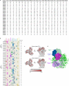
Extended Data Figure 1
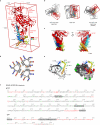
Extended Data Figure 1. Antibody-mediated crystallization and antibody-induced conformation
a, Atomic-level structures for HIV-1-Env regions determined in complex with HIV-1-neutralizing antibodies. Neutralizing antibodies generally recognize the prefusion conformation of HIV-1 Env. Structures highlighted here display a cumulative sum total of prefusion HIV-1-Env structural information. Env residues are numbered according to standard HX numbering (from PDBs). One structure, for antibody D5 (blue), is in the postfusion gp41 conformation, and is not included in the sum total. Regions of other structures (purple), did not define sequence register, and were also not included in the sum total. Reference listed here are cited elsewhere in the manuscript, except for Rini et al., 1993, Stanfield et al., 1999,, Ofek et al., 2004, Cardoso et al., 2005, Luftig et al., 2006, Cardoso et al., 2007. b, Antibody-induced conformation of HIV-1 Env in the context of infectious JR-FL virions as assessed by smFRET. HIV-1JR-FL gp160 was labelled with fluorescent dyes in variable regions, V1 and V4, at positions that did not interfere with Env function (see methods), and virus was surface immobilized for imaging via total internal reflection fluorescence microscopy. smFRET trajectories were compiled into histograms for the HIV-1JR-FL Env trimer, either unliganded or after pre-incubation for 30 min with 0.1 mg/ml PGT122, 35O22, or both PGT122 and 35O22 prior to imaging. Resultant Env conformational landscapes could be deconvoluted into three gaussian distributions: a low-FRET population that predominated for the prefusion mature unliganded state, and intermediate- and high-FRET populations, which predominated in the presence of CD4 receptor and CD4-induced antibody. smFRET trajectories are shown for the unliganded HIV-1JR-FL Env trimer as well as in the presence of PGT122, 35O22, and both PGT122 and 35O22. The concordance between conformational ensembles indicates unliganded and PGT122+35O22-bound conformation to be similar (Spearman correlation coefficient of 0.988). Interestingly, the presence of just one of the antibodies (PGT122) appeared to reduce the high FRET population, an effect not observed in the presence of both antibodies; this suggests that the antibody-induced stability of a particular state is not solely additive, and that antibodies can both induce a particular conformational state as well as alter the transition dynamics from that state.
Extended Data Figure 2
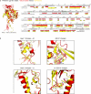
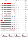
Extended Data Figure 2. HIV-1 subunit interactions: principle component analysis and interface contacts
a, Minimum-bounding box, generated by principle component analysis, encasing 90% of the HIV-1-Env gp120-gp41 protomer. Each gp120-gp41 blade forms a rectangle of height of ~100 Å, width of ~65 Å, and thickness of ~35 Å. Subunits are displayed in ribbon representation with gp41 colored rainbow and gp120 colored and labeled red. As previously visualized,, the membrane-distal portion of the rectangle is made up of the gp120-outer and -inner domains, with the central 7-stranded β-sandwich of the inner domain occupying the trimer-distal, membrane-proximal portion of gp120. We have now resolved the rest of the spike: the membrane-proximal portion of the rectangle is made up of gp41, with the membrane-distal portion of gp41 closest to the molecular 3-fold axis occupied by helix α7 (which corresponds in register to the C-terminal portion of the postfusion HR1 helix of gp41), and the rest of gp41 folding around N- and C-termini-strands of gp120, which extend over 20 Å toward the viral membrane. b, Different views of trimeric protomer association. The protomer association at the membrane-distal trimer apex occurs through the corners of the minimum-bounding box, whereas the association at the membrane-proximal region occurs with substantial interpenetration of the minimum-bounding box; these interaction differences and the protruding nature of the gp120 outer domain result in the overall mushroom shape of the trimer. c, gp120-gp41 interface. Ribbon representation of gp120 (red) and gp41 (rainbow from blue N terminus to orange C terminus), with gp120 residues that interact with gp41 shown in surface representation and gp41 residues that interact with gp120 shown in semitransparent surface. A complete list of subunits interactions is provided in Supplementary Table 1. Membrane-proximal interactions are further stabilized by hydrophobic interactions, which gp41 makes with the N and C termini of gp120 –such as between Trp35gp120 and Pro609gp41 and between Trp610gp41 and Pro498gp120. d, Wheel diagram representation of α7 coiled-coil in the prefusion mature closed conformation of gp41 as generated by DrawCoil 1.0: http://www.grigoryanlab.org/drawcoil/. e, gp41-trimer interfaces as viewed from the viral membrane in ribbon and surface representation (90° rotation from Fig. 2c). f, BG505 SOSIP.664 sequence with residues identified by mutagenesis- to be important for gp120/gp41 association underlined. Residues that were found to interact between gp120 and gp41 by examination of the crystal structure are indicated in red (intra-protomer interactions) and in brown (inter-protomer interactions). Sites of N-linked glycosylation are shown in green; glycan N88 is shown in red because it is part of the gp120/gp41 interactions; no density was observed for potential N-linked glycans at residues 185, 398, 406, 411, 462 and 625. Residues that were disordered in the crystal structure are gray. SOS (A501C/T605C) and IP (I559P) mutations are labeled in bold and italics. Dots indicate residues not present in the BG505 sequence.
Extended Data Figure 3
Extended Data Figure 3. Modeling of gp41: prefusion α6-to-α7 density, HIV-1/SIV postfusion chimera, and liganded interactions
a, Modeling of gp41 residues 548-568. At low contour, suggestive density is observed that might correspond to the connection between α6 and α7 helices. To investigate the degree to which a model for this region might be defined, we built and refined two different models for this region: electron density (blue) shown for 2F0-Fc density at 1σ contour; gp41 (rainbow color from blue to orange) shown in ribbon representation with side chains; gp120 (red) shown in ribbon representation. The location of the I559P mutation is indicated. b, The two models from panel a are superimposed and shown in perpendicular orientations. c, HIV-1-SIV postfusion chimera. Sequences of HIV-1 gp41 from prefusion structure (BG505 strain, PDB ID: 4TVP), postfusion structure (HIVpost, PDB ID: 2X7R) and SIV gp41 postfusion structure (SIVpost, PDB ID: 2EZO) are aligned with secondary structure indicated. Residues that were used to make the postfusion HIV-1/SIV chimera used in Figure 3 are highlighted in red. d, Binding residues of representative fusion-intermediate entry inhibitors or antibodies mapped onto the structure of prefusion HIV-1-Env spike-. (top) Ribbon representation of prefusion envelope protomer A (gp120 in red and gp41 in blue) at two orientations, with the binding residues of the fusion-intermediate inhibitors 5-helix,T20, and of monoclonal antibody D5 shown in orange, green, and yellow, respectively. (bottom) Surface representation of the prefusion envelope trimer, with inhibitor and antibody binding residues mapped onto the surfaces of all protomers. gp120 is colored gray and gp41 is colored in shades of blue, depending on protomer. Binding residues of fusion-intermediate inhibitors 5-helix, T20, and monoclonal antibody D5 are shown in same color shades as in the top panels. e. 5-helix, T20 and D5 Fab (all colored magenta and gray) docked onto a model of fusion-intermediate gp41 (colored as in d). f, A previously defined binding pocket on postfusion gp41 is the target of prefusion gp41 tryptophan-clasp residues Trp628 and Trp631. Shown is a surface representation of gp41 5-helix protein (left, with N-heptad repeat (NHR) helices colored in shades of green and C-heptad repeat (CHR) helices colored in shades of orange). The footprint of gp41 tryptophan-clasp residues Trp628 and Trp631 is shown in magenta (middle) and that of a representative NHR-specific neutralizing antibody, D5, in yellow ,, (right).
Extended Data Figure 4
Extended Data Figure 4. Conformational changes between prefusion mature closed state and CD4-bound state of gp120
a, Overall structure and sequence comparison. gp120 is shown in ribbon representation in prefusion mature closed (red) and CD4-bound (yellow, PDB ID: 3JWD) conformation. V1V2 (PDB ID: 3U2S) has been modeled onto the CD4-bound conformation. Secondary structure is defined for prefusion and CD4-bound conformation on the BG505 sequence, with cylinders representing α-helix and arrows β-strands. Disordered residues are indicated by “X”. Residues that move more than 3 Å between the mature closed and the CD4-bound gp120 conformations are shown with grey shadows. Sites of N-linked glycosylation are shown in green. b. Details of conformational changes between the mature closed (red) and the CD4-bound conformations (yellow) of gp120 (shown in ribbon): regions highlighted cover layer 1 with changes at α0 (we note that density in this region is not well defined), layer 2 with changes at α1 and β20-21 rearrangements. All atoms rmsd are: residues 54-74gp120, rmsd = 4.759 Å; residues 98-117 gp120, rmsd = 0.497 Å; 424-436 gp120, rmsd = 3.196 Å.
Extended Data Figure 5
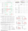
Extended Data Figure 5. Antigenic profiles of HIV-1 envelope conformational states
a, Qualitative recognition of HIV-1 envelope by diverse antibodies is shown for five conformational states. Green bars indicate reported recognition, red bars no recognition, and absence of a bar indicates that recognition is undefined. The compiled data is from both cited references and experiments described in this figure. b, Octet Biosensorgrams of BG505 SOSIP.664 (left) and BG505 gp120 (right) binding to human monoclonal IgGs. The dotted line indicates the beginning of the dissociation phase and the maximal specific binding after 300 s reported in the table (− <0.05 RU, + 0.05 RU to 0.25 RU, ++ 0.25 RU to 0.5 RU, and +++ >0.5 RU). BG505gp120 did not contain the T332N mutation (no glycan at that position). Both proteins were made in GnTi−/−. We note that antigenicity of the BG505 SOSIP.664 and BG505gp120 protein varied depending on the assay done. Thus, using surface plasmon resonance, no CD4i antibodies binding was detected while some binding could be observed using biolayer interferometry. Although PG9 bound BG505gp120 in ELISA, it did not bind in biolayer interferometry format. We observed 447-52D binding while it was not observed in ELISA. c, SPR binding affinities of 35O22, PGT151 and PGT145 to BG505 SOSIP.664 and influence of sCD4. d, Estimation of binding stoichiometry for 35O22, PGT151, and PGT145 to trimeric BG505 SOSIP.664 by SPR and comparison to published data,,. e, Effect of sCD4 and sCD4/17b on binding of antibodies 35O22 and PGT151 to BG505 SOSIP.664 by SPR. The structure of a prefusion mature closed state of HIV-1 provides a critical addition to the pantheon of HIV-1 Env structures with atomic-level detail. Moreover, antibodies 35O22 and PGT151, which bind specifically to the trimeric prefusion conformation of gp41, provide new tools by which to assess the conformational state of gp41,,. The binding of antibodies 35O22 and PGT151 to BG505 SOSIP.664 trimer was tested in the presence of the CD4 receptor and the 17b antibody (a co-receptor surrogate which recognizes a bridging sheet epitope that overlaps the site of co-receptor recognition). In the case of antibody 35O22, CD4 binding to the BG505 SOSIP.664 trimer impacted the kinetics, affinity and stoichiometry of binding. 35O22 bound to BG505 SOSIP.664 with an 8.4-fold reduced affinity, primarily contributed by an increased rate of dissociation. The overall binding level (Rmax) normalized to the average level of trimer captured (see also panel d) was lower suggesting substoichiometric binding. Capturing the trimer on a CD4-Ig surface reduced normalized Rmax for PGT151 compared to the 2G12 capture format, suggesting reduced stoichiometry for PGT151 binding to trimer pre-bound with CD4, although kinetics and affinity of interaction were similar. A BG505 SOSIP.664 trimer + sCD4 complex captured onto a 17b surface bound 35O22 but showed no detectable binding to PGT151.
Extended Data Figure 6
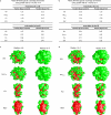
Extended Data Figure 6. N-Linked glycan occlusion of type I fusion machines
The prefusion mature closed conformation of HIV-1 Env evades the humoral immune response with a fully assembled glycan shield. Here we calculate and display the solvent-accessible surface of glycan and protein for HIV-1 Env, influenza virus hemagglutinin and RSV fusion glycoprotein. Calculations of the percentage coverage of the protein surface were determined for trimeric type I fusion machines based on two probe sizes of 1.4 Å (solvent radius) and 10.0 Å (the estimated steric footprint of an antibody combining region). Surface area calculations were carried out according to Kong et. al, and images were generated using Grasp v1.3. All models were refined using Amber with the GLYCAM force field (see Methods for details). The PDB IDs associated with the glycosylated models are: 4TVP (HIV-1), 2YP7 (Flu) and 4JHW (RSV). The strains associated with the PDB IDs are: BG505.SOSIP.664 (HIV-1), H3N2 A/Hong Kong/4443/2005 (Flu) and A/A2/61 (RSV). The solvent-accessible protein surface is shown in red, and N-linked glycans are shown in green. a, Estimated Man9 glycan coverage. b, Estimated Man5 glycan coverage. c, Visualization of Man9 N-linked glycan coverage for two probe radii. d, Visualization of Man5 N-linked glycan coverage for two probe radii.
Extended Data Figure 7
Extended Data Figure 7. Glycan shield and sequence variability for HIV-1 prefusion mature closed and CD4-bound conformations
Many conformations of HIV-1 Env divert the immune response. Thus for example, shed gp120 and post-fusion gp41 represent dominant viral antigens; however these forms of Env are not functional, and antibodies that only target them are not neutralizing. Functional conformations, however, may be significantly shielded from the neutralizing antibody. The CD4-bound conformation of HIV-1 Env, for example, is only functionally present when the viral and target-cell membranes are in close proximity, and the exposed co-receptor binding site (including V3- and CD4-induced epitopes) is spatially occluded from neutralizing antibody. Here we provide models for the prefusion closed state versus the CD4-bound conformation, which display the fully assembled glycan shield and surface Env variability. Env N-linked glycans are depicted in light green (conserved; greater than 90% conservation) or dark green (variable; less than 90% conservation) on the mature closed Env structure and modeled CD4-bound conformation. Env sequence variability is shown from white to purple (conserved to variable). A conserved glycan at residue 241gp120 not present in the BG505 sequence is shown in yellow-green. As can be seen, the prefusion closed state has few glycan-free surfaces, whereas the CD4-bound state exposes substantial glycan-free conserved surface.
Extended Data Figure 8
Extended Data Figure 8. Prevalence of neutralizing responses identified serologically from cohorts from 2-3 years and 5+ years post infection
a, Serum neutralization on 21-strain virus panel. ID50s are shown for serum (rows) titrated against HIV-1 viral strains (columns). b, For each serum, the predicted neutralization prevalence for each of 12 antibody specificities is shown based on neutralization of 21 diverse HIV-1 strains. c, Prevalence of antibody specificities onto the HIV-1-Env colored as indicated in the bar graph. d, The antibody specificities for high serum prevalence in the 5+ years cohort are depicted by Fabs of representative antibodies (surface transparency proportional to prevalence) binding the BG505 SOSIP.664 Env trimer, shown in grey ribbon representation, with glycans as green sticks. Note that while prevalence is highly correlated, there were notable differences, for example between PGT151 at 2-3 years and 5+ years in this study, as well as between the cohorts analyzed here and in ref. .
Extended Data Figure 9
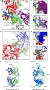
Extended Data Figure 9. Antibodies 35O22 and PGT122: interface with HIV-1 Env and comparison of bound and unbound Fab conformations
Despite the substantial immune evasion protecting the mature unliganded state from humoral recognition, after several years of infection, the human immune system does generate broadly neutralizing antibodies. 35O22 and PGT122 are two of these antibodies, which neutralize 62% and 65% of HIV-1 isolates at a median IC50 of 0.033 and 0.05 μg/ml, respectively, . Here we provide additional details on 35O22 and PGT122 recognition. a, 35O22 Fab is shown in ribbon representation (purple (heavy chain) and white (light chain)). The gp120 subunit is shown in red, the gp41 subunit in rainbow (from blue N terminus to orange C terminus), and glycans in green sticks. Complementary determining regions (CDRs) are labeled, and interactive HIV-1-Env residues highlighted in semi-transparent surface representation. At the membrane-distal surface of 35O22, an extended framework 3 region (FW3) of the heavy chain (resulting from an insertion of 8 residues) interacts with strand β1 of the 7-stranded inner domain sandwich of gp120. The heavy chain-CDRs form extensive contacts with the N-linked glycan extending from residue 88gp120. In addition to glycan contacts, the CDR H3 of 35O22 interacts with the α9 helix of gp41. Helix α9 interactions are also made by the FW3 of the light chain (a complete list of contacts is provided in Supplementary Table 3). Overall, 35O22 buries 1,105 Å2 solvent surface on gp120 (including 793 Å2 with the Asn88gp120 glycan) and 594 Å2 solvent surface on gp41 (including 127 Å2 with the Asn618gp41 glycan). Despite residue 625gp41 being part of the glycan sequon “NMT”, no glycan is observed; indeed, the side-chain amide of residue 625gp41 hydrogen bonds with the side-chain oxygen of Tyr32 in the 35O22 heavy chain, and the presence of an N-linked glycan at residue 625gp41 is difficult to reconcile with 35O22 recognition. b, Same colors as a, with 35O22 Fab shown in surface representation. c, Same colors as a, with 2Fo-Fc at 1σ contour (blue density) shown around glycan 88 of gp120. Antibody 35O22 employs a novel mechanism of glycan-protein recognition, combining a protruding FW3 with CDR H1, H2 and H3 to form a “bowl” that holds glycan. FW3 and CDR H3 provide the top edges of the bowl and interact with the protein surface of gp120, whereas CDR H1 and H2 are recessed and hold/recognize glycan. This structural mechanism of recognition contrasts with the extended CDR H3-draping glycan observed with other antibodies that penetrate the glycan shield such as PG9 and PGT128. d, PGT122 interface details. Ribbon representation of PGT122 Fab in blue (heavy chain) and light blue (light chain) interacting with one gp120 subunit shown in red with glycans in green sticks. Complementary determining regions (CDRs) are labeled, and interactive HIV-1-Env residues highlighted in surface representation. Primary contacts between antibody PGT122 and N-linked glycan involve N137 and N332, with minor contact with N156. Although portions of glycan N301 can be observed in the electron density, no direct contacts with PGT122 are observed; a complete list of contacts between PGT122 and BG505 SOSIP.664 is provided in Supplementary Table 4. e, Same colors as d, with PGT122 Fab shown in surface representation, f, Same colors as d, with 2Fo-Fc at 1σ contour (grey density) shown around glycan 332 of gp120. g,Comparison of bound and unbound Fab conformations. Unbound and HIV-1-Env bound Fabs were superimposed, and ribbon representations and rmsds are displayed. (Left) Unbound 35O22 Fab is colored cyan (heavy chain) and green (light chain) and bound 35O22 Fab deep purple (heavy chain) and white (light chain). (Right) Unbound PGT122 Fab is colored cyan, and bound PGT122 Fab blue (heavy chain) and light blue (light chain). Regions which showed conformational changes are highlighted with black dotted lines. We note that in the 35O22 bound conformation, density is poor and/or sparse for the Fc portion of the Fab.
Extended Data Figure 10
Extended Data Figure 10. Structural implementation of HIV-1 molecular trickery
The prefusion HIV-1-Env trimer (left) is displayed with evasion mechanisms and their structural implementation (right). The gp120 subunit is shown in red, the gp41 subunit in rainbow (from blue N terminus to orange C terminus), and crystallographically defined glycans in green. One protomer is shown with Cα trace and glycans in stick representation, a second protomer is shown in ribbon representation with secondary structure elements labeled, and the third protomer is shown in light grey surface. The MPER region for each protomer is shown as a stylized helix associated with the viral membrane. The location of secondary structural elements, termini, and residues called in the text has been labeled (red font for gp120 and black font for gp41).
Figure 1
Figure 1. Structure of a prefusion HIV-1-Env trimer bound by PGT122 and 35O22 antibodies
One protomer and associated Fabs is shown in ribbon and stick representation, a second protomer in surface representation, and the third protomer in gray. Residues comprising the refined HIV-1-Env model are displayed on the bar, with beginning and final ordered residue of each segment labeled; vertical lines demark termini of the mature ectodomain subunits; unmodeled regions, residues not present in the BG505 SOSIP.664 construct, and disordered glycans are shown in gray. 35O22 and PGT122 interactions with the HIV-1-Env trimer are shown in Extended Data Fig. 9a-f, and bound versus unbound Fabs are shown in Extended Data Fig. 9g.
Figure 2
Figure 2. Prefusion structure of gp41
a, gp41 forms a 4-helix collar, which wraps around extended N and C termini of gp120. Both gp120 (red) and gp41 (rainbow from blue to orange) are depicted in ribbon representation, with select residues and secondary structure labeled (additional labels are shown in Extended Data Fig.10). The location of the trimer axis is indicated with triangle-surround “3”. The orientation shown here is similar to that of Fig. 1, with perpendicular orientations provided in b and c. (zoom insert) The gp41 collar is clasped by the insertion of Met530gp41 into a tryptophan sandwich and by the complementary dipoles of helices α6 and α8. 2Fo-Fc electron density for clasp residues is depicted at 1σ. b, gp41 holds the N and C termini of gp120 in its hydrophobic core. Coloring and representation are the same as in a, excepted that hydrophobic side chains are shown in stick representation and the orientation is rotated 90°, to depict the view from the viral membrane. c, gp41-trimer interfaces as viewed from side in ribbon and surface representation. Overall, the prefusion structure of gp41 and its trimeric arrangement appear to have no close structural relatives in the PDB (Supplementary Table 2).
Figure 3
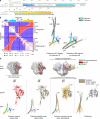
Figure 3. Entry rearrangements of HIV-1 Env
a, BG505 sequence of gp41, with prefusion and postfusion secondary structure. Fusion peptide (FP) is underlined and labeled green. Several postfusion gp41 structures have been determined ranging from a minimal, protease-treated, crystal structure (residues 556gp41-581gp41; 628gp41-661gp41; PDB ID: 1AIK) with 80% sequence identity to BG505 to a more complete gp41 structure (residues 531gp41-581gp41; 624-681gp41; PDB ID: 2X7R) and an NMR structure that includes the cysteine loop (residues 539gp41-665gp41; PDB ID: 2EZO) of the simian immunodeficiency virus (SIV), which shares 48% sequence identity with BG505 and is substantially similar to the HIV-1 structures (less than 1-Å Cα rmsd between overlapping residues of 1AIK and 2EZO). The postfusion structure utilized here for comparisons was constructed from a chimera of HIV-1/SIV structures (Extended Data Fig. 3c). b, Difference distance analysis of prefusion BG505 and postfusion HIV-1/SIV chimeric gp41. Secondary structure is indicated, along with missing residues of BG505 (548-568) and of SIV (611-614). c, Superposition of postfusion gp41 (grey) onto prefusion gp41 (rainbow) for α7 (left) and α9 (right) prefusion helices. d, HIV-1-Env entry rearrangements. EM reconstructions (top row) with gp120 (middle) and gp41 (bottom) rearrangements between each conformational state highlighted with orange lines depicting movement of each Cα between conformations. Subunit models are shown in gray with modeling parameters and references provided in Extended Data Table 2. Antigenic recognition of each of these states is shown in Extended Data Fig. 5.
Figure 4
Figure 4. Prefusion HIV-1 gp120-gp41 structure shares conserved structural and topological features with other type I fusion machines
a, Prefusion (left) and postfusion (right) structures. The prefusion structures are shown for a single protomer in ribbon-representation with gp120-equivalent subunits in red, and gp41-equivalent subunits in rainbow (blue to orange). The trimeric postfusion structures are shown with one subunit in rainbow (blue to orange), and the other in light and dark gray. b, The C-terminal portion of the preformed interior helix of postfusion coiled-coil from a is shown, with fusion peptides (FP) and N and C terminal residues of postfusion coiled-coils labeled, and the distance the inner coiled-coil extends between prefusion and postfusion conformations indicated. c, The gp41-equivalents encircle extended β-strands of their gp120-equivalent partners. Ribbon representations are shown looking towards the viral membrane. With influenza, it is only the N terminus of the gp120-equivalent (HA1) that is wrapped by the gp41-equivalent (HA2), with the N terminus of HA2 completing about 20% more than a single encirclement. With RSV, it is also only the N terminus of the gp120 equivalent (F2) that is wrapped by the gp41-equivalent (F1), and the termini do not have to be expelled to transition to the postfusion form. With Ebola, the gp41-equivalent (gp2) wraps both N and C termini-strands of the gp120-equivalent (gp1), completing about 70% of a single encirclement. Such encirclement likely helps capture the energy of prefusion folding, which is released during the postfusion transition to power membrane fusion.
Figure 5
Figure 5. Fully assembled shield revealed by prefusion HIV-1 gp120-gp41 trimer
a, Glycan shield. Env N-linked glycans are depicted in light green (conserved; greater than 90% conservation) or dark green (variable; less than 90% conservation) on the prefusion mature closed Env structures for BG505 strain of HIV-1 (left), influenza virus H3 hemagglutinin (PDB ID: 2YP7) (middle), and RSV fusion glycoprotein subtype A (PDB ID: 4JHW) (right). A conserved glycan at residue 241gp120 not present in the BG505 sequence is shown in yellow-green. b, Sequence variability.
Figure 6
Figure 6. Location and prevalence on the HIV-1-Env spike of neutralizing responses identified serologically from cohorts, 2-3 and 5+ years post-infection
a, The location of the neutralization epitopes for broadly neutralizing antibodies is depicted on the prefusion mature closed Env spike with red for CD4-binding-site-directed antibody specificities (VRC01-, b12-, CD4-, and HJ16-like), purple for 8ANC195-like, green for V1V2-directed (PG9-like), blue for glycan-V3 specificities (PGT128- and 2G12-like), orange for 35O22-like specificities, and green-yellow for PGT151-like specificities. b, (top) Broadly neutralizing epitopes on influenza virus hemagglutinin (left, PDB ID: 2YP7) and RSV fusion glycoprotein (right, PDB ID: 4JHW). (bottom) Glycan-surface area and residue entropy of antibody epitopes for HIV-1, influenza, and RSV with bars colored according to epitopes shown in a and b (except for epitopes not present in SOSIP.664 or where there is no atomic level definition). c, Neutralization fingerprint. For each serum, the predicted neutralization prevalence for each of the 12 antibody specificities is shown based on neutralization of 21 diverse HIV-1 strains (Extended Data Fig. 8).

Comment in

  • HIV: A stamp on the envelope.
    Sanders RW, Moore JP. Sanders RW, et al. Nature. 2014 Oct 23;514(7523):437-8. doi: 10.1038/nature13926. Epub 2014 Oct 8. Nature. 2014. PMID: 25296251 Free PMC article.

Similar articles

Cited by

References

    1. The Joint United Nations Programme on HIV/AIDS 2013 Report on the global AIDS epidemic. 2013
    1. Wyatt R, Sodroski J. The HIV-1 envelope glycoproteins: fusogens, antigens, and immunogens. Science. 1998;280:1884–1888. - PubMed
    1. Wei X, et al. Antibody neutralization and escape by HIV-1. Nature. 2003;422:307–312. - PubMed
    1. Leonard CK, et al. Assignment of intrachain disulfide bonds and characterization of potential glycosylation sites of the type 1 recombinant human immunodeficiency virus envelope glycoprotein (gp120) expressed in Chinese hamster ovary cells. The Journal of biological chemistry. 1990;265:10373–10382. - PubMed
    1. Cimbro R, et al. Tyrosine sulfation in the second variable loop (V2) of HIV-1 gp120 stabilizes V2-V3 interaction and modulates neutralization sensitivity. Proceedings of the National Academy of Sciences of the United States of America. 2014;111:3152–3157. - PMC - PubMed

Publication types

MeSH terms

Associated data