Review
. 2014 Sep 15;5(17):7285-302.
doi: 10.18632/oncotarget.2439.

The structural basis for cancer treatment decisions

Affiliations
Review

The structural basis for cancer treatment decisions

Ruth Nussinov et al. Oncotarget. .

Abstract

Cancer treatment decisions rely on genetics, large data screens and clinical pharmacology. Here we point out that genetic analysis and treatment decisions may overlook critical elements in cancer development, progression and drug resistance. Two critical structural elements are missing in genetics-based decision-making: the mechanisms of oncogenic mutations and the cellular network which is rewired in cancer. These lay the foundation for the structural basis for cancer treatment decisions, which is rooted in the physical principles of the molecular conformational behavior of single molecules and their interactions. Improved tumor mutational analysis platforms and knowledge of the redundant pathways which can take over in cancer, may not only supplement known actionable findings, but forecast possible cancer progression and resistance. Such forward-looking can be powerful, endowing the oncologist with mechanistic insight and cancer prognosis, and consequently more informed treatment options. Examples include redundant pathways taking over after inhibition of EGFR constitutive activation, mutations in PIK3CA p110α and p85, and the non-hotspot AKT1 mutants conferring constitutive membrane localization.


Figures
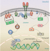
Figure 1
Figure 1. Ras signaling pathways
Ras signaling is involved in numerous cellular functions, including cell proliferation, apoptosis, migration, fate specification, and differentiation. A key Ras effector pathway is the mitogen-activated protein kinase (MAPK), Raf-MEK-ERK pathway. EGF binds to the extracellular domain of the epidermal growth factor receptor (EGFR), a receptor tyrosine kinase (RTK). The signal is transmitted through the transmembrane domain resulting in EGFR dimerization and activation. Activated EGFR recruits the son of sevenless (SOS), a guanine nucleotide exchange factor (GEF), to its phosphorylated C-terminal tail via the adaptor proteins, SH2-adaptor protein (SHC) and growth factor receptor-bound protein 2 (Grb2). GEF exchanges GDP by GTP, activating Ras. Active, GTP-loaded Ras dimerizes and binds Raf, thereby promoting Raf dimerization and activation. Active Raf dimer phosphorylates and activates mitogen-activated protein kinase kinase 1 and 2 (MEK1/2), which induces ERK1/2 activation. Transcription factor Elk-1 is among ERK1/2 many downstream phosphorylation targets. Elk-1 binds to its cofactor, a dimer of serum response factor (SRF), leading to transcription activation and cell proliferation. Active GTP-bound Ras regulates a number of signaling pathways; among these is phosphatidylinositol 3-kinase (PI3K). PI3K is a heterodimer with a regulatory (p85) and catalytic (p110) subunits (not shown here). RTKs recruit the p85 subunit of PI3K. Ras activates p110 independently of p85 [172]. PI3K phosphorylates phosphatidylinositol-4,5-bisphosphate (PIP2) to phosphatidylinositol-3,4,5-trisphosphate (PIP3), a process which can be reversed by the action of phosphatase and tensin homologue (PTEN). PIP3 recruits Phosphoinositide-dependent kinase-1 (PDK1) that phosphorylates a serine/threonine kinase, Akt (also known as PKB, protein kinase B) in the plasma membrane. This further induces the activation of mammalian target of rapamycin (mTOR) complex, one of the major pathways leading to cell growth. This pathway plays important roles in Ras-mediated cell survival and proliferation.
Figure 2
Figure 2. The structural basis of an oncogenic mutation
The figure illustrates how an oncogenic mutation can work on the structural level. Ras the most common mutated oncogene in cancer is shown as an example. Wild type Ras acts as a binary signal switch cycling between active and inactive states. Ras only binds its effectors in its GTP-bound active state. Ras is activated by the son of sevenless (SOS) nucleotide exchange factor (GEF). In contrast, the GTP→GDP hydrolysis, helped by GTPase-activating protein (GAP) inactivates Ras. A key oncogenic mechanism aborts the hydrolysis reaction, keeping Ras in a constitutively active GTP-bound state. Residues most prone to these mutations are G12, G13 and Q61. Mutation of G12 K-Ras is most prevalent and oncogenic in colon cancer [173]. G12C and G12V K-Ras mutants activate the Ral guanine nucleotide dissociation stimulator (RaLGDS) pathway, whereas G12D preferentially activates the PI3K and MAPK signaling pathways [105, 106]. The reason for this differential preferred activation is still unclear. Figure (A) sketches Ras regulation under normal conditions (on the left hand-side) and constitutive activation (right hand-side). The constitutively active conformation of Ras harboring these mutations does not permit formation of the transition state required for catalysis upon binding to GAP. Under normal conditions, the flexibility of G12 allows the Arg789 side-chain (Arg finger) on the finger loop of GAP to insert into Ras active site. However, G12 mutants with bulkier or charged residue prevent the Arg finger insertion, blocking the transition state with GAP [174]. It is also likely that these mutations allosterically differentially affect the effector binding sites (not shown). (B) This panel illustrates native Ras in complex with GAP, poised for the catalytic reaction. The crystal structure of GDP-H-Ras/RasGAP complex (PDB code 1WQ1) is remodeled with the GTP-K-Ras crystal structure (PDB code 3GFT). The finger loop of RasGAP is in blue (taken from the complex, PDB code 1WQ1). The Arg finger is highlighted as a blue stick, positioned at the Ras active site, near the G12 residue in green mesh. (C) This panel clarifies why mutation of G12 prevents hydrolysis through a steric clash mechanism in which the G12C residue in green mesh prevents the insertion of Arg finger. Crystal structure of G12C GTP-H-Ras mutant (PDB code 4L9W) is remodeled to G12C GTP-K-Ras mutant.
Figure 3
Figure 3. A structural view of redundant pathways taking over during drug resistance
Ras is normally activated in response to the binding of extracellular ligands to various receptors. Among these is epidermal growth factor (EGF) binding to its cognate receptor EGFR, as shown in Figure 1. Upon EGF binding to the extracellular domain of EGFR, the intracellular domain of EGFR forms an asymmetric dimer in the cytosol. EGFR and its ERBB receptor family members can form homo- or hetero-dimers. Downstream signaling proceeds through Ras in the Raf-MEK-ERK and/or PI3K-Akt-mTOR pathways. The figure provides a sequence of events induced by a constitutive mutation taking place in EGFR, keeping it in an active state even in the absence of its ligand. Drug treatment abolishes EGFR signaling; however, a drug resistant mutation leads to overexpression of another receptor, populating an otherwise low-activity second receptor. (A) L858R mutation in EGFR kinase (the circled R) causes non-small cell lung cancer (NSCLC) by constitutively activating its kinase domain [175]. Under normal conditions EGFR largely populates its inactive state. The mutation shifts the free energy landscape of EGFR stabilizing its active with respect to its inactive conformation even in the absence of a bound EGF. (B) Drugs such as the 4-anilinoquinazolines gefitinib (Iressa) [176] and erlotinib (Tarceva) [177] can inhibit the activity of EGFR L858R mutant. (C) However, tumors develop resistance, in this case one possibility is through overexpression of MET [178]. Overexpressed MET leads to phosphorylation of ERBB3 which interacts with ERBB2. The ERBB2/ERBB3 receptor can activate Ras and thus its PI3K-Akt signaling pathway, independent of EGFR. A key question is how the blockage of an addicted growth pathway is able to rewire the oncogenic cellular network within a short period, leading to MET's overexpression and ERBB3 activation.

Similar articles

Cited by

References

    1. Yakhini Z, Jurisica I. Cancer computational biology. BMC Bioinformatics. 2011;12:120. - PMC - PubMed
    1. Hogeweg P. The roots of bioinformatics in theoretical biology. PLoS Comput Biol. 2011;7:e1002021. - PMC - PubMed
    1. Winter C, Kristiansen G, Kersting S, Roy J, Aust D, Knosel T, Rummele P, Jahnke B, Hentrich V, Ruckert F, Niedergethmann M, Weichert W, Bahra M, et al. Google goes cancer: Improving outcome prediction for cancer patients by network-based ranking of marker genes. PLoS Comput Biol. 2012;8:e1002511. - PMC - PubMed
    1. Mathew JP, Taylor BS, Bader GD, Pyarajan S, Antoniotti M, Chinnaiyan AM, Sander C, Burakoff SJ, Mishra B. From bytes to bedside: Data integration and computational biology for translational cancer research. PLoS Comput Biol. 2007;3:e12. - PMC - PubMed
    1. Bilal E, Dutkowski J, Guinney J, Jang IS, Logsdon BA, Pandey G, Sauerwine BA, Shimoni Y, Moen Vollan HK, Mecham BH, Rueda OM, Tost J, Curtis C, et al. Improving breast cancer survival analysis through competition-based multidimensional modeling. PLoS Comput Biol. 2013;9:e1003047. - PMC - PubMed

Publication types