

p-21 activated kinase 4 promotes proliferation and survival of pancreatic cancer cells through AKT- and ERK-dependent activation of NF-κB pathway
- PMID: 25238288
- PMCID: PMC4226721
- DOI: 10.18632/oncotarget.2398
p-21 activated kinase 4 promotes proliferation and survival of pancreatic cancer cells through AKT- and ERK-dependent activation of NF-κB pathway
Abstract
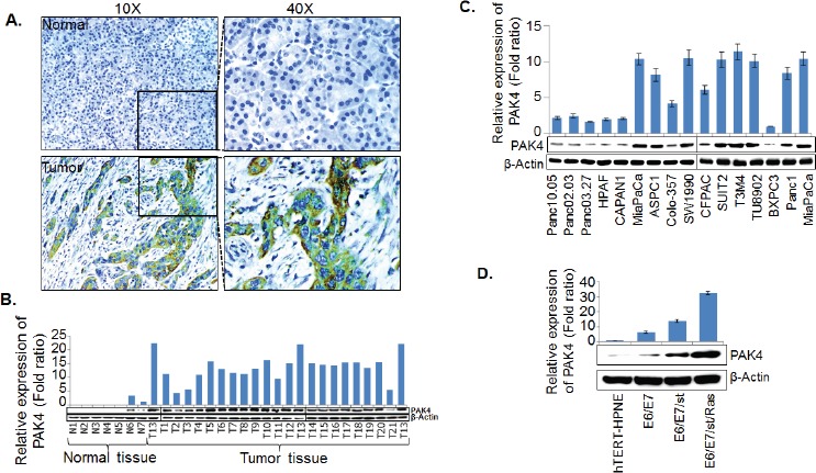
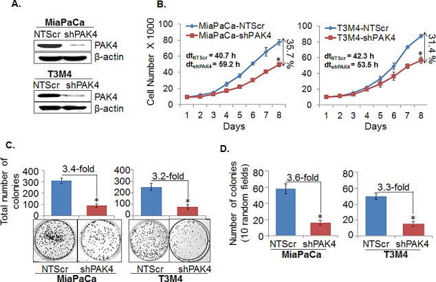
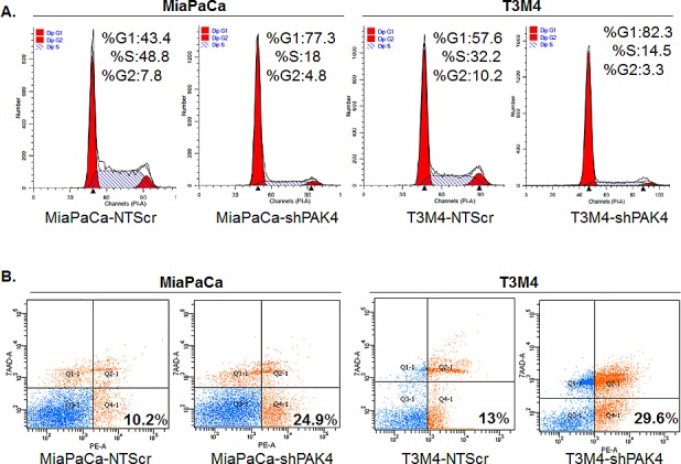
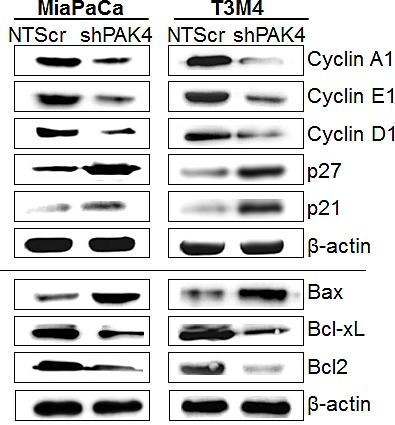
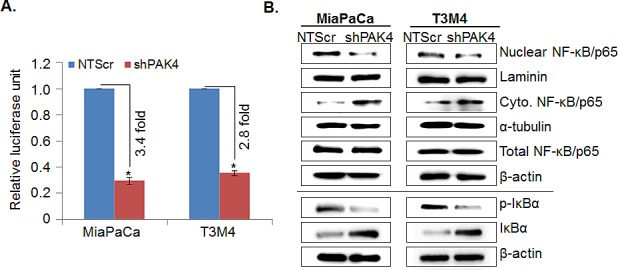
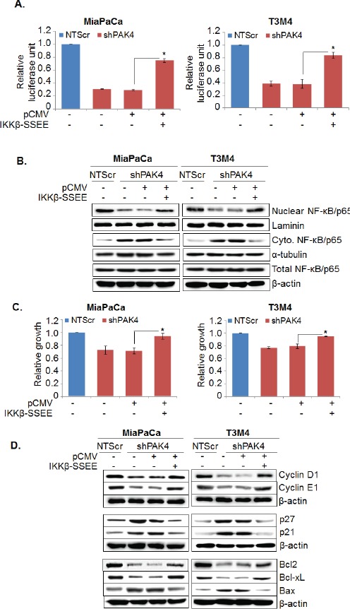
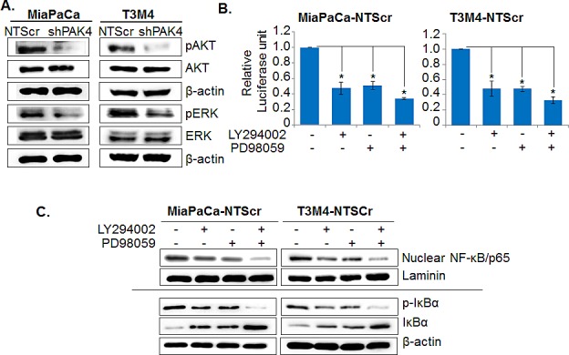
Identification of novel molecular targets and understanding the mechanisms underlying the aggressive nature of pancreatic cancer (PC) remain prime focus areas of research. Here, we investigated the expression and pathobiological significance of p21-activated kinase 4 (PAK4), a gene that was earlier shown to be amplified in a sub-set of PC. Our data demonstrate PAK4 overexpression in PC tissues and cell lines with little or no expression in the normal pancreas. PAK4 silencing in two PC cell lines, MiaPaCa and T3M4, by RNA interference causes suppression of growth and clonogenic ability due to decreased cell cycle progression and apoptosis-resistance. PAK4-silenced PC cells exhibit altered expression of proliferation- and survival-associated proteins. Moreover, we observe decreased nuclear accumulation and transcriptional activity of NF-κB in PAK4-silenced PC cells associated with stabilization of its inhibitory protein, IκBα. Transfection of PAK4-silenced PC cells with constitutively-active mutant of IKKβ, an upstream kinase of IκBα, leads to restoration of NF-κB activity and PC cell growth. Furthermore, we show that PAK4-induced NF-κB activity is mediated through activation and concerted action of ERK and Akt kinases. Together, these findings suggest that PAK4 is a regulator of NF-κB pathway in PC cells and can serve as a novel target for therapy.
Conflict of interest statement
No potential conflict of interest to disclose.
Figures
Similar articles
-
An undesired effect of chemotherapy: gemcitabine promotes pancreatic cancer cell invasiveness through reactive oxygen species-dependent, nuclear factor κB- and hypoxia-inducible factor 1α-mediated up-regulation of CXCR4.J Biol Chem. 2013 Jul 19;288(29):21197-21207. doi: 10.1074/jbc.M113.484576. Epub 2013 Jun 5. J Biol Chem. 2013. PMID: 23740244 Free PMC article.
-
Mesothelin confers pancreatic cancer cell resistance to TNF-α-induced apoptosis through Akt/PI3K/NF-κB activation and IL-6/Mcl-1 overexpression.Mol Cancer. 2011 Aug 31;10:106. doi: 10.1186/1476-4598-10-106. Mol Cancer. 2011. PMID: 21880146 Free PMC article.
-
Zic2 promotes tumor growth and metastasis via PAK4 in hepatocellular carcinoma.Cancer Lett. 2017 Aug 28;402:71-80. doi: 10.1016/j.canlet.2017.05.018. Epub 2017 Jun 1. Cancer Lett. 2017. PMID: 28577975
-
Drug discovery targeting p21-activated kinase 4 (PAK4): a patent review.Expert Opin Ther Pat. 2021 Nov;31(11):977-987. doi: 10.1080/13543776.2021.1944100. Epub 2021 Aug 9. Expert Opin Ther Pat. 2021. PMID: 34369844 Review.
-
The role of PAK4 in the immune system and its potential implication in cancer immunotherapy.Cell Immunol. 2021 Sep;367:104408. doi: 10.1016/j.cellimm.2021.104408. Epub 2021 Jul 1. Cell Immunol. 2021. PMID: 34246086 Review.
Cited by
-
miR-199a-3p increases the anti-tumor activity of palbociclib in liver cancer models.Mol Ther Nucleic Acids. 2022 Jul 20;29:538-549. doi: 10.1016/j.omtn.2022.07.015. eCollection 2022 Sep 13. Mol Ther Nucleic Acids. 2022. PMID: 36035756 Free PMC article.
-
Resistin and interleukin-6 exhibit racially-disparate expression in breast cancer patients, display molecular association and promote growth and aggressiveness of tumor cells through STAT3 activation.Oncotarget. 2015 May 10;6(13):11231-41. doi: 10.18632/oncotarget.3591. Oncotarget. 2015. PMID: 25868978 Free PMC article.
-
Off-target toxicity is a common mechanism of action of cancer drugs undergoing clinical trials.Sci Transl Med. 2019 Sep 11;11(509):eaaw8412. doi: 10.1126/scitranslmed.aaw8412. Sci Transl Med. 2019. PMID: 31511426 Free PMC article.
-
PAK4 inhibition significantly potentiates Gemcitabine activity in PDAC cells via inhibition of Wnt/β-catenin, p-ERK/MAPK and p-AKT/PI3K pathways.Biochem Biophys Rep. 2023 Sep 9;35:101544. doi: 10.1016/j.bbrep.2023.101544. eCollection 2023 Sep. Biochem Biophys Rep. 2023. PMID: 37720313 Free PMC article.
-
Functional role and therapeutic targeting of p21-activated kinase 4 in multiple myeloma.Blood. 2017 Apr 20;129(16):2233-2245. doi: 10.1182/blood-2016-06-724831. Epub 2017 Jan 17. Blood. 2017. PMID: 28096095 Free PMC article.
References
-
- Siegel R, Ma J, Zou Z, Jemal A. Cancer statistics, 2014. CA Cancer J Clin. 2014;64:9–29. - PubMed
-
- Siegel R, Naishadham D, Jemal A. Cancer statistics, 2013. CA Cancer J Clin. 2013;63:11–30. - PubMed
-
- Kumar R, Gururaj AE, Barnes CJ. p21-activated kinases in cancer. Nat Rev Cancer. 2006;6:459–71. - PubMed
Publication types
MeSH terms
Substances
Grants and funding
LinkOut - more resources
Full Text Sources
Other Literature Sources
Medical
Research Materials
Miscellaneous
