. 2014 May 15;9(10):1003-13.
doi: 10.4103/1673-5374.133159.

Intraspinal transplantation of motoneuron-like cell combined with delivery of polymer-based glial cell line-derived neurotrophic factor for repair of spinal cord contusion injury

Affiliations

Intraspinal transplantation of motoneuron-like cell combined with delivery of polymer-based glial cell line-derived neurotrophic factor for repair of spinal cord contusion injury

Alireza Abdanipour et al. Neural Regen Res. .

Abstract

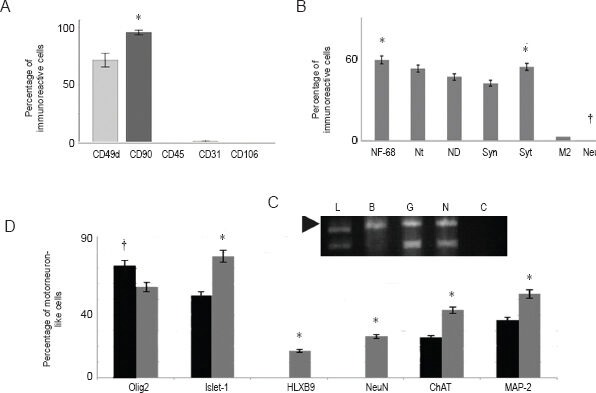
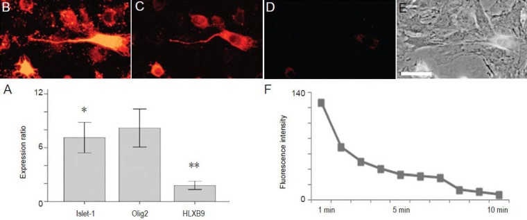
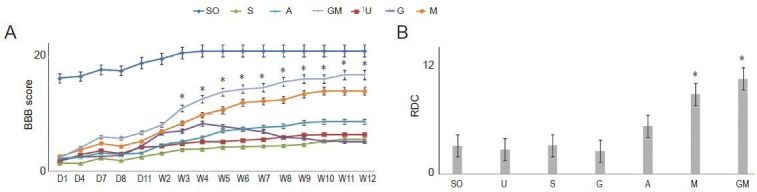
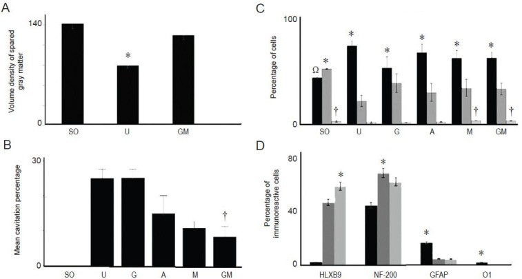
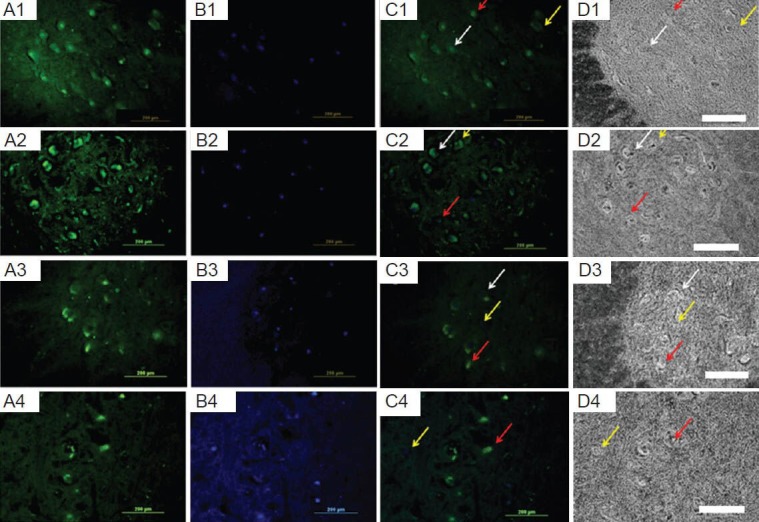
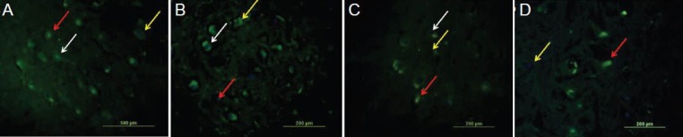
To evaluate the effects of glial cell line-derived neurotrophic factor transplantation combined with adipose-derived stem cells-transdifferentiated motoneuron delivery on spinal cord contusion injury, we developed rat models of spinal cord contusion injury, 7 days later, injected adipose-derived stem cells-transdifferentiated motoneurons into the epicenter, rostral and caudal regions of the impact site and simultaneously transplanted glial cell line-derived neurotrophic factor-gelfoam complex into the myelin sheath. Motoneuron-like cell transplantation combined with glial cell line-derived neurotrophic factor delivery reduced cavity formations and increased cell density in the transplantation site. The combined therapy exhibited superior promoting effects on recovery of motor function to transplantation of glial cell line-derived neurotrophic factor, adipose-derived stem cells or motoneurons alone. These findings suggest that motoneuron-like cell transplantation combined with glial cell line-derived neurotrophic factor delivery holds a great promise for repair of spinal cord injury.

Keywords: adipose-derived stem cells; cell transplantation; glial cell line-derived neurotrophic factor; motoneurons; nerve regeneration; neural regeneration; neurotrophic factor; spinal cord contusion injury; spinal cord injury.


Conflict of interest statement

Conflicts of interest: None declared.

Figures

Figure 1
Figure 1
The percentage of immunoreactive cells to different markers of adipose-derived stem cells, pre-induced cells and motoneurons-like cells. The data in the histogram were presented as mean ± SEM. Statistical analysis was done using one-way analysis of variance (ANOVA) and Tukey's test (A, B and D) and reverse transcriptase polymerase chain reaction of pre-induced cells (C). Each experiment was performed five times. (A) A histogram representing the percentages of adipose-derived stem cells that were immunoreactive to different CD markers including CD 49d (a specific marker for fat cells), CD 90 (a marker of mesenchymal stem cells), CD 45 (a hematopoietic cell marker), CD 31 (an endothelial cell marker) and CD 106 (a marker of mesenchymal stem cells derived from bone marrow stromal cells). The “*” indicates that the expression is significantly higher than that of the other markers. (B) A histogram representing the percentages of cells pre-induced with selegiline (10−9 mmol/L) that were immunoreactive to different markers including neurofilament 68 (NF-68: a proneural marker), nestin (Nt: a neural stem cell marker), Neuro D (ND: a proneural marker), synapsin (Syn: a neuronal marker), synaptophysin (Syt: a neuronal marker), microtubule-associated protein 2 (M2: a neuronal marker) and NeuN (a neuronal marker). The “*” indicates that the expression is significantly higher than that of the other markers; “†” indicates that the expression is significantly lower than that of the other markers. (C) The electrophorogram of reverse transcriptase polymerase chain reaction of pre-induced cells for B (brain derived neurotrophic factor), G (glial cell derived neurotrophic factor, N (neurotrophin-3) and C (negative control). L is DNA ladder. (D) A histogram representing the percentages of motoneuron-like cells at day 2 (solid black column) and day 7 (solid gray column) that were immunoreactive to different markers including Olig2 (a motoneuron lineage marker), Islet-1 (a motoneuron lineage marker), HLXB9 (a differentiated motoneuron lineage marker), neuronal nuclei (NeuN; a neuronal marker), choline acetyl transferase (ChAT: a cholinergic neuron marker) and microtubule-associated protein 2 (MAP-2; a neuronal marker). The “*” indicates that the expression is significantly higher on day 7 than on day 2.
Figure 2
Figure 2
The cellular and molecular functionality of motoneuron-like cells. (A) A histogram representing the fold change ratios of expression of Islet-1 (a motoneuron lineage marker), Olig2 (a motoneuron lineage marker) and HLXB9 (a differentiated motoneuron lineage marker). The mRNA was extracted from motoneuron-like cells derived from the adipose-derived stem cells using pre-induction (using selegiline) and induction (using 1 μg/mL sonic hedgehog and 2 × 10⁻⁸ mol/L all-trans retinoic acid for 2 days) steps. The fold change ratio was assessed using quantitative real-time polymerase chain reaction. The “*” indicates that difference with Islet 1 was not significant, and “**” indicates that the difference with the other genes was significant (P < 0.05). The data in the histogram were presented as mean ± SEM. Statistical analysis was done using one-way analysis of variance and Tukey's post hoc test. The data were obtained from three experiments. Functionality assay using staining and destaining of neuron-like cells differentiating into motoneuron-like cells exposed for 120 seconds to FM1-43 fluorochrome at 1 (B), 5 (C), and 10 minutes (min; D) after the start of the Potassium ion stimulation. (E) A phase-contrast image. Scale bar: 25 μm. (F) The total number of pixels in the time course (1–10 min) used in the study during which the motoneuron-like cells were stained with FM1-43 fluorochrome and then destained following the release of the synaptic vesicles. The bar graphs indicate the mean ± SEM.
Figure 3
Figure 3
Results of open field behavioral test (BBB score test) and RDC of BBB score test. (A) Represents the time course of BBB locomotor scores. The scores were recorded from the 1st day to the end of the 12th week post-injury. The histogram represents the sham operated (laminoectomy: SO), untreated spinal cord contusion injury (U), contused, injected with normal saline (S), GDNF-treated (G), ADSC-treated (A), MNLC-treated (M), and MNLC plus GDNF-treated ones (GM). Eight animals per group were used and the data were presented as mean ± SEM. Statistical analysis was done using repeated-measures analysis of variance and Tukey's post hoc test. BBB in the M and GM groups significantly differed from those of the other experimental groups. The “*” represents significant differences between the M and GM groups and the other groups (P < 0.05). (B) A histogram of the RDC in BBB scores between weeks 1 and 12 after injury in each group including the SO, U, G, A, M and GM groups. The data in the histogram were presented as mean ± SEM. One-way ANOVA followed by Tukey's post hoc test was used. The “*” represents significant differences between the GM group and the other groups, except the G group (P < 0.05). BBB: Basso-Beattie-Bresnahan; RDC: relative difference coefficient; GDNF: glial cell line-derived neurotrophic factor; ADSC: adipose-derived stem cell; MNLC: motoneuron-like cell.
Figure 4
Figure 4
Morphometric analysis of spinal tissues. Eight animals per group were used and the data were presented as mean ± SEM. Statistical analysis was performed using one-way analysis of variance and Tukey's post hoc test. (A) The mean percentage of the spared gray matter in the sham-operated (SO), untreated (U) and MNLC plus GDNF-treated (GM) groups. The “*” indicates that the percentage is significantly lower than that of the other groups. (B) The mean cavitation percentages in the SO, U, GDNF-treated (G), ADSC-treated (A), MNLC-treated (M), and GM groups. The “†” indicates that the percentage in the GM group is significantly lower than that of the other groups, except the M group. (C) The mean percentages of glial cells (solid black column), neurons (solid dark gray column), and MNLC (solid light gray column) in the SO, U, G, A, M and GM groups. The “*” indicates that the percentage of the cells is significantly higher than that of the other cells in the same group (P < 0.05). The “†” indicates that the percentage of cells is significantly higher than that in the U, A and G groups (P < 0.05). The “Ω” indicates that the percentage of cells is significantly lower than that in the other groups, except the G group (P < 0.05). (D) The mean percentage of the cells in the A (solid black column), M (solid dark gray column), and GM (solid light gray column) groups. The cells were immunoreactive to HLXB9 (a marker of differentiated motoneuron), NF-200 (a neuronal marker), glial fibrillary acidic protein (an astrocyte marker), and O1 (an oligodendrocyte marker). The “*” indicates that the percentage of the immunoreactive cells in a given group is signifi-cantly higher than that of the other groups (P < 0.05). GDNF: Glial cell line-derived neurotrophic factor; ADSC: adipose-derived stem cell; MNLC: motoneuron-like cell; NF-200: neurofilament-200.
Figure 5
Figure 5
The fate of the transplanted cells in tissue sections from the injured spinal cord from the animal group treated with motoneuron-like cells with GDNF. Photomicrographs represent the immunoreactivity of the transplanted cells to HLXB9 (A1, B1, C1 and D1), NF-200 (A2, B2, C2 abd D2), GFAP (A3, B3, C3 and D3) and O1 (A4, B4, C4 and D4) in the GM group. The cells were immunostained with relevant primary antibodies and labeled with FITC-conjugated secondary antibody (green) and Hoechst 33342 stain (a nuclear stain; blue). (A) Cells immunoreactive to HLXB9, NF-200, GFAP or O1 (A1, A2, A3 or A4, respectively). (B) Cells labeled with Hoechst 33342 stain (all panels). (C) A merged image of A and B (all panels), and a phase-contrast photomicrograph from the same field in A and B (all panels). The white, yellow, and red arrows indicate immunoreactive transplanted cells, non-immunoreactive transplanted cells, and immunoreactive host cells, respectively (scale bar: 200 μm). GDNF: Glial cell line-derived neurotrophic factor; NF-200: neurofilament-200; GFAP: glial fibrillary acidic protein; GM: motoneuron-like cell plus GDNF-treated.
Figure 6
Figure 6
The fate of the transplanted cells in tissue sections from the injured spinal cord. Merged photomicrographs represent the immunoreactivity of the transplanted cells to HLXB9 (A), NF-200 (B), GFAP (C), and O1 (D) in the GM group at higher magnification than figure 5. The cells were immunostained with relevant primary antibodies and labeled with FITC-conjugated secondary antibody (green) and Hoechst 33342 stain (a nuclear stain; blue). The white, yellow, and red arrows indicate immunoreactive transplanted, non-immunoreactive transplanted, and immunoreactive host cells, respectively (scale bars: 200 μm). NF-200: Neurofilament-200; GFAP: glial fibrillary acidic protein; GM: motoneuron-like cell plus GDNF-treated.

Similar articles

Cited by

References

    1. Abdanipour A, Tiraihi T. Induction of adipose-derived stem cell into motoneuron-like cells using selegiline as preinducer. Brain Res. 2012;1440:23–33. - PubMed
    1. Abdanipour A, Tiraihi T, Delshad A. Trans-differentiation of the adipose tissue-derived stem cells into neuron-like cells expressing neurotrophins by selegiline. Iran Biomed J. 2011;15:113–121. - PMC - PubMed
    1. Alexanian AR, Crowe MJ, Kurpad SN. Efficient differentiation and integration of lineage-restricted neural precursors in the traumatically injured adult cat spinal cord. J Neurosci Methods. 2006;150:41–46. - PubMed
    1. Arboleda D, Forostyak S, Jendelova P, Marekova D, Amemori T, Pivonkova H, Masinova K, Sykova E. Transplantation of predifferentiated adipose-derived stromal cells for the treatment of spinal cord injury. Cell Mol Neurobiol. 2011;31:1113–1122. - PubMed
    1. Basso DM, Beattie MS, Bresnahan JC. A sensitive and reliable locomotor rating scale for open field testing in rats. J Neurotrauma. 1995;12:1–21. - PubMed