. 2014 Nov:468-470:256-264.
doi: 10.1016/j.virol.2014.08.007. Epub 2014 Sep 7.

Lung epithelial cells resist influenza A infection by inducing the expression of cytochrome c oxidase VIc which is modulated by miRNA 4276

Affiliations

Lung epithelial cells resist influenza A infection by inducing the expression of cytochrome c oxidase VIc which is modulated by miRNA 4276

Sreekumar Othumpangat et al. Virology. 2014 Nov.

Abstract

Influenza virus infection induces several changes in host miRNA profile, host cell death and tissue damage. Cytochrome c is a regulator of the intrinsic apoptotic pathway and is altered during viral infections. Within the first 3h of infection with influenza virus, significant down-regulation of hsa-miRNA-4276 (miRNA-4276) is followed by a 2-fold increase in cytochrome c oxidase VIC (COX6C) mRNA was found to occur in human alveolar and bronchial epithelial cells. Expression of caspase-9 also increased within the first 3h of infection, but subsequently decreased. Modulation of miR-4276 using mimic and inhibitor oligonucleotides showed significant down-regulation or up-regulation, respectively, of COX6C expression. Our data suggests that on initial exposure to influenza virus, host cells upregulate COX6C mRNA expression through silencing miR-4276 and repressed viral replication by inducing the apoptotic protein caspase-9. Taken together, these data suggest that miR-4276 may be an important regulator of the early stages of infection by influenza.

Keywords: COX6C; Influenza virus; Lung epithelial cells; miRNA.


Figures

Fig. 1
Fig. 1
Influenza virus infection induced changes in miRNA expression: A) Cluster analysis of influenza virus altered miRNA expression in A549 cells. Microarray analysis for miRNA was performed with RNA extracts from influenza virus infected A549 cells for 3 h. The expression index or color key with the ROW-Z-score is on the top representing degree of abundance in each cluster. Green shaded areas indicates higher expression of the specific miRNA, whereas the red shaded areas indicates lower expression and black denotes the relatively similar expression between infected and mock infected cells. HEK 10 07 virus1 & 2 are the replicates of cells infected with influenza virus. B) Scatter plot showing the expression profile of PCR array analysis for Mitochondrial energy metabolism (RT2 Profiler™ PCR Array) was performed with RNA extracts from influenza virus inflected A549 cells. The fold change is given in brackets. MitoH1-Polycistronic H1, MitoH2-12106 - Polycistronic_H2_12106, MitoH2_14573 - Polycistronic H2_57263, MitoH2-5726 - Polycistronic_H2200-5726, MitoH2_4162 - Polycistronic _H2_4162, ARRDC3- Arrestin domain containing 3, COX6C- cytochrome c oxidase subunit VI C, SDHC-Succinate dehydrogenase complex. C) PCR array analysis (RT2 Profiler™ PCR Array) was performed with RNA extracts from influenza virus infected A549 cells. GAPDH was used as the internal control. Data for cytochrome c oxidase subunits are shown with the p Values.
Fig. 2
Fig. 2
miRNA 4276 targets COX6C expression: A) A549 cells were infected with influenza virus with MOI of 3 for 9 h were sampled every 3 h, miRNAs extracted and then analyzed by qPCR. Let-7 was used as the internal control. Data from three independent experiments. *=p<0.05, **=p<0.01. B) A549 cells were infected with influenza virus for 9 h and samples were withdrawn at the indicated time points. cDNA synthesized from 1 mg of extracted RNA and used for COX6C qPCR analysis. Data was normalized to GAPDH. Bar graphs represent data from three independent experiments (n=3). *=p<0.05, **=p<0.01. C) Matrix gene copy number was analyzed from the RNA extracted from A549cells infected with influenza virus, (n=3).
Fig. 3
Fig. 3
Changes in miR-4276 levels alter COX6C mRNA expression: A) miRNA4276 response to increasing MOIs of influenza virus. A549 cells were infected for 3 h with increasing MOIs of influenza. Let-7 was used as the internal control. Data from three independent experiments. ***=p<0.001. B) COX6C expression in response to increasing MOIs of influenza in A549 cells. GAPDH was used as the internal control. Data from three independent experiments. **=p<0.01, ***=p<0.001, (n=3).
Fig. 4
Fig. 4
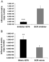
Down-regulation of miRNA 4276 induces COX6C expression: A) A549 cells were transfected for 48 h with 25 nM miRNA-4276 mimic or a scrambled oligonucleotide (SCR control). The transfected cells were then either mock infected or infected with 1 MOI influenza for an additional 3 h. Let-7 was used as the internal control. Data are expressed as7SEM, ***=p<0.001, (n=3). B) A549 cells transfected with mimic or SCR control were infected with 1 MOI of influenza virus for additional 3 h. The relative abundance of COX6C was measured in infected cells and the data normalized to the abundance of GAPDH. Data are expressed as±SEM, *=p<0.05, **=p<0.01 compared the cells transfected with the scrambled oligonucleotide, (n=3) C) A549 cells were transfected with 25 nM of either miRNA 4276 inhibitor or a SCR control. Relative expression of miRNA4276 was analyzed by qPCR. Data are expressed as±SEM, ***=p<0.001 compared the cells transfected with the scrambled oligonucleotide, (n=3). D) After 48 h, the transfected cells were infected with 1 MOI of influenza virus. The relative COX6C expression was measured in both infected and uninfected cells; GAPDH was used as the internal control. Data are expressed as7SEM, **=p<0.01 compared the cells transfected with the scrambled oligonucleotide, (n=3).
Fig. 5
Fig. 5
Immunofluorescence of influenza A virus infected cells: A549 cells were transfected with either the scrambled oligonucleotide (SCR) (top panel), miRNA-4276 inhibitor (middle panel), or miRNA-4276 mimic (bottom panel). Transfected cells were infected with influenza virus and expression of COX6C protein (green) and influenza nucleoprotein (red) was determined by immunofluorescence. DAPI (blue) represents the stained nucleus of the cells.
Fig. 6
Fig. 6
Abundance of COX6C reduced viral replication: A) Matrix copy number in cells transfected with either miRNA-4276 inhibitor or scrambled oligonucleotide is shown. Data are expressed as±SEM, **=p<0.001 (n=4). B) Matrix copy number in cells transfected with either miRNA-4276 mimic or scrambled oligonucleotide is shown. Data are expressed as±SEM, **=p<0.01 compared the cells transfected with the scrambled oligonucleotide, (n=3).
Fig. 7
Fig. 7
Specificity of miR-4276 to COX6C: Other cytochrome oxidase family of proteins was not affected by miR4276. RNA extracted from A549 cells transfected with mimic, inhibitor or a scrambled oligonucleotide used for the PCR array (RT2 Profiler™ PCR Array Human Mitochondrial Energy Metabolism) containing 11 cytochrome family protein was analyzed by qPCR (SYBR green).
Fig. 8
Fig. 8
Influenza virus H1N1 and H3N2 alter miR-4276 levels in HBEpC cells and increase COX6C and Caspase-9 expression. A) HBEpC cells were transfected with 25 nM of either miRNA 4276 inhibitor or a SCR control. Relative expression of miRNA4276 was analyzed by qPCR. Data are expressed as±SEM, ***=p<0.001 compared the cells transfected with the scrambled oligonucleotide, (n=3). B) HBEpC cells were transfected for 48 h with 25 nM miRNA-4276 mimic or a scrambled oligonucleotide (SCR control). The transfected cells were then either mock infected or infected with 1 MOI influenza for an additional 3 h. Let-7 was used as the internal control. Data are expressed as±SEM, ***=p<0.001, (n=3). C) Primary human bronchial epithelial cells grown to 80% confluency were infected with influenza virus H1N1 or H3N2 of MOI of 1, for 3 h. COX6C expression was analyzed and normalized to GAPDH, Data are expressed as±SEM, *=p<0.05 and **=p<0.01, n=3 independent experiments. D) HBEpC cells grown to 80% confluency were infected with influenza virus H1N1 or H3N2 of MOI of 1, for 3 h, p-caspase-9 expression was analyzed and normalized to GAPDH, Data are expressed as±SEM, *=p<0.05 and **=p<0.01, n=3 independent experiments analyzed in duplicates.
Fig. 9
Fig. 9
COX6C expression alters the expression of apoptotic protein caspase-9. A) HBEpCs were exposed influenza virus for a time course and the expression of Cox6C and p-caspase-9 were analyzed using Western blot analysis. Primary human bronchial epithelial cells grown to 80% confluency were infected with influenza virus H1N1 or H3N2 of MOI of 1, for 3 h. pro-caspase 9 expression was analyzed and normalized to GAPDH, Data are expressed as±SEM, *=p<0.05 and **=p<0.01, n=3 independent experiments. B) Primary human bronchial epithelial cells grown to 80% confluence were transfected with mimic-4276, inhibitor or SCR control for 48 h and the pattern of p-caspase-9 analyzed by Western immunoblot analysis. GAPDH was used as a loading control.

Similar articles

Cited by

References

    1. Bartel DP. MicroRNAs: genomics, biogenesis, mechanism, and function. Cell. 2004;116:281–297. - PubMed
    1. Blachere FM, Cao G, Lindsley WG, Noti JD, Beezhold DH. Enhanced detection of infectious airborne influenza virus. J. Virol. Methods. 2011;176:120–124. - PubMed
    1. Blachere FM, Lindsley WG, Pearce TA, Anderson SE, Fisher M, Khakoo R, Meade BJ, Lander O, Davis S, Thewlis RE, Celik I, Chen BT, Beezhold DH. Measurement of airborne influenza virus in a hospital emergency department. Clin. Infect. Diseases: an Official Publication Infect. Dis. Soc Am. 2009;48:438–440. - PubMed
    1. Brydon EW, Morris SJ, Sweet C. Role of apoptosis and cytokines in influenza virus morbidity. FEMS Microbiol. Rev. 2005;29:837–850. - PubMed
    1. Collins M. Potential roles of apoptosis in viral pathogenesis. Am. J. Respir. Crit. Care Med. 1995;152:S20–S24. - PubMed

MeSH terms

LinkOut - more resources