. 2014 Oct 24;289(43):30063-74.
doi: 10.1074/jbc.M114.593616. Epub 2014 Sep 8.

The LIMP-2/SCARB2 binding motif on acid β-glucosidase: basic and applied implications for Gaucher disease and associated neurodegenerative diseases

Affiliations

The LIMP-2/SCARB2 binding motif on acid β-glucosidase: basic and applied implications for Gaucher disease and associated neurodegenerative diseases

Benjamin Liou et al. J Biol Chem. .

Abstract

The acid β-glucosidase (glucocerbrosidase (GCase)) binding sequence to LIMP-2 (lysosomal integral membrane protein 2), the receptor for intracellular GCase trafficking to the lysosome, has been identified. Heterologous expression of deletion constructs, the available GCase crystal structures, and binding and co-localization of identified peptides or mutant GCases were used to identify and characterize a highly conserved 11-amino acid sequence, DSPIIVDITKD, within human GCase. The binding to LIMP-2 is not dependent upon a single amino acid, but the interactions of GCase with LIMP-2 are heavily influenced by Asp(399) and the di-isoleucines, Ile(402) and Ile(403). A single alanine substitution at any of these decreases GCase binding to LIMP-2 and alters its pH-dependent binding as well as diminishing the trafficking of GCase to the lysosome and significantly increasing GCase secretion. Enterovirus 71 also binds to LIMP-2 (also known as SCARB2) on the external surface of the plasma membrane. However, the LIMP-2/SCARB2 binding sequences for enterovirus 71 and GCase are not similar, indicating that LIMP-2/SCARB2 may have multiple or overlapping binding sites with differing specificities. These findings have therapeutic implications for the production of GCase and the distribution of this enzyme that is delivered to various organs.

Keywords: Enterovirus 71; Enzyme Kinetics; Glucocerebrosidase; Inborn Error of Metabolism; Intracellular Trafficking; Lysosomal Storage Disease; Lysosomal Targeting; Protein Sorting.


Figures

FIGURE 1.
FIGURE 1.
GCase-GFP transfection constructs for Gba1 null/null cells. On the left are the resultant GCases following the various deletions, indicated as GCaseZZZ to show the encoded mature amino acid contents of the expressed enzymes. The constructs are labeled GCase-XX or -XXX to designate the number of amino acids that were deleted from the -COOH end of the mature 497-amino acid sequence of the WT GCase. GFP was cloned in frame with the WT or GCase-XX or -XXX. These constructs were subsequently transfected into Gba1−/− fibroblasts.
FIGURE 2.
FIGURE 2.
Co-localization of GCase-XX- or GCase-XXX-GFP with LysoTracker. Pearson indices were obtained (see “Methods”) for co-localization of the WT or truncated GCases using LysoTracker to label the lysosomes. The construct containing only GFP (N-GFP) showed no co-localization. Compared with the WT sequence, GCase-23 and -75 showed only small decreases in lysosomal localization. GCase-150 and -225 showed decreases in co-localization with LysoTracker to nearly background (N-GFP) levels. GCase-150 and -225 showed much more extensive retention in the ER and Golgi compared with WT, GCase-23, or GCase-75 (data not shown). Error bars, S.E.
FIGURE 3.
FIGURE 3.
Mature WT GCase amino acid sequence highlighting the regions targeted for mutagenesis, expression, and binding analyses. The amino acid sequence of GCase is marked with various dark red arrows indicating the number of amino acids deleted from the COOH-terminal end of the enzyme for the GFP reporter. Shown in orange is a typical dileucine sequence for indirect targeting of membrane-bound lysosomal proteins. Underlined in red are the amino acids for targeted mutagenesis and binding studies to ldLIMP-2. These sequences have very high conservation across species (data not shown).
FIGURE 4.
FIGURE 4.
Structure of WT GCase highlighting the potential ldLIMP2 binding sequence localization. The two amino acid sequences from Fig. 3 were mapped to the crystal structure of WT human GCase. The orientation of the sequences is indicated by the amino acid numbers in the highlighted (yellow or orange) regions. The domains of GCase are shown in various indicated colors as are the disulfides. The potential ldLIMP-2 binding (yellow, Asp399–Asp409) sequence forms a surface-accessible loop in domain I (black, with the motif in yellow). The DSPIIVDITKD sequence and the DDQRLLL (orange, Asp282–Leu288 in Domain III) sequence are highly conserved in GCases from insects to humans (data not shown). The acid-base (Glu235) and nucleophile (Glu340) in catalysis and the N370S and L444P common mutations causal to Gaucher disease are shown in red (5).
FIGURE 5.
FIGURE 5.
Co-localization (Pearson) indices of GCases with specific alanine substitutions in the WT TKD sequence. GCase/mutant constructs were transfected into Gba1−/− fibroblasts, and co-localization was evaluated with directly labeled anti-mouse calreticulin (rhodamine; red) antibody for ER/Golgi, directly labeled Lamp1 (rhodamine; red) for lysosome (Lyso), and directly labeled anti-human GCase (FITC; green) for GCase. The alanine (A) substitutions are as altered from the WT TKD sequence. For the AAA and AKA GCases, the majority of the intracellular enzymes co-localized to the ER/Golgi (ER), whereas the TAD GCase showed about 50% of the total intracellular enzyme localized to the lysosome (Lyso). Co-localization indices (Pearson) were calculated (n = 5) and presented as the mean ± S.E. (error bars)
FIGURE 6.
FIGURE 6.
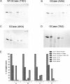
Immunoprecipitation of the alanine-substituted GCases in the TKD sequence with ldLIMP-2. In A–D, lanes 1–6 correspond to the band densities indicated by the bars in E. The panels show that the WT sequence (A) had nearly complete binding with ldLIMP-2 (i.e. retention on the beads to which ldLIMP-2 was bound (lane 4) and no GCase in the wash (lane 5)). In comparison, the triple alanine mutant (B) shows essentially no binding to ldLIMP-2 (i.e. all of the GCase is in the wash (lane 5)). The AKA GCase mutant (C) showed about equal amounts of GCase in lanes 4 and 5, or about 50% binding to ldLIMP-2. The AKD GCase mutant (D) had a binding pattern that was similar to the WT sequence but with more GCase in the wash (lane 5) (i.e. somewhat less binding). The quantitative results are shown in E, in which 1, 2, 3, and 4 correspond to WT, AAA, AKA, and AKD, respectively. The results are typical of multiple experiments.
FIGURE 7.
FIGURE 7.
Binding and competition of fluorescence-labeled or unlabeled DSPIIV GCase peptides to ldLIMP-2. The change (δmP) in fluorescence polarization is plotted on the ordinate, and the increasing concentrations of the various peptides are shown on the abscissa. A, the concentration of ldLIMP-2 (50 nm) was fixed. With the WT (DSPIIV) peptide (● and ▿, duplicate experiments), saturation kinetics with about half-maximal binding to ldLIMP-2 was observed at 50 nm. The DSPAIV and DSPIAV mutants did not show saturation up to 250 nm, indicating their poor interaction with ldLIMP-2. The ASPAAP peptide showed background changes. The ASPIIV peptide showed δmP values slightly above background, indicating little binding to ldLIMP-2. B, to ensure that the label did not interfere/promote binding, similar studies were conducted using fluorescently labeled peptides as binders and their respective unlabeled peptides as competitors. Each of the unlabeled peptides “competed” with the labeled peptides in the expected ratios. C, labeled (WT*) and unlabeled (WT) peptides were used in complementary competition studies. Either the labeled or unlabeled WT peptides equally competed for binding to ldLIMP-2, showing that both were equally effective in binding and that the label did not change the properties of the peptide-ldLIMP-2 interaction. The inset indicates 1:1 stoichiometry and tight binding properties of DSPIIV and ldLIMP-2. D, the δmP of the WT (DDQRLLL) or variously alanine-substituted labeled peptides were plotted against the corresponding unlabeled peptide competitors. Note that the fixed ldLIMP-2 concentration was 1000 nm, or 20 times that used in Fig. 7, A and B. For all peptides, the δmP values were near or at background levels. E, binding δmP for the various peptides as in D. The signals were near background levels for all peptides using concentrations 5–10 times that for DSPIIV and with a 20 times greater LIMP-2 fixed concentration.
FIGURE 8.
FIGURE 8.
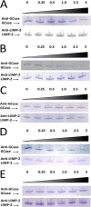
Immunoprecipitation of GCase in the presence of ldLIMP-2 and increasing molar ratios of the WT (DSPIIV) peptide. A, purified WT GCase was preincubated with purified ldLIMP-2 in a molar ratio of 1:1. Then unlabeled DSPIIV was added in varying molar excesses (0–5×; top) over GCase, incubated, and then immunoprecipitated with protein G-coupled anti-LIMP-2 antibody beads. The beads were then eluted, and the eluants were analyzed for GCase and ldLIMP-2 on Western blots using the specified antibodies. The results show decreasing amounts of bound GCase with increasing peptide molar ratios (i.e. peptide DSPIIV competed bound GCase off of ldLIMP-2. In the bottom panel, ldLIMP-2 recovery was the same at all peptide ratios. B, purified ldLIMP-2 was preincubated with unlabeled WT peptide at various molar ratios. Then, purified WT GCase was added in a 1:1 molar ratio with ldLIMP-2, incubated, and then immunoprecipitated and processed as in A. The results show that the peptide prevents the binding of ldLIMP-2 and WT GCase. C, purified WT GCase was preincubated with purified ldLIMP-2 in a molar ratio of 1:1. Then peptide DDQRLLL, a potential candidate for the GCase ligand for ldLIMP-2, was added in varying molar excesses over WT GCase, incubated, and then immunoprecipitated and processed as in A. The results show that peptide DDQRLLL did not compete GCase off of ldLIMP-2, thereby showing that this peptide did not have specificity for the GCase binding site on ldLIMP-2. DDQRLLL is a dileucine peptide outside of the localized targeting region of DSPIIVDITKD (see Fig. 3). D and E show the immunoprecipitation of GCase in the presence of ldLIMP-2 and increasing molar ratios of the WT (DDQRLLL) peptide. In D and E, ldLIMP-2 and DDQRLLL were incubated before (D) or together with (E) GCase and analyzed as in A–C. Under either condition, the peptide did not complete with GCase for ldLIMP-2 binding.
FIGURE 9.
FIGURE 9.
Effect of pH on binding WT and alanine-substituted DSPIIV peptides to ldLIMP-2. The binding of the different peptides to ldLIMP-2 showed little effect of alanine substitutions at pH 5.8. In comparison, incremental decreases were evident in ldLIMP-2 binding at pH 6.8, the approximate pH of the ER/cis-Golgi, as follows: the triple mutant (ASPAAV) having the lowest binding (<10% of WT), the DSPAIV (I402A) and DSPIAV (I403A) mutants being intermediate (∼40–50% of WT), and ASPIIV (D399A) having the least change (∼50–60% of WT), relative to WT. Error bars, S.E.
FIGURE 10.
FIGURE 10.
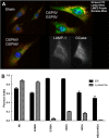
Intracellular localization of GCase DSPIIV variants in Gba1−/− cells. A, typical examples of immunofluorescence localization of DSPIIV-substituted GCase variants following transient transfections. The DSPIIV (WT) and ESPIIV show co-localization of GCase (FITC) with either Lamp1 (red) or LIMP-2 (purple) (top right), indicating that the retention of charge by Glu399 does not impact localization. The cells are typical for either Asp399 (DSPIIV) or Glu399 (ESPIIV). The DSPAIV (I402A) or DSPIAV (I403A) mutant shows no co-localization with ldLIMP-2 or Lamp1 (bottom left) (i.e. no binding to ldLIMP-2 in the cell and no localization to the lysosome (Lamp1/ldLIMP2)). The black and white images in the bottom right indicate the relative localization of LAMP-1 (lysosomes) and WT GCase (ER/Golgi and lysosomes) in Gba1−/− fibroblasts for comparison. Sham control is shown in the top left. B, Pearson indices for co-localization of the various mutants in the ER/Golgi (black) or lysosome (hatched). The substitution of Glu for Asp (WT) at 399 (ESPIIV) did not alter the co-localization compared with WT. In comparison, ASPIIV (D399A) and the double mutant, DSPAAV (I402A/I403A) showed little co-localization to the lysosome but significant retention in the ER/Golgi. The single mutants, DSPAIV (I402A) and DSPIAV (I403A), showed partial co-localization to the lysosome but greater retention in the ER/Golgi. Error bars, S.E.
FIGURE 11.
FIGURE 11.
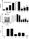
Secretion and retention of GCase with alanine variants in the TDK (A) or DSPIIV (B and C) sequences. A, left, activities in the medium of Gba1−/− fibroblasts following transient transfections of each of the specified WT or alanine-substituted GCases in the TDK region. Increases of 4–8-fold in GCase activities were found in the medium, with AKD showing the greatest increases and AAA the least. Right, CRIM-specific activity of WT and these same mutant GCases, demonstrating that the mutations decreased this activity relative to WT as indicated above the bars. B, left, activities in the medium following transient transfections of various DSPIIV alanine-substituted GCases and the percentage of GCase secreted. This was calculated as (ng of GCase (in medium)/(ng of GCase (in medium) + ng of GCase (in lysates))) × 100. The ESPIIV GCase did not alter secretion (i.e. little enzyme in the medium). All of the alanine substitutions in the DSPIIV sequence resulted in an ∼20–25-fold increase in GCase activity in the medium (solid bars). The transfections alone (Null Lipo) had no effect. In comparison, the amount of secreted GCase protein varied between 22 and 83% (hatched bars). Right, CRIM-specific activity of WT and these same mutant GCases, demonstrating that the mutations decreased this activity relative to WT as indicated above the bars. C, cellular lysate GCase activities from the various alanine-substituted GCases in the DSPIIV sequence as indicated. The WT and ESPIIV had equal intracellular activities of GCase, whereas the singly alanine-substituted GCases had ∼30% of WT levels. The doubly alanine-substituted GCase had little intracellular activity (i.e. nearly all of the GCase was secreted into the medium). All results are from n = 3 determinations. Error bars, S.E.

Similar articles

Cited by

References

    1. Brady R. O., Kanfer J. N., Bradley R. M., Shapiro D. (1966) Demonstration of a deficiency of glucocerebroside-cleaving enzyme in Gaucher's disease. J. Clin. Invest. 45, 1112–1115 - PMC - PubMed
    1. Brady R. O., Kanfer J. N., Shapiro D. (1965) Metabolism of glucocerebrosides. II. Evidence of an enzymatic deficiency in Gaucher's disease. Biochem. Biophys. Res. Commun. 18, 221–225 - PubMed
    1. Grabowski G. A., Petsko G. A., Kolodny E. (2010) Gaucher disease. in The Metabolic and Molecular Bases of Inherited Disease (Valle D., Beaudet A. L., Vogelstein B., Kinzler K. W., Antonarakis S. E., Ballabio A., Sly W. S., eds) McGraw-Hill, New York
    1. Grabowski G. A., Kolodny E. H., Weinreb N. J., Rosenbloom B. E., Prakash-Cheng A., Kaplan P., Charrow J., Pastores G. M., Mistry P. K. (2006) Gaucher disease: phenotypic and genetic variation. in The Metabolic and Molecular Bases of Inherited Diseases (Scriver C., Beaudet A., Sly W., Valle D., eds) McGraw-Hill, New York
    1. Liou B., Kazimierczuk A., Zhang M., Scott C. R., Hegde R. S., Grabowski G. A. (2006) Analyses of variant acid β-glucosidases: effects of Gaucher disease mutations. J. Biol. Chem. 281, 4242–4253 - PubMed

Publication types

MeSH terms