. 2014 Oct;124(10):4549-63.
doi: 10.1172/JCI76602. Epub 2014 Aug 26.

CD4⁺ and CD8⁺ T cell-dependent antiviral immunity requires STIM1 and STIM2

CD4⁺ and CD8⁺ T cell-dependent antiviral immunity requires STIM1 and STIM2

Patrick J Shaw et al. J Clin Invest. 2014 Oct.

Abstract

Calcium signaling is critical for lymphocyte function, and intracellular Ca2+ concentrations are regulated by store-operated Ca2+ entry (SOCE) through Ca2+ release-activated Ca2+ (CRAC) channels. In patients, loss-of-function mutations in CRAC channel components ORAI1 and STIM1 abolish SOCE and are associated with recurrent and chronic viral infections. Here, using mice with conditional deletion of Stim1 and its homolog Stim2 in T cells, we determined that both components are required for the maintenance of virus-specific memory CD8+ T cells and recall responses following secondary infection. In the absence of STIM1 and STIM2, acute viral infections became chronic. Early during infection, STIM1 and STIM2 were required for the differentiation of naive CD8+ T cells into fully functional cytolytic effector cells and mediated the production of cytokines and prevented cellular exhaustion in viral-specific CD8+ effector T cells. Importantly, memory and recall responses by CD8+ T cells required expression of STIM1 and STIM2 in CD4+ T cells. CD4+ T cells lacking STIM1 and STIM2 were unable to provide "help" to CD8+ T cells due to aberrant regulation of CD40L expression. Together, our data indicate that STIM1, STIM2, and CRAC channel function play distinct but synergistic roles in CD4+ and CD8+ T cells during antiviral immunity.


Figures
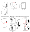
Figure 6
Figure 6. STIM1 and STIM2 in CD4+ T cells are essential for recall responses to viral reinfection.
WT and DKO mice (AC) or Cd8a–/– chimeras (DF) were infected with LCMVARM for 60 days, then reinfected with LCMVCL13, and analyzed 7 days p.r.i. (AC) Impaired recall response in DKO mice. Representative contour plots (A) and total number (mean ± SEM in B) of LCMV-specific CD8+ T cells per spleen before and 7 days p.r.i. with LCMVCL13. Numbers in B indicate fold increase in LCMV-specific CD8+ T cells p.r.i. (C) Serum LCMV titers in WT (n = 7) and DKO (n = 6) mice 7 days p.r.i. Each dot represents 1 mouse; horizontal lines show the mean viral titers. (DF) The presence of WT CD4+ T cells in chimeric mice restored recall responses by DKO CD8+ T cells. (D) Total number of LCMV-specific WT and DKO CD8+ T cells in the spleens of WT:Cd8a–/– (n = 4) and DKO:Cd8a–/– (n = 4) chimeras analyzed before and p.r.i. with LCMVCL13. Numbers indicate the fold increase of cell numbers 7 days p.r.i. (E and F) Expression of KLRG1 and CD127 (E) and T-bet and Eomes (F) on splenic LCMV-specific CD8+ T cells of WT or DKO origin in chimeras 7 days p.r.i. Line and bar graphs in D and E show the mean ± SEM of LCMV-specific CD8+ T cells from 4 mice per group. Statistical significance was calculated by Student’s t test (*P < 0.05; **P < 0.01; ***P < 0.001). Numbers in dot plots in A and E represent the percentages of cells in gates.
Figure 5
Figure 5. STIM1 and STIM2 control the maintenance of CD8 memory and generation of LCMV-specific antibodies by regulating CD40L expression on CD4+ T cells.
(A and B) Impaired CD40L expression on DKO CD4+ T cells from LCMVARM-infected mice. (A) Total cellular CD40L in unstimulated splenic CD4+CD44+ T cells 8 days p.i. Bar graphs represent the mean MFI ± SEM of CD40L expression (5 mice per group). (B) Surface expression of CD40L on splenic CD4+ T cells 8 days p.i. and following stimulation with GP61–80 peptide in vitro. Bar graphs represent the means ± SEM of 3 repeat experiments. (C) MHC class II expression on splenic CD11c+ DCs 8 days p.i. Each dot represents 1 mouse; horizontal lines show the mean MFI. (D) Generation of WT:Cd40l–/– and DKO:Cd40l–/– chimeras used in EH. (E and F) Frequencies (E) and absolute numbers (F) of LCMV-specific KLRG1CD127+ memory CD8+ T cells 80 days p.i. with LCMVARM. Bar graphs in F show the mean ± SEM of cell numbers from 4 WT:Cd40l–/– and 4 DKO:Cd40l–/– chimeras. (G) Relative levels of LCMV-specific serum IgG (means ± SEM; 4–5 mice per group). (H) Impaired plasma membrane expression of CD40L on CD4+ T cells from a STIM1-deficient patient (PAT) (13) compared with that in a healthy donor (HD). CD4+ T cells were left unstimulated or stimulated with PMA/ionomycin for 5 hours. Representative histograms and mean MFI ± SEM from 3 experiments. Statistical significance was calculated by Student’s t test (*P < 0.05; **P < 0.01; ***P < 0.001). Numbers in FACS plots represent percentages.
Figure 4
Figure 4. STIM1 and STIM2 are required for CD4+ T cell help to maintain memory CD8+ T cells.
(AD) WT CD4+ T cells restore the maintenance of DKO memory CD8+ T cells. (A) Generation of WT:Cd8a–/– and DKO:Cd8a–/– chimeras. (BD) Chimeras were infected with LCMVARM and analyzed 60 days p.i. for the frequencies (B) and absolute numbers (C) of LCMV-specific CD8+ T cells (mean ± SEM of cells from 6 WT and 6 DKO chimeras). (D) Intracellular cytokine staining for IL-2 and IFN-γ in splenic CD8+ T cells isolated from chimeras 60 days p.i. and restimulated with GP33–41 peptide for 5 hours in vitro. (EL) WT CD8+ T cells require STIM1/2-dependent CD4+ T cell help for memory maintenance. (E) 5 × 10⁴ WT P14 T cells (Thy1.1) were adoptively transferred into congenic WT or DKO mice and simultaneously infected with LCMVARM. (FH) Frequencies of Thy1.1+KLRG1CD127+ memory P14 T cells in the blood 8–60 days p.i. Bar graphs show the means ± SEM. (I and J) Frequencies (I) and total numbers (J) of splenic effector and memory subsets of Thy1.1+ WT P14 T cells 60 days p.i. (K) Serum LCMV titers. Each circle represents 1 mouse; horizontal lines represent the mean viral titers. (L) Tim-3 and PD-1 expression on splenic P14 T cells. Data in FL are from 6 WT and 5 DKO mice. Statistical significance was calculated by Student’s t test (**P < 0.01; ***P < 0.001). Numbers in FACS plots represent percentages. Green boxes in B and FH highlight memory CD8+ T cell populations.
Figure 3
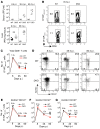
Figure 3. STIM1 and STIM2 regulate CD8 memory in a non-CD8+ T cell–intrinsic manner.
(AE) Increased expression of cell death, proliferation, exhaustion, and memory differentiation markers in DKO CD8+ T cells after LCMVARM infection. WT and DKO mice were analyzed for the frequencies of apoptotic (annexin V+ in A), proliferating (Ki67+ in B), and exhausted (Tim-3+, PD-1+ in C) splenic CD8+ T cells. Line graphs show the mean ± SEM of all DbNP396–404 tetramer+ CD8+ T cells over the course of LCMV infection in WT and DKO mice (3–6 per group). (D) Impaired IL-2 and IFN-γ production by splenic DKO CD8+ T cells after in vitro stimulation with GP33–41 peptide for 6 hours. Representative dot plots (left) and mean percentages (right) of IL-2+ cells among IFN-γ+ CD8+ T cells. (E) T-bet and Eomes expression (ratio of MFI values) in DbNP396–404 tetramer+ CD8+ T cells from 3 WT and 3 DKO mice analyzed by flow cytometry. (FK) Frequencies (F) and total numbers (G) of DbNP396–404 tetramer+ CD8+ T cells in 5 WT:DKO chimeras. Green boxes highlight memory CD8+ T cells. (HJ) Frequencies of apoptotic (annexin V+ in H), proliferating (Ki67+ in I), and exhausted (Tim-3+, PD-1+ in J) splenic memory CD8+ T cells of WT and DKO origin. (K) T-bet/Eomes ratio in memory CD8+ T cells from 5 WT:DKO chimeras. Statistical significance was calculated using Student’s t test (*P < 0.05; **P < 0.01; ***P < 0.001). Bar graphs show the means ± SEM. Numbers in D and F represent the percentages of cells in gates.
Figure 2
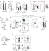
Figure 2. STIM1 and STIM2 regulate the function and differentiation of effector CD8+ cells.
(A) CD8+ cytotoxic CTLs from DKO P14 or WT P14 mice were cocultured with peptide-pulsed EL-4 cells; apoptosis was detected by annexin V staining. (B) Splenic CD8+ T cells from LCMVARM-infected WT (n = 6) and DKO (n = 5) mice were stimulated with PMA/ionomycin (iono) or GP33–41 peptide for 6 hours and IFN-γ production determined by flow cytometry. (C) Apoptosis and proliferation of LCMV-specific WT (n = 6) and DKO (n = 5) CD8+ T cells analyzed by annexin V and Ki67 staining. (D) T-bet and Eomes expression in CD8+ T cells from 3 mice per group. (EH) Mixed BM chimeras were generated by reconstituting Rag2–/– mice with BM from WT (CD45.1) and DKO (CD45.2) mice and infected with LCMVARM (E). Frequency (F and G) and total number (H) of splenic effector CD8+ T cells of WT or DKO origin. Each dot in G represents 1 WT:DKO chimera; horizontal lines represent mean cell percentages. (I and J) Congenic CD45.1 WT mice were infected with LCMVARM and injected with 5 × 10⁴ CD8+ T cells from DKO P14 and WT P14 mice. (J) Left panels show percentages of DbGP33–41 tetramer+ CD8+ T cells; right panels show percentages of transferred versus host cells among LCMV-specific cells. Plots are representative of 4 mice per group. Statistical significance was calculated using Student’s t test (*P < 0.05; **P < 0.01; ***P < 0.001). Bar graphs in AD and H represent the means ± SEM. Numbers in F and J represent the percentage of cells.
Figure 1
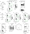
Figure 1. STIM1 and STIM2 in T cells control immunity to acute LCMV infection and the maintenance of virus-specific memory CD8+ T cells.
Stim1fl/fl Stim2fl/fl Cd4-Cre (DKO) and WT mice were infected with LCMVARM (2 × 10⁵ PFU; i.p.). (A) Viral titers in the sera and livers of mice. Each dot represents 1 mouse; horizontal lines show the means of the viral titers. ND, not detectable. (B) Representative flow cytometric plots of splenic CD8+ T cells from WT and DKO mice analyzed 8 days p.i. using DbNP396–404 and Db-GP33–41 tetramers. (C) Total numbers of LCMV-specific (DbNP396–404 tetramer+) CD8+ T cells in the spleens of WT (n = 5–9) and DKO (n = 7–9) mice at days 8, 35, and 60 p.i. (DG) Expression of KLRG1 and CD127 (IL-7Rα) on DbNP396–404 tetramer+ splenic CD8+ T cells from WT (n = 5–9) and DKO (n = 7–9) mice (D). Total numbers of LCMV-specific terminal effector (KLRG1+CD127 in E) and memory CD8+ T cell populations (KLRG1+CD127+ in F, KLRG1CD127+ in G). Numbers in EG indicate fold change differences between WT and DKO mice. Numbers in dot plots in B and D represent the percentages of cells in gates. Statistical significance was calculated by Student’s t test (*P < 0.05; **P < 0.01; ***P < 0.001).

Similar articles

Cited by

References

    1. Feske S. Calcium signalling in lymphocyte activation and disease. Nat Rev Immunol. 2007;7(9):690–702. - PubMed
    1. Hogan PG, Lewis RS, Rao A. Molecular basis of calcium signaling in lymphocytes: STIM and ORAI. Annu Rev Immunol. 2010;28:491–533. doi: 10.1146/annurev.immunol.021908.132550. - DOI - PMC - PubMed
    1. Feske S, Skolnik EY, Prakriya M. Ion channels and transporters in lymphocyte function and immunity. Nat Rev Immunol. 2012;12(7):532–547. - PMC - PubMed
    1. Lewis RS. Calcium signaling mechanisms in T lymphocytes. Annu Rev Immunol. 2001;19:497–521. doi: 10.1146/annurev.immunol.19.1.497. - DOI - PubMed
    1. Macian F. NFAT proteins: key regulators of T-cell development and function. Nat Rev Immunol. 2005;5(6):472–484. - PubMed

Publication types

MeSH terms