Review
. 2014 Aug 21;6(8):2483-540.
doi: 10.3390/toxins6082483.

Channel-forming bacterial toxins in biosensing and macromolecule delivery

Affiliations
Review

Channel-forming bacterial toxins in biosensing and macromolecule delivery

Philip A Gurnev et al. Toxins (Basel). .

Abstract

To intoxicate cells, pore-forming bacterial toxins are evolved to allow for the transmembrane traffic of different substrates, ranging from small inorganic ions to cell-specific polypeptides. Recent developments in single-channel electrical recordings, X-ray crystallography, protein engineering, and computational methods have generated a large body of knowledge about the basic principles of channel-mediated molecular transport. These discoveries provide a robust framework for expansion of the described principles and methods toward use of biological nanopores in the growing field of nanobiotechnology. This article, written for a special volume on "Intracellular Traffic and Transport of Bacterial Protein Toxins", reviews the current state of applications of pore-forming bacterial toxins in small- and macromolecule-sensing, targeted cancer therapy, and drug delivery. We discuss the electrophysiological studies that explore molecular details of channel-facilitated protein and polymer transport across cellular membranes using both natural and foreign substrates. The review focuses on the structurally and functionally different bacterial toxins: gramicidin A of Bacillus brevis, α-hemolysin of Staphylococcus aureus, and binary toxin of Bacillus anthracis, which have found their "second life" in a variety of developing medical and technological applications.


Figures

Figure 1
Figure 1
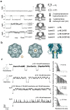
(a) Side and end views of the bilayer-spanning gramicidin A (GrA) channel. The energy-minimized structure represents a composite consistent with several NMR structures (PDB 1GRM, 1MAG); (b) GrA channels formed by the transbilayer dimerization of two β-helical subunits; (c) Single-channel current trace obtained with GrA in diphytanoylphosphatidylcholine bilayer [34]. Reprinted and modified with permission from reference [34].
Figure 2
Figure 2
(a) Typical recording of a single αHL channel reconstituted into diphytanoyl-phosphatidylcholine membrane. Applied voltage is 100 mV. Channel current corresponds to ~100 pA in 1M KCl, pH 7.4; (b) Crystal structure of the αHL heptamer (top and side views are shown) (PDB 7AHL) [78]. The mushroom-shaped complex is approximately 100 Å tall and up to 100 Å in diameter, and the stem domain measures about 52 Å in height and 26 Å in diameter.
Figure 3
Figure 3
(a) A schematic model of Bacillus anthracis toxins cell entry; (b) Side and top views of anthrax toxin PA63 component (the symmetric model, PDB:1V36) [101]. The Phe427s are marked because of their importance in the transport properties; (c) Conductance of a single PA63 channel reconstituted into planar lipid membranes demonstrate fast flickering between open and closed states at 1-ms time resolution [102]. Reprinted with permission from references [86,101,102]. Copyright 2011 Wiley, 2012 Elsevier.
Figure 4
Figure 4
(a) Model representation of the engineered GrA-ethylenediamine channel in a lipid bilayer [49]. At both entrances of the channels, an ionizable site (the primary amino group in gram-ethylenediamine; the imidazole group in gramicidin-histamine) is connected to the gramicidin backbone via a carbamate linkage). Representative single-channel currents of gramicidin-ethylenediamine (b) and gramicidin-histamine (c) at different pH values [49]. The closed channel states are seen as the zero current levels. The currents of the trans (It) and cis (Ic) isomeric states of the channel are indicated. Reprinted and modified with permission from reference [49]. ScienceDirect open archive.
Figure 5
Figure 5
Monitoring activity of phospholipase D (PLD) in the model lipid bilayers by changes in single channel conductance of GrA pores [52]. (a) As PLD hydrolyzes electrically neutral PC lipids and produces negatively charged PA lipids, the membrane negative charge is generated. It induces accumulation of the cations close to the membrane surface, leading to a significant increase in channel conductance. Negative charges are shown in red, and positive ions are shown in blue; (b) GrA unitary current vs. time recordings before and after addition of PLD. Reprinted with permission from reference [52]. Copyright 2009 American Chemical Society.
Figure 6
Figure 6
(a) Bilayer recordings showing the interaction of a single αHL pore with β-cyclodextrin and model analytes 2-adamantanamine and 1-adamantanecarboxylic acid; (b) Molecular graphics representation of the interaction between αHL and cyclodextrin; (c) Analysis of drug molecules by stochastic sensing with αHL and a βCD or γCD adapter. Reprinted and modified with permission from reference [139]. Copyright 1999 Nature Publishing Group.
Figure 7
Figure 7
(a) A scheme of single-stranded DNA characterization with an ion channel. The ion current through a single unoccupied pore (illustrated by the channel at the left) is reduced as a single-stranded DNA molecule begins its passage through the pore (illustrated by the channel at the right) [182]; (b) Schematic representation of DNA sequencing model by the ion channel method. The individual DNA bases (G, A, T, and C) interfere sequentially and differentially with the flow of small ions through the pore which leads to the discrete conductance levels characteristic of the G, A, T, and C bases. The order of appearance of the conductance levels sequentially corresponds to the order of bases in the DNA [182]; (c) Oligomers of poly[U] cause transient blockades in the single αHL channel current [182]. Reprinted and modified with permission from reference [182].
Figure 8
Figure 8
A αHL nanopore can be used to detect the single nucleotides in single-stranded DNA or RNA molecules. (a) A homopolymeric DNA oligonucleotide (blue circles) can be immobilized inside the αHL pore with a biotin (yellow)/streptavidin (red) linkage [199]; (b) Detection of the individual nucleotides cleaved from a single stranded RNA by polynucleotide phosphorylase [205]. Schematic representation of RNA oligonucleotide (circles) digested by polynucleotide phosphorylase (PNPase, green), one base at a time. The unbound nucleotides (rNDPs) are detected by the mutant M113R αHL (mutation highlighted in blue) pore equipped with a cyclodextrin adapter (orange). Reprinted with permission from references [199,205].
Figure 9
Figure 9
Detecting intrinsically disordered amyloid peptides, Ab42 [231] and α-synuclein [232] with αHL nanopore. (a) Aβ42 in the presence and absence of β-cyclodextrin (Aβ42-CD) or Congo Red (Aβ42-CR) added from the cis-side chamber (top). Representation of the blockage behavior of the translocation event of Aβ42-CR (bottom); “i” is equal to the difference between the open pore current and the average amplitude of the blockage current, “t” is the duration time of the blockage [231]; (b) Schematic model of α-synuclein interaction with the αHL channel pore [232]. While the membrane-bound helical part of α-synuclein stays on one side of the membrane, the highly negatively charged C-terminal tail of the protein (red/solid line) enters the αHL pore from the trans-side and goes past the channel constriction under the externally applied electric field. Reprinted with permission from references [231,232].
Figure 10
Figure 10
The interaction of LFN with the PA63 multichannel membrane [239]. (a) After the macroscopic PA63-induced current had reached a steady state at +20 mV, ~3 nM LFN was added to the cis compartment of the chamber, which resulted in a rapid fall in current. The cis compartment was then perfused of unbound LFN. At time zero, the voltage was increased to +50 mV, and LFN translocation kinetics through the PA63 channels was indicated by the increase in conductance vs. time; (b) Kinetic transients for LFN translocation at +60 mV and the indicated symmetrical pH values; (c) The rate of LFN translocation is controlled by the magnitude and sign of the transmembrane ΔpH (pHtrans > pHcis); (d) A schematic illustration of the tandem Brownian ratchet translocation mechanism shows a model of several partially unfolded intermediates of LFN during translocation [239]. The scheme demonstrates how the hydrophobic, ϕ-clamp ratchet and the protonation-state ratchet may work together facilitating translocation of LFN. Reprinted with permission from reference [239].
Figure 11
Figure 11
(a) A schematic illustration of two proposed mechanisms for PA63 channel-mediated lethal factor (LF) and edema factor (EF) transport into the cytoplasm. The channel-mediated translocation model suggests that LF and EF pass through the pore [118,239]. The recent membrane rupturing model suggests that the enzymatically active anthrax toxin complexes (namely, LF or EF bound to the PA63 channel) rupture membranes [18]. Time course of the PA63 channel conductance with (b) LF removed before or (c) maintained at 1 nM during cis-side acidification. (Top rows) ~60 PA63 channels were reconstituted into a planar bilayer membrane at pHcis|trans 7.2|7.2 (black) and, 1 nM LF was added to the cis chamber (blue). (Middle rows) The pHcis|trans 5.5|7.2 gradient was formed by perfusing the cis chamber with pH 5.5 buffer that contained either [LF] = 0 (red) or [LF] = 1 nM (green). (Bottom rows) The neutral pH condition (pHcis|trans 7.2|7.2) (black) was restored by perfusing the cis chamber with pH 7.2 buffer. If LF was present, the cis chamber was first perfused with pH 5.5 buffer (red) then pH 7.2 buffer (black). Reprinted with permission from reference [18].
Figure 12
Figure 12
The receptor-based approach to re-engineer PA by disrupting the native receptor-binding function of the toxin and specific linking of the mutated protein to a heterologous receptor-binding protein [268]. Composite representation of the heptameric prepore formed by PA63 (PDB: 1TZO) with EGF (PDB: 1JL9) linked to the C-terminus. An axial view of the heptameric prepore is shown, with domains 1, 2, 3, and 4 in a single subunit of PA63 colored magenta, green, gold, and purple, respectively. EGF is in red. Broken lines represent an 8-amino-acid linker (SPGHKTQP) connecting the N terminus of EGF to the C terminus of PA63. Reprinted with permission from reference [268].
Figure 13
Figure 13
(a) Scheme for reengineering anthrax toxin protective antigen (PA) to modify its action towards two distinct proteolytic activities overexpressed by the cancer cells [281]; (b) Scheme for discovery of PA mutants that exclusively form octamers [282]; (c) EM images of heptameric and octameric PA species [282]. Reprinted with permission from references [281,282].

Similar articles

Cited by

References

    1. Parker M.W., Feil S.C. Pore-forming protein toxins: From structure to function. Prog. Biophys. Mol. Biol. 2005;88:91–142. - PubMed
    1. Geny B., Popoff M.R. Bacterial protein toxins and lipids: Pore formation or toxin entry into cells. Biol. Cell. 2006;98:667–678. doi: 10.1042/BC20050082. - DOI - PubMed
    1. Van der Goot G., editor. Pore-Forming Toxins. Springer-Verlag; New York, NY, USA: 2001. p. 168.
    1. Los F.C., Randis T.M., Aroian R.V., Ratner A.J. Role of pore-forming toxins in bacterial infectious diseases. Microbiol. Mol. Biol. Rev. 2013;77:173–207. doi: 10.1128/MMBR.00052-12. - DOI - PMC - PubMed
    1. Alouf J.E. Pore-forming bacterial protein toxins: An overview. In: Gisou van der Goot F., editor. Pore-Forming Toxins. Springer-Verlag Berlin Heidelberg; Berlin, Germany: 2001. pp. 1–14. - PubMed

Publication types

MeSH terms

LinkOut - more resources