Zygotic genome activation during the maternal-to-zygotic transition
- PMID: 25150012
- PMCID: PMC4303375
- DOI: 10.1146/annurev-cellbio-100913-013027
Zygotic genome activation during the maternal-to-zygotic transition
Abstract
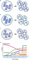
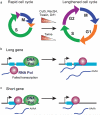
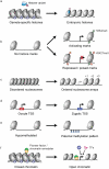
Embryogenesis depends on a highly coordinated cascade of genetically encoded events. In animals, maternal factors contributed by the egg cytoplasm initially control development, whereas the zygotic nuclear genome is quiescent. Subsequently, the genome is activated, embryonic gene products are mobilized, and maternal factors are cleared. This transfer of developmental control is called the maternal-to-zygotic transition (MZT). In this review, we discuss recent advances toward understanding the scope, timing, and mechanisms that underlie zygotic genome activation at the MZT in animals. We describe high-throughput techniques to measure the embryonic transcriptome and explore how regulation of the cell cycle, chromatin, and transcription factors together elicits specific patterns of embryonic gene expression. Finally, we illustrate the interplay between zygotic transcription and maternal clearance and show how these two activities combine to reprogram two terminally differentiated gametes into a totipotent embryo.
Keywords: cellular reprogramming; embryogenesis; maternal clearance; pioneer factors; pluripotency.
Figures


Similar articles
-
The conserved regulatory basis of mRNA contributions to the early Drosophila embryo differs between the maternal and zygotic genomes.PLoS Genet. 2020 Mar 30;16(3):e1008645. doi: 10.1371/journal.pgen.1008645. eCollection 2020 Mar. PLoS Genet. 2020. PMID: 32226006 Free PMC article.
-
Regulatory principles governing the maternal-to-zygotic transition: insights from Drosophila melanogaster.Open Biol. 2018 Dec;8(12):180183. doi: 10.1098/rsob.180183. Open Biol. 2018. PMID: 30977698 Free PMC article. Review.
-
The maternal-to-zygotic transition revisited.Development. 2019 Jun 12;146(11):dev161471. doi: 10.1242/dev.161471. Development. 2019. PMID: 31189646 Review.
-
A story of birth and death: mRNA translation and clearance at the onset of maternal-to-zygotic transition in mammals†.Biol Reprod. 2019 Sep 1;101(3):579-590. doi: 10.1093/biolre/ioz012. Biol Reprod. 2019. PMID: 30715134 Review.
-
Post-translational regulation of the maternal-to-zygotic transition.Cell Mol Life Sci. 2018 May;75(10):1707-1722. doi: 10.1007/s00018-018-2750-y. Epub 2018 Feb 9. Cell Mol Life Sci. 2018. PMID: 29427077 Free PMC article. Review.
Cited by
-
Epigenetic manipulation to improve mouse SCNT embryonic development.Front Genet. 2022 Aug 30;13:932867. doi: 10.3389/fgene.2022.932867. eCollection 2022. Front Genet. 2022. PMID: 36110221 Free PMC article. Review.
-
Metabolic control of histone acetylation for precise and timely regulation of minor ZGA in early mammalian embryos.Cell Discov. 2022 Sep 27;8(1):96. doi: 10.1038/s41421-022-00440-z. Cell Discov. 2022. PMID: 36167681 Free PMC article.
-
The Dynamics of Histone Modifications during Mammalian Zygotic Genome Activation.Int J Mol Sci. 2024 Jan 25;25(3):1459. doi: 10.3390/ijms25031459. Int J Mol Sci. 2024. PMID: 38338738 Free PMC article. Review.
-
The gene expression network regulating queen brain remodeling after insemination and its parallel use in ants with reproductive workers.Sci Adv. 2020 Sep 16;6(38):eaaz5772. doi: 10.1126/sciadv.aaz5772. Print 2020 Sep. Sci Adv. 2020. PMID: 32938672 Free PMC article.
-
Herpesviruses mimic zygotic genome activation to promote viral replication.Res Sq [Preprint]. 2023 Dec 13:rs.3.rs-3125635. doi: 10.21203/rs.3.rs-3125635/v1. Res Sq. 2023. PMID: 38168299 Free PMC article. Preprint.
References
-
- Adenot PG, et al. Differential H4 acetylation of paternal and maternal chromatin precedes DNA replication and differential transcriptional activity in pronuclei of 1-cell mouse embryos. Development (Cambridge, England) 1997;124(22):4615–4625. - PubMed
Publication types
MeSH terms
Substances
Grants and funding
- R01-GM103789-2/GM/NIGMS NIH HHS/United States
- R01-GM081602-06A1/GM/NIGMS NIH HHS/United States
- R01-GM102251-2/GM/NIGMS NIH HHS/United States
- R01 GM103789/GM/NIGMS NIH HHS/United States
- T32GM007499/GM/NIGMS NIH HHS/United States
- R01 GM101108/GM/NIGMS NIH HHS/United States
- R01-HD074078-3/HD/NICHD NIH HHS/United States
- R01 GM102251/GM/NIGMS NIH HHS/United States
- R01 GM081602/GM/NIGMS NIH HHS/United States
- F32HD071697-03/HD/NICHD NIH HHS/United States
- R01 HD074078/HD/NICHD NIH HHS/United States
- R21-HD073768-2/HD/NICHD NIH HHS/United States
- R01-GM101108-3/GM/NIGMS NIH HHS/United States
LinkOut - more resources
Full Text Sources
Other Literature Sources
Molecular Biology Databases

