Comparison of mammographic density assessed as volumes and areas among women undergoing diagnostic image-guided breast biopsy
- PMID: 25139935
- PMCID: PMC4337788
- DOI: 10.1158/1055-9965.EPI-14-0257
Comparison of mammographic density assessed as volumes and areas among women undergoing diagnostic image-guided breast biopsy
Abstract
Background: Mammographic density (MD), the area of non-fatty-appearing tissue divided by total breast area, is a strong breast cancer risk factor. Most MD analyses have used visual categorizations or computer-assisted quantification, which ignore breast thickness. We explored MD volume and area, using a volumetric approach previously validated as predictive of breast cancer risk, in relation to risk factors among women undergoing breast biopsy.
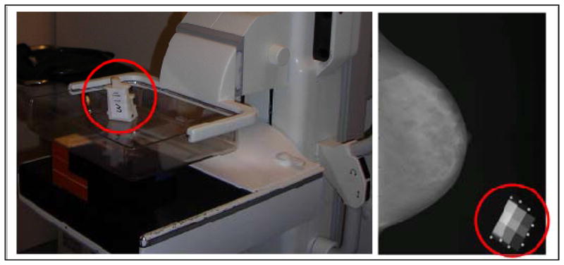
Methods: Among 413 primarily white women, ages 40 to 65 years, undergoing diagnostic breast biopsies between 2007 and 2010 at an academic facility in Vermont, MD volume (cm(3)) was quantified in craniocaudal views of the breast contralateral to the biopsy target using a density phantom, whereas MD area (cm(2)) was measured on the same digital mammograms using thresholding software. Risk factor associations with continuous MD measurements were evaluated using linear regression.
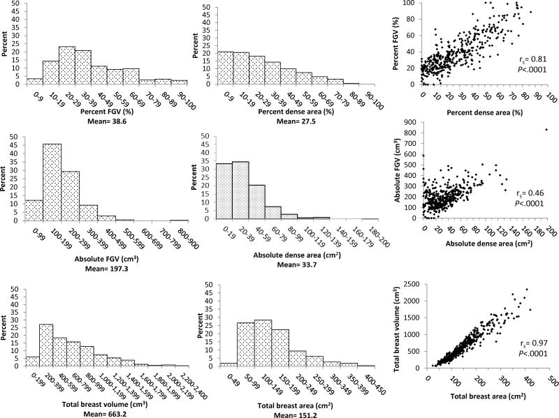
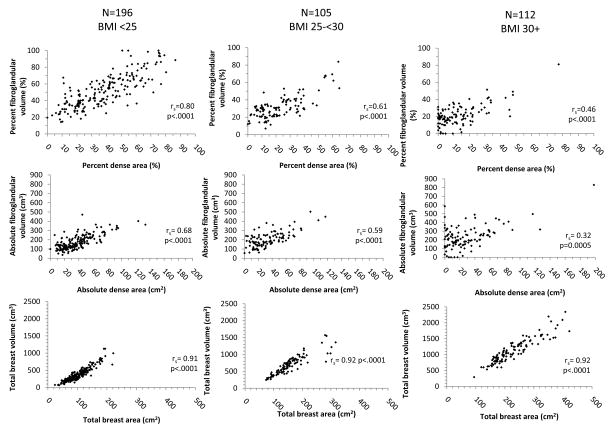
Results: Percent MD volume and area were correlated (r = 0.81) and strongly and inversely associated with age, body mass index (BMI), and menopause. Both measures were inversely associated with smoking and positively associated with breast biopsy history. Absolute MD measures were correlated (r = 0.46) and inversely related to age and menopause. Whereas absolute dense area was inversely associated with BMI, absolute dense volume was positively associated.
Conclusions: Volume and area MD measures exhibit some overlap in risk factor associations, but divergence as well, particularly for BMI.
Impact: Findings suggest that volume and area density measures differ in subsets of women; notably, among obese women, absolute density was higher with volumetric methods, suggesting that breast cancer risk assessments may vary for these techniques.
©2014 American Association for Cancer Research.
Conflict of interest statement
Figures
Similar articles
-
Leukocyte telomere length and its association with mammographic density and proliferative diagnosis among women undergoing diagnostic image-guided breast biopsy.BMC Cancer. 2015 Oct 30;15:823. doi: 10.1186/s12885-015-1860-2. BMC Cancer. 2015. PMID: 26519084 Free PMC article.
-
Relationship of circulating insulin-like growth factor-I and binding proteins 1-7 with mammographic density among women undergoing image-guided diagnostic breast biopsy.Breast Cancer Res. 2019 Jul 23;21(1):81. doi: 10.1186/s13058-019-1162-8. Breast Cancer Res. 2019. PMID: 31337427 Free PMC article.
-
Relationship of Terminal Duct Lobular Unit Involution of the Breast with Area and Volume Mammographic Densities.Cancer Prev Res (Phila). 2016 Feb;9(2):149-58. doi: 10.1158/1940-6207.CAPR-15-0282. Epub 2015 Dec 8. Cancer Prev Res (Phila). 2016. PMID: 26645278 Free PMC article.
-
Mammographic density and risk of breast cancer.Am Soc Clin Oncol Educ Book. 2013. doi: 10.14694/EdBook_AM.2013.33.e57. Am Soc Clin Oncol Educ Book. 2013. PMID: 23714456 Review.
-
Mammographic density is not a worthwhile examination to distinguish high cancer risk women in screening.Eur Radiol. 2014 Oct;24(10):2412-6. doi: 10.1007/s00330-014-3278-7. Epub 2014 Jun 28. Eur Radiol. 2014. PMID: 24972955 Review.
Cited by
-
Relation of Serum Estrogen Metabolites with Terminal Duct Lobular Unit Involution Among Women Undergoing Diagnostic Image-Guided Breast Biopsy.Horm Cancer. 2016 Dec;7(5-6):305-315. doi: 10.1007/s12672-016-0265-2. Epub 2016 May 2. Horm Cancer. 2016. PMID: 27138982 Free PMC article.
-
Relationships between mammographic density, tissue microvessel density, and breast biopsy diagnosis.Breast Cancer Res. 2016 Aug 23;18(1):88. doi: 10.1186/s13058-016-0746-9. Breast Cancer Res. 2016. PMID: 27552842 Free PMC article.
-
Ages at menarche- and menopause-related genetic variants in relation to terminal duct lobular unit involution in normal breast tissue.Breast Cancer Res Treat. 2016 Jul;158(2):341-50. doi: 10.1007/s10549-016-3859-z. Epub 2016 Jun 24. Breast Cancer Res Treat. 2016. PMID: 27342457 Free PMC article.
-
Adiposity, breast density, and breast cancer risk: epidemiological and biological considerations.Eur J Cancer Prev. 2017 Nov;26(6):511-520. doi: 10.1097/CEJ.0000000000000310. Eur J Cancer Prev. 2017. PMID: 27571214 Free PMC article. Review.
-
Leukocyte telomere length and its association with mammographic density and proliferative diagnosis among women undergoing diagnostic image-guided breast biopsy.BMC Cancer. 2015 Oct 30;15:823. doi: 10.1186/s12885-015-1860-2. BMC Cancer. 2015. PMID: 26519084 Free PMC article.
References
-
- Boyd NF, Lockwood GA, Byng JW, Tritchler DL, Yaffe MJ. Mammographic densities and breast cancer risk. Cancer Epidemiol Biomarkers Prev. 1998;7:1133–44. - PubMed
-
- McCormack VA, dos Santos Silva I. Breast Density and Parenchymal Patterns as Markers of Breast Cancer Risk: A Meta-analysis. Cancer Epidemiol Biomarkers Prev. 2006;15:1159–69. - PubMed
-
- D’Orsi CJ, Bassett LW, Berg WA, et al. Breast Imaging Reporting and Data System: ACR BI-RADS-Mammography. 4. Reston, VA: American College of Radiology; 2003.
-
- Highnam R, Pan X, Warren R, Jeffreys M, Davey Smith G, Brady M. Breast composition measurements using retrospective standard mammogram form (SMF) Phys Med Biol. 2006;51:2695–713. - PubMed
Publication types
MeSH terms
Grants and funding
LinkOut - more resources
Full Text Sources
Other Literature Sources
Medical