Effect of ERK1/2 signaling pathway in electro-acupuncture mediated up-regulation of heme oxygenase-1 in lungs of rabbits with endotoxic shock
- PMID: 25139460
- PMCID: PMC4144948
- DOI: 10.12659/MSM.890736
Effect of ERK1/2 signaling pathway in electro-acupuncture mediated up-regulation of heme oxygenase-1 in lungs of rabbits with endotoxic shock
Abstract
Background: The anti-oxidative and anti-inflammatory activities of electro-acupuncture (EA), a traditional clinical method, are widely accepted, but its mechanisms are not yet well defined. In this study, we investigated the role of extracellular signal-regulated kinases1/2 (ERK1/2) pathways on electro-acupuncture - mediated up-regulation of heme oxygenase-1 (HO-1) in rabbit lungs injured by LPS-induced endotoxic shock.
Material/methods: Seventy rabbits were randomly divided into 7 groups: group C, group M, group D, group SEAM, group EAM, group EAMPD, and group PD98059. Male New England white rabbits were given EA treatment on both sides once a day on days 1-5, and then received LPS to replicate the experimental model of injured lung induced by endotoxic shock. Then, they were killed by exsanguination at 6 h after LPS administration. The blood samples were collected for serum examination, and the lungs were removed for pathology examination, determination of wet-to-dry weight ratio, MDA content, SOD activity, serum tumor necrosis factor-α, determination of HO-1 protein and mRNA expression, and determination of ERK1/2 protein.
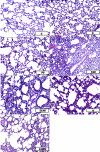
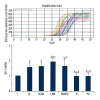
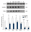
Results: The results revealed that after EA treatment, expression of HO-1and ERK1/2 was slightly increased compared to those in other groups, accompanied with less severe lung injury as indicated by lower index of lung injury score, lower wet-to-dry weight ratio, MDA content, and serum tumor necrosis factor-α levels, and greater SOD activity (p<0.05 for all). After pretreatment with ERK1/2 inhibitor PD98059, the effect of EA treatment and expression of HO-1 were suppressed (p<0.05 for all).
Conclusions: After electro-acupuncture stimulation at ST36 and BL13, severe lung injury during endotoxic shock was attenuated. The mechanism may be through up-regulation of HO-1, mediated by the signal transductions of ERK1/2 pathways. Thus, the regulation of ERK1/2 pathways via electro-acupuncture may be a therapeutic strategy for endotoxic shock.
Figures


Similar articles
-
Role of HO-1 in protective effect of electro-acupuncture against endotoxin shock-induced acute lung injury in rabbits.Exp Biol Med (Maywood). 2013 Jun;238(6):705-12. doi: 10.1177/1535370213489487. Exp Biol Med (Maywood). 2013. PMID: 23918882
-
Role of Nrf2/ARE pathway in protective effect of electroacupuncture against endotoxic shock-induced acute lung injury in rabbits.PLoS One. 2014 Aug 12;9(8):e104924. doi: 10.1371/journal.pone.0104924. eCollection 2014. PLoS One. 2014. PMID: 25115759 Free PMC article.
-
Electroacupuncture Ameliorates Acute Renal Injury in Lipopolysaccharide-Stimulated Rabbits via Induction of HO-1 through the PI3K/Akt/Nrf2 Pathways.PLoS One. 2015 Nov 2;10(11):e0141622. doi: 10.1371/journal.pone.0141622. eCollection 2015. PLoS One. 2015. PMID: 26524181 Free PMC article.
-
Protective effects of heme-oxygenase expression against endotoxic shock: inhibition of tumor necrosis factor-alpha and augmentation of interleukin-10.J Trauma. 2006 Nov;61(5):1078-84. doi: 10.1097/01.ta.0000239359.41464.ef. J Trauma. 2006. PMID: 17099512
-
[The role of hydrogen sulfide in acute lung injury during endotoxic shock and its relationship with nitric oxide and carbon monoxide].Zhonghua Yi Xue Za Zhi. 2008 Aug 19;88(32):2240-5. Zhonghua Yi Xue Za Zhi. 2008. PMID: 19087669 Chinese.
Cited by
-
CircMAPK1 induces cell pyroptosis in sepsis-induced lung injury by mediating KDM2B mRNA decay to epigenetically regulate WNK1.Mol Med. 2024 Sep 19;30(1):155. doi: 10.1186/s10020-024-00932-6. Mol Med. 2024. PMID: 39300342 Free PMC article.
-
Acupoint Catgut Embedding Improves the Lipopolysaccharide-Induced Acute Respiratory Distress Syndrome in Rats.Biomed Res Int. 2020 May 2;2020:2394734. doi: 10.1155/2020/2394734. eCollection 2020. Biomed Res Int. 2020. PMID: 32566670 Free PMC article.
-
Revealing the biological mechanism of acupuncture in alleviating excessive inflammatory responses and organ damage in sepsis: a systematic review.Front Immunol. 2023 Sep 11;14:1242640. doi: 10.3389/fimmu.2023.1242640. eCollection 2023. Front Immunol. 2023. PMID: 37753078 Free PMC article. Review.
-
Electro-acupuncture Pretreatment at Zusanli (ST36) Acupoint Attenuates Lipopolysaccharide-Induced Inflammation in Rats by Inhibiting Ca2+ Influx Associated with Cannabinoid CB2 Receptors.Inflammation. 2019 Feb;42(1):211-220. doi: 10.1007/s10753-018-0885-5. Inflammation. 2019. PMID: 30168040
-
Cytotoxic Effects of Tropodithietic Acid on Mammalian Clonal Cell Lines of Neuronal and Glial Origin.Mar Drugs. 2015 Nov 27;13(12):7113-23. doi: 10.3390/md13127058. Mar Drugs. 2015. PMID: 26633426 Free PMC article.
References
-
- Yang RH, Yang LN, Shen XC, et al. Suppression of NF-κB pathway by crocetin contributes to attenuation of lipopolysaccharide-induced acute lung injury in mice. Eur J Pharmacol. 2012;674:391–96. - PubMed
-
- Holst LB, Haase N, Wetterslev J, et al. Transfusion requirements in septic shock (TRISS) trial – comparing the effects and safety of liberal versus restrictive red blood cell transfusion in septic shock patients in the ICU: protocol for a randomised controlled trial. Trials. 2013;14:150. - PMC - PubMed
-
- Chon TY, Lee MC. Acupuncture. Mayo Clin Proc. 2013;10:1141–46. - PubMed
-
- Huang CL, Huang CJ, Tsai PS, et al. Acupuncture stimulation of ST-36 (Zusanli) significantly mitigates acute lung injury in lipopolysaccharide-stimulated rats. Acta Anaesthesiol Scand. 2006;50:722–30. - PubMed
Publication types
MeSH terms
Substances
LinkOut - more resources
Full Text Sources
Other Literature Sources
Miscellaneous

