. 2015 Feb 1;211(3):462-71.
doi: 10.1093/infdis/jiu460. Epub 2014 Aug 19.

BB0323 and novel virulence determinant BB0238: Borrelia burgdorferi proteins that interact with and stabilize each other and are critical for infectivity

Affiliations

BB0323 and novel virulence determinant BB0238: Borrelia burgdorferi proteins that interact with and stabilize each other and are critical for infectivity

Toru Kariu et al. J Infect Dis. .

Abstract

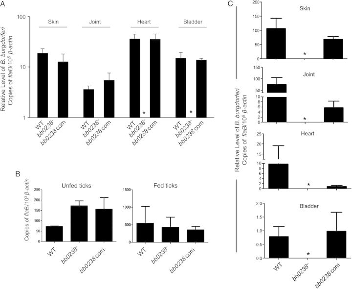
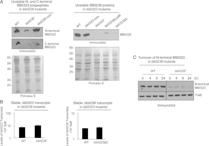
We have shown that Borrelia burgdorferi gene product BB0323 is essential for cell fission and pathogen persistence in vivo. Here we describe characterization of a conserved hypothetical protein annotated as BB0238, which specifically interacts with the N-terminal region of BB0323. We show that BB0238 is a subsurface protein, and similar to BB0323, exists in the periplasm and as a membrane-bound protein. Deletion of bb0238 in infectious B. burgdorferi did not affect microbial growth in vitro or survival in ticks, but the mutant was unable to persist in mice or transmit from ticks--defects that are restored on genetic complementation. Remarkably, BB0238 and BB0323 contribute to mutual posttranslational stability, because deletion of one causes dramatic reduction in the protein level of the other partner. Interference with the function of BB0238 or BB0323 and their interaction may provide novel strategies to combat B. burgdorferi infection.

Keywords: BB0238; BB0323; Borrelia burgdorferi; pathogen persistence; posttranslational stability; protein–protein interaction.


Figures

Figure 1.
Figure 1.
A, B, BB0238 interacts with BB0323. Far-Western blot analyses demonstrate specific binding of BB0238 to BB0323 (A) or binding of BB0323 to BB0238 (B). The recombinant proteins (either BB0238 or BB0323) were separated by sodium dodecyl sulfate–polyacrylamide gel electrophoresis (SDS-PAGE) and incubated with glutathione S-transferase (GST)–fused proteins (either BB0323 or BB0238) or GST (control). Bound proteins were detected by anti-GST antibodies conjugated to horseradish peroxidase. Arrows denote the position of the band specifically detected by the recombinant protein; asterisk, either a nonspecific band or a degraded form of BB0323. Migration of protein standards is shown to the left in kilodaltons. C, Schematic representation of various truncated recombinant BB0323 proteins representing N- and C-terminal polypeptides. Truncated polypeptides were produced with or without GST tags. D, BB0238 binds to the N-terminal BB0323. Left panel denotes far-Western analysis; recombinant BB0238 proteins were separated by SDS-PAGE, blotted onto nitrocellulose membranes, and incubated with GST-fused N1 (GST-N1), GST-fused N3 (GST-N3), GST, or GST-fused C-terminal polypeptide (GST-C). Bound proteins were detected with immunoblotting using anti-GST antibodies. Only N-terminal polypeptides exhibit detectable interaction in the assay. Equal protein loading is indicated by immunoblotting using BB0238 antibody (right panel).
Figure 2.
Figure 2.
BB0238 is a subsurface protein that exists in the periplasm and as a membrane-bound form and is produced throughout the tick-mouse infection cycle of Borrelia burgdorferi. A, BB0238 is not exposed on the spirochete surface. B, burgdorferi cells were incubated with (+) or without (−) proteinase K for the removal of protease-sensitive surface proteins and then processed for immunoblot analysis using either BB0238 or control surface-exposed (OspA) and subsurface (FlaB) protein antibodies. B, BB0238 is undetectable in the isolated outer membrane vesicle fraction. B. burgdorferi lysates were separated into protoplasmic cylinder (PC) and outer membrane vesicle (OMV) fractions by sodium dodecyl sulfate–polyacrylamide gel electrophoresis and immunoblotted with antiserum against BB0238 or outer membrane protein (OspA). C, Detection of BB0238 in the periplasmic and spheroplasmic fractions of B. burgdorferi cells. Periplasmic or spheroplasmic proteins were extracted from cultured spirochetes and immunoblotted using BB0238 antibodies. D, Triton X-114 phase partitioning of B. burgdorferi proteins. Spirochete lysates were subjected to Triton X-114 phase partitioning of aqueous and detergent phases and immunoblotted using antibodies against BB0238, N-terminal BB0323, C-terminal BB0323, or a control (OspA) protein. E, Consistent expression of bb0238 during the enzootic life cycle of the spirochete. Total tissue RNA was isolated from representative stages of murine and tick infection, and bb0238 transcripts were measured using quantitative reverse-transcription polymerase chain reaction (qRT-PCR) and presented as copies of bb0238 transcript per copy of flaB transcript. Error bars show the means and standard errors of the mean from ≥3 independent qRT-PCR analyses from individual ticks.
Figure 3.
Figure 3.
Construction and analysis of the bb0238 mutant and complemented isolates of Borrelia burgdorferi. A, Schematic drawings of wild-type (WT) and bb0238-mutant (bb0238) isolates at the bb0238 locus. Genes bb0237–bb0241 (white arrows) and the kanamycin-resistance cassette driven by B. burgdorferi flaB promoter (flaB-kan) (black arrow) are indicated. Primers P1–P4 (arrowheads) were used to amplify 5′ and 3′ arms for homologous recombination, and regions flanking the bb0238 locus were ligated on either side of the flaB-kan cassette to obtain the mutagenic construct, as detailed in the text. B, Integration of the mutagenic construct, flaB-kan, in the intended genomic locus. Primers 5–10 (arrowheads) were employed for polymerase chain reaction (PCR) analysis, using DNA isolated from WT or mutant B. burgdorferi and subjected to agarose gel electrophoresis. The combination of primers used for PCR is indicated at the top, and migration of the DNA ladder is shown on the left. C, Reverse-transcription PCR assessment of bb0238 ablation and the polar effects of mutagenesis. Total RNA was isolated from WT or bb0238 mutant (bb0238), converted to complementary DNA, and used to amplify regions within bb0238, flaB, the kanamycin-resistance cassette, and genes surrounding the bb0238 locus (bb0237 and bb0239); the products were visualized on a gel. D, Schematic illustration of the bb0238-complemented construct for chromosomal integration of the bb0238 in the original gene locus of the spirochete chromosome. Construct was used for integration into B. burgdorferi genomic DNA via homologous recombination. E, Production of BB0238 by the complemented B. burgdorferi. Spirochete lysates were separated by sodium dodecyl sulfate–polyacrylamide gel electrophoresis and immunoblotted with anti-BB0238 (top) or anti-FlaB (bottom) antibodies. F, Growth curves for WT and genetically manipulated spirochetes. B. burgdorferi were diluted to a density of 105 (or 1.E+05) cells/mL and grown at 34°C in BSK-H medium. Samples were counted under a dark field microscope every 24 hours using a Petroff-Hausser cell counter. Differences between the numbers of WT and bb0238 or bb0238-complemented strains were not significant at all times (P > .05).
Figure 4.
Figure 4.
Deletion of bb0238 attenuates spirochete ability to infect mice and transmit from ticks to mice. A, bb0238 mutants failed to persist in mice. C3H/HeN mice were inoculated intradermally with equal numbers of wild-type (WT), bb0238 mutant (bb0238), or bb0238-complemented (bb0238 com) Borrelia burgdorferi (105 spirochetes per mouse, 3 animals per group). Infection was assessed by quantitative reverse-transcription polymerase chain reaction (qRT-PCR) analysis of pathogen burden in skin, joint, heart, and bladder samples obtained 2 weeks after infection. Asterisks indicate undetectable levels of bb0238 mutants. B, B. burgdorferi burdens in artificially infected ticks. Ixodes scapularis nymphs were microinjected with equal numbers of WT, bb0238 mutant, or bb0238-complemented B. burgdorferi and then placed on mice (5 ticks per mouse). B. burgdorferi levels in unfed and fed repleted ticks were analyzed with qRT-PCR, measuring the number of copies of B. burgdorferi flaB transcripts and normalizing against tick β-actin transcripts. C, bb0238 is required for spirochete transmission from ticks to mice. Ticks were microinjected with equal numbers WT, bb0238 mutant, or bb0238-complemented B. burgdorferi and allowed to engorge on mice (3 animals per group). Spirochete burdens in different tissues were analyzed with qRT-PCR after 2 weeks of infection by measuring copies of flaB and were normalized against mouse β-actin levels. Error bars represent means and standard errors of the mean for relative levels of pathogen in tissues from 3 independent animal infection experiments.
Figure 5.
Figure 5.
BB0238 and BB0323 contribute to mutual posttranslational stability. A, Deletion of bb0238 or bb0323 in spirochetes reduces protein levels of the respective partner, as revealed by Western blot analysis. Comparable amounts of lysates from wild-type (WT), bb0238 mutants (bb0238), bb0238-complemented isolates (bb0238 com), bb0323 mutants (bb0323), BB0323 C-terminal–deletion mutants (bb0323ΔC), and BB0323 LysM domain–deletion mutants (bb0323ΔLysM) were separated by sodium dodecyl sulfate–polyacrylamide gel electrophoresis (SDS-PAGE) and immunoblotted with specific antibodies against BB0238 or N- or C-terminal polypeptides of BB0323. Protein loading is indicated by staining with Ponceau S (bottom panels). Migration of protein standards is shown to the left in kilodaltons (left panel). B, Targeted loss of bb0238 or bb0323 in Borrelia burgdorferi did not alter transcript levels of the respective partner. The level of bb0323 transcripts in WT and bb0238 mutants (top panel) or bb0238 transcripts in WT and bb323 mutants (bb0323ΔC) (bottom panel) was measured with quantitative reverse-transcription polymerase chain reaction. C, Posttranslational stability of BB0323 in bb0238 mutant. Protein synthesis was inhibited by the addition of 100 µg/mL spectinomycin to growing culture of spirochetes, and levels of N-terminal BB0323 were determined with immunoblot analyses. At indicated time points, spirochetes were collected, and lysates were separated by SDS-PAGE and immunoblotted using anti-N-terminal BB0323 and anti-FlaB (loading control).

Similar articles

Cited by

References

    1. Piesman J, Eisen L. Prevention of tick-borne diseases. Annu Rev Entomol. 2008;53:323–43. - PubMed
    1. Piesman J, Gern L. Lyme borreliosis in Europe and North America. Parasitology. 2004;129(suppl)):S191–220. - PubMed
    1. Steere AC, Coburn J, Glickstein L. The emergence of Lyme disease. J Clin Invest. 2004;113:1093–101. - PMC - PubMed
    1. Barbour AG, Hayes SF. Biology of Borrelia species. Microbiol Rev. 1986;50:381–400. - PMC - PubMed
    1. Burgdorfer W, Barbour AG, Hayes SF, Benach JL, Grunwaldt E, Davis JP. Lyme disease—a tick-borne spirochetosis? Science. 1982;216:1317–9. - PubMed

Publication types