

Distinct roles of major peptidoglycan recycling enzymes in β-Lactamase production in Shewanella oneidensis
- PMID: 25136029
- PMCID: PMC4249409
- DOI: 10.1128/AAC.03238-14
Distinct roles of major peptidoglycan recycling enzymes in β-Lactamase production in Shewanella oneidensis
Abstract
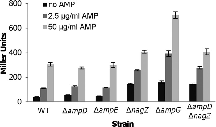
β-Lactam antibiotics were the earliest discovered and are the most widely used group of antibiotics that work by inactivating penicillin-binding proteins to inhibit peptidoglycan biosynthesis. As one of the most efficient defense strategies, many bacteria produce β-lactam-degrading enzymes, β-lactamases, whose biochemical functions and regulation have been extensively studied. A signal transduction pathway for β-lactamase induction by β-lactam antibiotics, consisting of the major peptidoglycan recycling enzymes and the LysR-type transcriptional regulator, AmpR, has been recently unveiled in some bacteria. Because inactivation of some of these proteins, especially the permease AmpG and the β-hexosaminidase NagZ, results in substantially elevated susceptibility to the antibiotics, these have been recognized as potential therapeutic targets. Here, we show a contrasting scenario in Shewanella oneidensis, in which the homologue of AmpR is absent. Loss of AmpG or NagZ enhances β-lactam resistance drastically, whereas other identified major peptidoglycan recycling enzymes are dispensable. Moreover, our data indicate that there exists a parallel signal transduction pathway for β-lactamase induction, which is independent of either AmpG or NagZ.
Copyright © 2014, American Society for Microbiology. All Rights Reserved.
Figures
Similar articles
-
PBP1a/LpoA but not PBP1b/LpoB are involved in regulation of the major β-lactamase gene blaA in Shewanella oneidensis.Antimicrob Agents Chemother. 2015;59(6):3357-64. doi: 10.1128/AAC.04669-14. Epub 2015 Mar 30. Antimicrob Agents Chemother. 2015. PMID: 25824223 Free PMC article.
-
Impact of Peptidoglycan Recycling Blockade and Expression of Horizontally Acquired β-Lactamases on Pseudomonas aeruginosa Virulence.Microbiol Spectr. 2022 Feb 23;10(1):e0201921. doi: 10.1128/spectrum.02019-21. Epub 2022 Feb 16. Microbiol Spectr. 2022. PMID: 35171032 Free PMC article.
-
The sentinel role of peptidoglycan recycling in the β-lactam resistance of the Gram-negative Enterobacteriaceae and Pseudomonas aeruginosa.Bioorg Chem. 2014 Oct;56:41-8. doi: 10.1016/j.bioorg.2014.05.011. Epub 2014 Jun 4. Bioorg Chem. 2014. PMID: 24955547 Free PMC article. Review.
-
Role of Pseudomonas aeruginosa low-molecular-mass penicillin-binding proteins in AmpC expression, β-lactam resistance, and peptidoglycan structure.Antimicrob Agents Chemother. 2015 Jul;59(7):3925-34. doi: 10.1128/AAC.05150-14. Epub 2015 Apr 20. Antimicrob Agents Chemother. 2015. PMID: 25896695 Free PMC article.
-
[Progress in regulatory mechanism for inducing β-lactamase in Gram-negative bacteria].Sheng Wu Gong Cheng Xue Bao. 2018 Aug 25;34(8):1288-1296. doi: 10.13345/j.cjb.180187. Sheng Wu Gong Cheng Xue Bao. 2018. PMID: 30152214 Review. Chinese.
Cited by
-
Complex Regulation Pathways of AmpC-Mediated β-Lactam Resistance in Enterobacter cloacae Complex.Antimicrob Agents Chemother. 2015 Dec;59(12):7753-61. doi: 10.1128/AAC.01729-15. Epub 2015 Oct 5. Antimicrob Agents Chemother. 2015. PMID: 26438498 Free PMC article.
-
PBP1a/LpoA but not PBP1b/LpoB are involved in regulation of the major β-lactamase gene blaA in Shewanella oneidensis.Antimicrob Agents Chemother. 2015;59(6):3357-64. doi: 10.1128/AAC.04669-14. Epub 2015 Mar 30. Antimicrob Agents Chemother. 2015. PMID: 25824223 Free PMC article.
-
Bacterial virulence regulation through soluble peptidoglycan fragments sensing and response: knowledge gaps and therapeutic potential.FEMS Microbiol Rev. 2023 Mar 10;47(2):fuad010. doi: 10.1093/femsre/fuad010. FEMS Microbiol Rev. 2023. PMID: 36893807 Free PMC article.
-
Positive regulation of the Shewanella oneidensis OmpS38, a major porin facilitating anaerobic respiration, by Crp and Fur.Sci Rep. 2015 Sep 18;5:14263. doi: 10.1038/srep14263. Sci Rep. 2015. PMID: 26381456 Free PMC article.
-
Deletion of Lytic Transglycosylases Increases Beta-Lactam Resistance in Shewanella oneidensis.Front Microbiol. 2018 Jan 22;9:13. doi: 10.3389/fmicb.2018.00013. eCollection 2018. Front Microbiol. 2018. PMID: 29403465 Free PMC article.
References
Publication types
MeSH terms
Substances
LinkOut - more resources
Full Text Sources
Other Literature Sources
Research Materials
