Comparative Study
. 2015 Sep;64(9):1368-78.
doi: 10.1136/gutjnl-2014-307020. Epub 2014 Aug 18.

A pro-inflammatory role for Th22 cells in Helicobacter pylori-associated gastritis

Affiliations
Comparative Study

A pro-inflammatory role for Th22 cells in Helicobacter pylori-associated gastritis

Yuan Zhuang et al. Gut. 2015 Sep.

Abstract

Objective: Helper T (Th) cell responses are critical for the pathogenesis of Helicobacter pylori-induced gastritis. Th22 cells represent a newly discovered Th cell subset, but their relevance to H. pylori-induced gastritis is unknown.

Design: Flow cytometry, real-time PCR and ELISA analyses were performed to examine cell, protein and transcript levels in gastric samples from patients and mice infected with H. pylori. Gastric tissues from interleukin (IL)-22-deficient and wild-type (control) mice were also examined. Tissue inflammation was determined for pro-inflammatory cell infiltration and pro-inflammatory protein production. Gastric epithelial cells and myeloid-derived suppressor cells (MDSC) were isolated, stimulated and/or cultured for Th22 cell function assays.

Results: Th22 cells accumulated in gastric mucosa of both patients and mice infected with H. pylori. Th22 cell polarisation was promoted via the production of IL-23 by dendritic cells (DC) during H. pylori infection, and resulted in increased inflammation within the gastric mucosa. This inflammation was characterised by the CXCR2-dependent influx of MDSCs, whose migration was induced via the IL-22-dependent production of CXCL2 by gastric epithelial cells. Under the influence of IL-22, MDSCs, in turn, produced pro-inflammatory proteins, such as S100A8 and S100A9, and suppressed Th1 cell responses, thereby contributing to the development of H. pylori-associated gastritis.

Conclusions: This study, therefore, identifies a novel regulatory network involving H. pylori, DCs, Th22 cells, gastric epithelial cells and MDSCs, which collectively exert a pro-inflammatory effect within the gastric microenvironment. Efforts to inhibit this Th22-dependent pathway may therefore prove a valuable strategy in the therapy of H. pylori-associated gastritis.

Keywords: GASTRITIS; HELICOBACTER PYLORI.


Figures

Figure 1
Figure 1
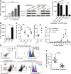
Th22 cells accumulated in gastric mucosa of Helicobacter pylori-infected patients and mice. (A) The percentage of T helper type 22 (Th22) cells in CD3+ cells in gastric mucosa of H. pylori-infected (n=22) and uninfected donors (n=12) was compared. Results are expressed as percentage of Th22 cells in CD3+ T cells. (B and C) Interleukin (IL)-22 mRNA expression (B) and IL-22 protein concentrations (C) in gastric mucosa of H. pylori-infected (n=46) and uninfected donors (n=27) were compared. (D) The correlation between IL-22 expression and H. pylori colonisation was analysed. (E) IL-22 mRNA expression in gastric mucosa of cagA+ H. pylori-infected (n=35), cagA H. pylori-infected (n=11) and uninfected donors (n=29) were compared. (F) Dynamic changes of Th22 cell response and IL-22 mRNA expression in wild-type (WT) H. pylori-infected, ΔcagA-infected and uninfected BALB/c mice. n=5 per group per time point in F. *p<0.05, **p<0.01 n.s. p>0.05 for groups connected by horizontal lines compared, or compared with uninfected mice. n.s. not significant.
Figure 2
Figure 2
Helicobacter pylori-stimulated DCs induce Th22 cell polarisation via IL-23. (A and B) Th22 cell polarisation was assessed by flow cytometry, as described in the Methods section, and statistically analysed. Results are expressed as percentage of Th22 cells in CD4+ T cells. (C) Concentrations of IL-22 protein in unstimulated, WT H. pylori-stimulated or ΔcagA-stimulated DCs derived from blood monocytes and in the DC supernatants were compared (n=3). (D) Th22 cell polarisation was assessed by flow cytometry, as described in the Methods section, and statistically analysed. Results are expressed as percentage of Th22 cells in CD4+ T cells. Results are representative of three independent experiments. (E and F) Th22 cell response in gastric mucosa of WT H. pylori-infected WT C57BL/6 and IL-23 KO (E) or WT H. pylori-infected BM chimaera mice (F) on day 35 postinfection were compared. *p<0.05, **p<0.01, n.s. p>0.05 for groups connected by horizontal lines compared. DC, dendritic cells; Th; helper T cells; WT; wild-type; KO, knockout; BM, bone marrow; MOI, multiplicity of infection; BMDC, bone marrow–derived dendritic cells; IL, interleukin; n.s., not significant.
Figure 3
Figure 3
IL-22 has pro-inflammatory effects during Helicobacter pylori infection and H. pylori induce gastric epithelial cells to upregulate IL-22R1. (A) IL-22 mRNA expression in gastric mucosa of H. pylori-infected patients with mild (n=12), moderate (n=10), severe inflammation (n=11) and uninfected donors with normal gastric histopathology (n=15) was compared. (B) Histological scores of inflammation in gastric antra of the WT H. pylori-infected WT BALB/c or IL-22 KO mice or WT H. pylori-infected WT BALB/c mice injected with IL-22 or Abs against IL-22 on day 49 postinfection were compared. H&E staining, scale bars: 100 μ. (C) Expression of IL-22 mRNA in gastric mucosa of H. pylori-infected patients (n=22) and uninfected donors (n=8) was compared. Dynamic change of IL-22R1 mRNA expression in WT H. pylori-infected, ΔcagA-infected and uninfected BALB/c mice. (D) Representative immunofluorescence staining images showed IL-22R1 expression in gastric mucosa of WT H. pylori-infected and ΔcagA-infected mice or cagA+ H. pylori-infected and cagA H. pylori-infected patients. Scale bars: 20 μ. (E) IL-22R1 mRNA expression or IL-22R1 protein in WT H. pylori-infected, ΔcagA-infected, and uninfected primary gastric epithelial cells from uninfected donors were compared (n=3) or analysed by western blot. *p<0.05, **p<0.01, n.s. p>0.05 for groups connected by horizontal lines compared. WT; wild-type; KO, knockout; IL, interleukin; MOI, multiplicity of infection; PBS, phosphate-buffered saline; GADPH, glyceraldehyde 3-phosphate dehydrogenase; Abs, antibodies; n.s., not significant.
Figure 4
Figure 4
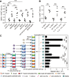
IL-22 promotes CXCL2 production in vivo and in vivo and CXCR2-expressing MDSCs accumulated in gastric mucosa during Helicobacter pylori infection. (A and B) AGS cells (A) and primary gastric epithelial cells (B) were pretreated and stimulated as described in Methods. CXC12 production was detected in cell supernatants by ELISA (n=3). STAT3 and p-STAT3 proteins were analysed by western blot. (C) Concentrations of CXCL2 protein in gastric mucosa of WT H. pylori-infected WT BALB/c mice injected with IL-22 or PBS control, or Abs against IL-22 or corresponding isotype control Ab, or of WT H. pylori-infected WT BALB/c and IL-22 KO mice on day 42 postinfection (p.i) were compared. (D) Dynamic change of MDSCs in WT H. pylori-infected, ΔcagA-infected and uninfected BALB/c mice. (E) Representative dot plots of MDSCs by gating on CD45+ cells and expression of Ly6C, Ly6G and CXCR2 on MDSCs in gastric mucosa of WT H. pylori-infected mice on day 49 p.i., and representative dot plots of MDSCs by gating on CD45+ cells and expression of CXCR2 on MDSCs in peripheral blood of H. pylori-infected and uninfected donors. Numbers indicate relative percentages in CD45+ cells. (F) MDSC level in peripheral blood of H. pylori-infected patients (n=22) and uninfected donors (n=21) was compared; n=5 per group per time point in D. *p<0.05, **p<0.01, n.s. p>0.05 for groups connected by horizontal lines compared, or compared with uninfected mice. WT; wild-type; KO, knockout; IL, interleukin; PBS, phosphate-buffered saline; GADPH, glyceraldehyde 3-phosphate dehydrogenase; Abs, antibodies; MDSC, myeloid-derived suppressor cell; DMSO, dimethylsulfoxide; n.s., not significant.
Figure 5
Figure 5
IL-22 promotes MDSC accumulation in gastric mucosa in vivo and migration in vitro during Helicobacter pylori infection by CXCL2-CXCR2 axis. (A) MDSC responses in gastric mucosa of WT H. pylori-infected WT BALB/c mice injected with IL-22 or PBS control, Abs against IL-22 (IgG2a), CXCR2 (IgG2a) and/or CXCL2 (IgG2b) or corresponding isotype control Abs, or SB225002 or DMSO control on day 49 p.i. were compared. (B) MDSC responses in gastric mucosa of WT H. pylori-infected WT BALB/c and IL-22 KO mice, or WT C57BL/6 and IL-23 KO mice, or WT H. pylori-infected IL-22 KO mice injected with IL-23 and IL-23 KO mice injected with IL-22 (B) on day 49 p.i. were compared. (C) MDSC migration was assessed by transwell assay, as described in Methods, and statistically analysed (n=3). *p<0.05, **p<0.01, n.s. p>0.05 for groups connected by horizontal lines compared. WT; wild-type; KO, knockout; IL, interleukin; PBS, phosphate-buffered saline; Abs, antibodies; MDSC, myeloid-derived suppressor cell; DMSO, dimethylsulfoxide; DC, dendritic cells; p.i., postinfection; n.s., not significant.
Figure 6
Figure 6
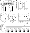
IL-22 induces proinflammatory proteins S100A8 and S100A9 production from MDSCs and regulates S100A8 and S100A9 in vivo, and IL-22-induced MDSCs suppress Th1 cell response in Helicobacter pylori infection. (A) S100A8 and S100A9 proteins in IL-22-stimulated human CD45+CD14+HLA-DRlow/− MDSCs for different time points or 24 h were analysed. (B and C) S100A8 and S100A9 protein in gastric mucosa of WT H pylori-infected WT BALB/c and IL-22 KO mice (B), or WT H. pylori-infected WT BALB/c mice injected with IL-22 or Abs against IL-22 (C) on day 49 p.i. were compared. (D and E) Th1 cell responses in gastric mucosa of H. pylori-infected WT BALB/c mice injected with IL-22 or PBS control, Abs against IL-22 (IgG2a), CXCR2 (IgG2a), and/or CXCL2 (IgG2b) or corresponding isotype control Abs, or SB225002 or DMSO control (D), or H pylori-infected WT BALB/c and IL-22 KO mice (E) on day 49 p.i. were compared. (F) T cell-MDSC coculture was assessed by flow cytometry as described in Methods and statistically analysed (n=3). Results are expressed as percentage of proliferated Th1 cells in CD4+ T cells. Results are representative of three independent experiments. *p<0.05, **p<0.01, n.s p>0.05 for groups connected by horizontal lines compared, or compared with uninfected mice. WT; wild-type; KO, knockout; IL, interleukin; PBS, phosphate-buffered saline; Abs, antibodies; MDSC, myeloid-derived suppressor cell; DMSO, dimethylsulfoxide; DC, dendritic cells; p.i., postinfection; GADPH, glyceraldehyde 3-phosphate dehydrogenase; IFN, interferon; CFSE, carboxyfluorescein diacetate succinimidyl ester; n.s., not significant.
Figure 7
Figure 7
A proposed model of cross-talk among Helicobacter pylori, dendritic cells (DC), gastric epithelial cells and myeloid-derived suppressor cells (MDSC) leading to Th22 cell differentiation and MDSC-mediated proinflammation in gastric mucosa during H. pylori infection. H. pylori stimulate DCs to secrete interleukin (IL)-23 and H. pylori induce gastric epithelial cells to express IL-22R1. Release of IL-23 induces the polarisation of Th22 cells. Polarised Th22 cells expand in gastric mucosa where they release cytokine IL-22 that stimulates gastric epithelial cells to secrete CXCL2. Responding to the CXCL2 chemokine gradient, myeloid progenitor cell-derived, CXCR2-expressing MDSCs migrate into gastric mucosa where they exert a proinflammatory effect by producing inflammatory proteins, S100A8 and S100A9, and suppressing T helper type 1 (Th1) cell response.

Similar articles

Cited by

References

    1. Suerbaum S, Michetti P. Helicobacter pylori infection. N Engl J Med 2002;347:1175–86. - PubMed
    1. Eaton KA, Mefford M, Thevenot T. The role of T cell subsets and cytokines in the pathogenesis of Helicobacter pylori gastritis in mice. J Immunol 2001;166:7456–61. - PubMed
    1. Zenewicz LA, Yancopoulos GD, Valenzuela DM, et al. Innate and adaptive interleukin-22 protects mice from inflammatory bowel disease. Immunity 2008;29:947–57. - PMC - PubMed
    1. Radaeva S, Sun R, Pan HN, et al. Interleukin 22 (IL-22) plays a protective role in T cell-mediated murine hepatitis: IL-22 is a survival factor for hepatocytes via STAT3 activation. Hepatology 2004;39:1332–42. - PubMed
    1. Munoz M, Heimesaat MM, Danker K, et al. Interleukin (IL)-23 mediates Toxoplasma gondii-induced immunopathology in the gut via matrixmetalloproteinase-2 and IL-22 but independent of IL-17. J Exp Med 2009;206:3047–59. - PMC - PubMed

Publication types

MeSH terms