Interferon regulatory factor 3 as key element of the interferon signature in plasmacytoid dendritic cells from systemic lupus erythematosus patients: novel genetic associations in the Mexican mestizo population
- PMID: 25130328
- PMCID: PMC4238870
- DOI: 10.1111/cei.12429
Interferon regulatory factor 3 as key element of the interferon signature in plasmacytoid dendritic cells from systemic lupus erythematosus patients: novel genetic associations in the Mexican mestizo population
Abstract
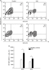
Many genetic studies have found an association between interferon regulatory factors (IRF) single nucleotide polymorphisms (SNPs) and systemic lupus erythematosus (SLE); however, specific dendritic cell (DC) alterations have not been assessed. The aim of the present study was to address the expression of IRF3 and IRF5 on different DC subsets from SLE patients, as well as their association with interferon (IFN)-α production and novel SNPs. For the genetic association analyses, 156 SLE patients and 272 healthy controls from the Mexican mestizo population were included. From these, 36 patients and 36 controls were included for functional analysis. Two IRF3 SNPs - rs2304206 and rs2304204 - were determined. We found an increased percentage of circulating pDC in SLE patients in comparison to controls (8.04 ± 1.48 versus 3.35 ± 0.8, P = 0.032). We also observed enhanced expression of IRF3 (64 ± 6.36 versus 36.1 ± 5.57, P = 0.004) and IRF5 (40 ± 5.25 versus 22.5 ± 2.6%, P = 0.010) restricted to this circulating pDC subset from SLE patients versus healthy controls. This finding was associated with higher IFN-α serum levels in SLE (160.2 ± 21 versus 106.1 ± 14 pg/ml, P = 0.036). Moreover, the IRF3 rs2304206 polymorphism was associated with increased susceptibility to SLE [odds ratio (OR), 95% confidence interval (CI) = 2.401 (1.187-4.858), P = 0.021] as well as enhanced levels of serum type I IFN in SLE patients who were positive for dsDNA autoantibodies. The IRF3 rs2304204 GG and AG genotypes conferred decreased risk for SLE. Our findings suggest that the predominant IRF3 expression on circulating pDC is a key element for the increased IFN-α activation based on the interplay between the rs2304206 gene variant and the presence of dsDNA autoantibodies in Mexican mestizo SLE patients.
Keywords: IRF3; dendritic cells; interferon; systemic lupus erythematosus.
© 2014 British Society for Immunology.
Figures


Similar articles
-
IRF5 haplotypes demonstrate diverse serological associations which predict serum interferon alpha activity and explain the majority of the genetic association with systemic lupus erythematosus.Ann Rheum Dis. 2012 Mar;71(3):463-8. doi: 10.1136/annrheumdis-2011-200463. Epub 2011 Nov 16. Ann Rheum Dis. 2012. PMID: 22088620 Free PMC article.
-
The IRF5 rs2004640 (G/T) polymorphism is not a genetic risk factor for systemic lupus erythematosus in population from south India.Indian J Med Res. 2018 Jun;147(6):560-566. doi: 10.4103/ijmr.IJMR_2025_16. Indian J Med Res. 2018. PMID: 30168487 Free PMC article.
-
Association of the IRF5 risk haplotype with high serum interferon-alpha activity in systemic lupus erythematosus patients.Arthritis Rheum. 2008 Aug;58(8):2481-7. doi: 10.1002/art.23613. Arthritis Rheum. 2008. PMID: 18668568 Free PMC article.
-
Interferon regulatory factors in human lupus pathogenesis.Transl Res. 2011 Jun;157(6):326-31. doi: 10.1016/j.trsl.2011.01.006. Epub 2011 Feb 8. Transl Res. 2011. PMID: 21575916 Free PMC article. Review.
-
Anti-interferon alpha treatment in SLE.Clin Immunol. 2013 Sep;148(3):303-12. doi: 10.1016/j.clim.2013.02.013. Epub 2013 Mar 1. Clin Immunol. 2013. PMID: 23566912 Review.
Cited by
-
STING-STAT6 Signaling Pathway Promotes IL-4+ and IFN-α+ Fibrotic T Cell Activation and Exacerbates Scleroderma in SKG Mice.Immune Netw. 2024 Oct 16;24(5):e37. doi: 10.4110/in.2024.24.e37. eCollection 2024 Oct. Immune Netw. 2024. PMID: 39513026 Free PMC article.
-
cFLIPL Interrupts IRF3-CBP-DNA Interactions To Inhibit IRF3-Driven Transcription.J Immunol. 2016 Aug 1;197(3):923-33. doi: 10.4049/jimmunol.1502611. Epub 2016 Jun 24. J Immunol. 2016. PMID: 27342840 Free PMC article.
-
LncRNA Malat1 inhibition of TDP43 cleavage suppresses IRF3-initiated antiviral innate immunity.Proc Natl Acad Sci U S A. 2020 Sep 22;117(38):23695-23706. doi: 10.1073/pnas.2003932117. Epub 2020 Sep 9. Proc Natl Acad Sci U S A. 2020. PMID: 32907941 Free PMC article.
-
Germline Genetic Variants of Viral Entry and Innate Immunity May Influence Susceptibility to SARS-CoV-2 Infection: Toward a Polygenic Risk Score for Risk Stratification.Front Immunol. 2021 Mar 8;12:653489. doi: 10.3389/fimmu.2021.653489. eCollection 2021. Front Immunol. 2021. PMID: 33763088 Free PMC article. Review.
-
Therapeutic Targeting of IRFs: Pathway-Dependence or Structure-Based?Front Immunol. 2018 Nov 20;9:2622. doi: 10.3389/fimmu.2018.02622. eCollection 2018. Front Immunol. 2018. PMID: 30515152 Free PMC article. Review.
References
-
- Banchereau J, Briere F, Caux C, et al. Immunobiology of dendritic cells. Annu Rev Immunol. 2000;18:767–811. - PubMed
-
- Liu YJ. IPC: professional type 1 interferon-producing cells and plasmacytoid dendritic cell precursors. Annu Rev Immunol. 2005;23:275–306. - PubMed
-
- Gottenberg JE, Chiocchia G. Dendritic cells and interferon-mediated autoimmunity. Biochimie. 2007;89:856–871. - PubMed
-
- Ytterberg SR, Schnitzer TJ. Serum interferon levels in patients with systemic lupus erythematosus. Arthritis Rheum. 1982;25:401–406. - PubMed
Publication types
MeSH terms
Substances
LinkOut - more resources
Full Text Sources
Other Literature Sources
Medical

