

Paramyxovirus glycoprotein incorporation, assembly and budding: a three way dance for infectious particle production
- PMID: 25105277
- PMCID: PMC4147685
- DOI: 10.3390/v6083019
Paramyxovirus glycoprotein incorporation, assembly and budding: a three way dance for infectious particle production
Abstract
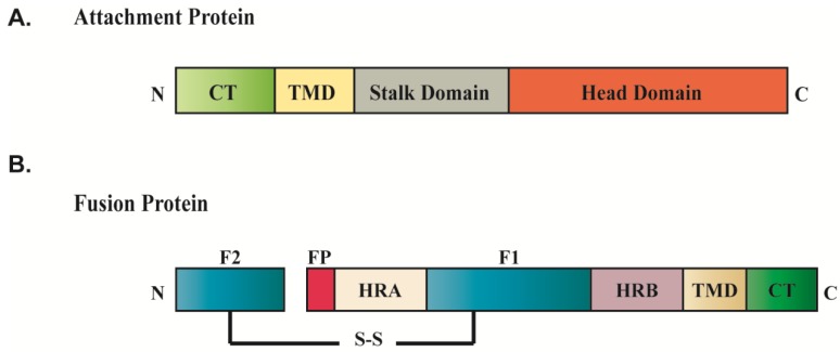
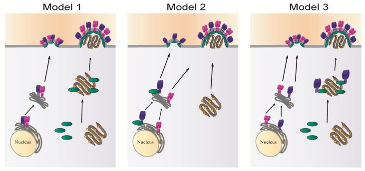
Paramyxoviruses are a family of negative sense RNA viruses whose members cause serious diseases in humans, such as measles virus, mumps virus and respiratory syncytial virus; and in animals, such as Newcastle disease virus and rinderpest virus. Paramyxovirus particles form by assembly of the viral matrix protein, the ribonucleoprotein complex and the surface glycoproteins at the plasma membrane of infected cells and subsequent viral budding. Two major glycoproteins expressed on the viral envelope, the attachment protein and the fusion protein, promote attachment of the virus to host cells and subsequent virus-cell membrane fusion. Incorporation of the surface glycoproteins into infectious progeny particles requires coordinated interplay between the three viral structural components, driven primarily by the matrix protein. In this review, we discuss recent progress in understanding the contributions of the matrix protein and glycoproteins in driving paramyxovirus assembly and budding while focusing on the viral protein interactions underlying this process and the intracellular trafficking pathways for targeting viral components to assembly sites. Differences in the mechanisms of particle production among the different family members will be highlighted throughout.
Figures


Similar articles
-
Cytoplasmic Motifs in the Nipah Virus Fusion Protein Modulate Virus Particle Assembly and Egress.J Virol. 2017 Apr 28;91(10):e02150-16. doi: 10.1128/JVI.02150-16. Print 2017 May 15. J Virol. 2017. PMID: 28250132 Free PMC article.
-
C-Terminal DxD-Containing Sequences within Paramyxovirus Nucleocapsid Proteins Determine Matrix Protein Compatibility and Can Direct Foreign Proteins into Budding Particles.J Virol. 2016 Jan 20;90(7):3650-60. doi: 10.1128/JVI.02673-15. J Virol. 2016. PMID: 26792745 Free PMC article.
-
Paramyxovirus assembly and budding: building particles that transmit infections.Int J Biochem Cell Biol. 2010 Sep;42(9):1416-29. doi: 10.1016/j.biocel.2010.04.005. Epub 2010 Apr 14. Int J Biochem Cell Biol. 2010. PMID: 20398786 Free PMC article. Review.
-
Assembly and budding of influenza virus.Virus Res. 2004 Dec;106(2):147-65. doi: 10.1016/j.virusres.2004.08.012. Virus Res. 2004. PMID: 15567494 Free PMC article. Review.
-
The Role of Phlebovirus Glycoproteins in Viral Entry, Assembly and Release.Viruses. 2016 Jul 21;8(7):202. doi: 10.3390/v8070202. Viruses. 2016. PMID: 27455305 Free PMC article. Review.
Cited by
-
Role of hydrogen sulfide in paramyxovirus infections.J Virol. 2015 May;89(10):5557-68. doi: 10.1128/JVI.00264-15. Epub 2015 Mar 4. J Virol. 2015. PMID: 25740991 Free PMC article.
-
Host cytoskeleton in respiratory syncytial virus assembly and budding.Virol J. 2016 Sep 26;13(1):161. doi: 10.1186/s12985-016-0618-z. Virol J. 2016. PMID: 27670781 Free PMC article. Review.
-
Newcastle Disease Virus Entry into Chicken Macrophages via a pH-Dependent, Dynamin and Caveola-Mediated Endocytic Pathway That Requires Rab5.J Virol. 2021 Jun 10;95(13):e0228820. doi: 10.1128/JVI.02288-20. Epub 2021 Jun 10. J Virol. 2021. PMID: 33762417 Free PMC article.
-
Nipah and Hendra Viruses: Deadly Zoonotic Paramyxoviruses with the Potential to Cause the Next Pandemic.Pathogens. 2022 Nov 25;11(12):1419. doi: 10.3390/pathogens11121419. Pathogens. 2022. PMID: 36558753 Free PMC article. Review.
-
A key region of molecular specificity orchestrates unique ephrin-B1 utilization by Cedar virus.Life Sci Alliance. 2019 Dec 20;3(1):e201900578. doi: 10.26508/lsa.201900578. Print 2020 Jan. Life Sci Alliance. 2019. PMID: 31862858 Free PMC article.
References
-
- Lamb R.A., Parks G.D. In: Paramyxoviridae: The Viruses and Their Replication. 5th ed. Knipe D.M., Howley P.M., editors. Volume 1 Lippincott Williams & Wilkins; Philadelphia, PA, USA: 2007. Fields Virology.
-
- Murray K., Selleck P., Hooper P., Hyatt A., Gould A., Gleeson L., Westbury H., Hiley L., Selvey L., Rodwell B., et al. A morbillivirus that caused fatal disease in horses and humans. Science. 1995;268:94–97. - PubMed
-
- Alexander D.J.S.D. Newcastle Disease, Other Avian Paramyxoviruses, and Pneumovirus Infections. In: Saif Y.M., Glisson J.R., McDougald L.R., Nolan L.K., Swayne D.E., editors. Diseases of Poultry. 12th ed. Blackwell Publishing; Ames, IA, USA: 2008.
Publication types
MeSH terms
Substances
Grants and funding
LinkOut - more resources
Full Text Sources
Other Literature Sources
Miscellaneous
