. 2014 Aug:462-463:1-13.
doi: 10.1016/j.virol.2014.05.022. Epub 2014 Jun 13.

The RNA binding of protein A from Wuhan nodavirus is mediated by mitochondrial membrane lipids

Affiliations

The RNA binding of protein A from Wuhan nodavirus is mediated by mitochondrial membrane lipids

Yang Qiu et al. Virology. 2014 Aug.

Abstract

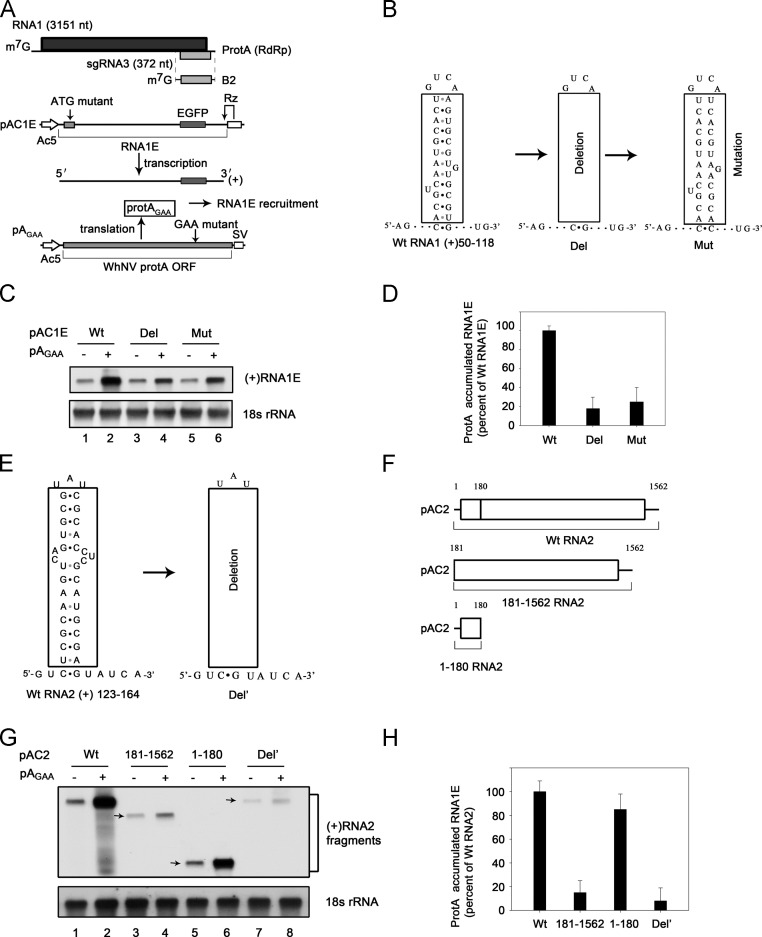
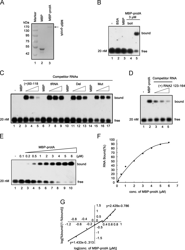
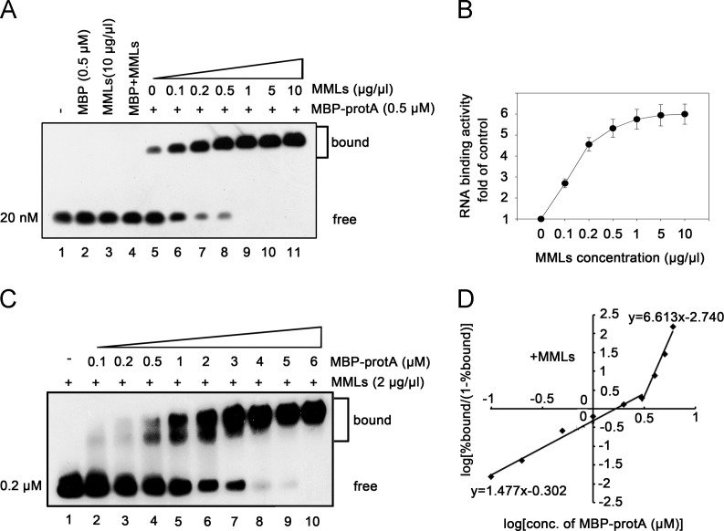
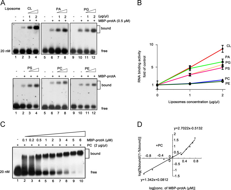
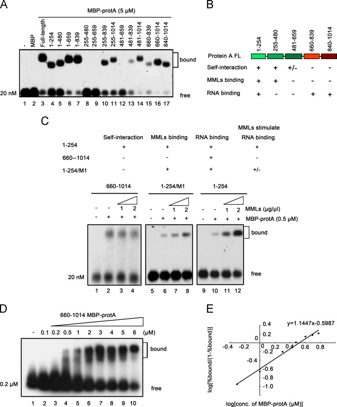
RNA replication of positive-strand (+)RNA viruses requires the lipids present in intracellular membranes, the sites of which viral replicases associate with. However, the direct effects of membrane lipids on viral replicases are still poorly understood. Wuhan nodavirus (WhNV) protein A, which associates with mitochondrial membranes, is the sole replicase required for RNA replication. Here, we report that WhNV protein A binds to RNA1 in a cooperative manner. Moreover, mitochondrial membrane lipids (MMLs) stimulated the RNA binding activity and cooperativity of protein A, and such stimulations exhibited strong selectivity for distinct phospholipids. Interestingly, MMLs stimulated the RNA-binding cooperativity only at higher protein A concentrations. Further investigation showed that MMLs stimulate the RNA binding of protein A by promoting its self-interaction. Finally, manipulating MML metabolism affected the protein A-induced RNA1 recruitment in cells. Together, our findings reveal the direct effects of membrane lipids on the RNA binding activity of a nodaviral replicase.

Keywords: Mitochondrial membrane lipids; Protein A; RNA binding; Wuhan Nodavirus.


Figures

Fig. 1
Fig. 1
Characterization of the RNA probe used for determining the RNA binding of WhNV protein A in vitro. (A) Schematic of plasmids used for protein AGAA (prot AGAA) and (+)RNA1E expression. RNA1E templates with authentic viral 5′ and 3′ termini of WhNV RNA1 and an inserting EGFP sequence were generated from pAC1E by precisely placing the Ac5 promoter start site and a hepatitisδribozyme (Rz), respectively, and by mutating the start codon at the indicated location to disrupt translation. The Ac5 promoter and SV40 polyadenylation signal (SV) flanking the protein A ORF in pAGAA thereof disrupt its activity as a viral RNA replication template and mutating the replication GDD sites into GAA but maintain its activity to recruit RNA (Qiu et al., 2014). pAGAA-derived protein AGAA subsequently directs (+)RNA1E recruitment from (+)RNA1E template transcribed from pAC1E. (B) The secondary structure predicted for RNA1 nt 50–118 [RNA1(50–118)]. Del represents removing the RNA sequences formed the helices structure and Mut represents destroying the base pairing in the helices section. (C) RNA1(50–118) mediates RNA1 recruitment in cells. Pr-E cells were transfected with the indicated plasmids, including pAC1E wt, Del or Mut (as shown in B) in the absence or in the presence of pAGAA (protein AGAA). After transfection for 36 h, total RNA was extracted and analyzed by Northern blot with the probes against EGFP and 18S rRNA, respectively. (D) The levels of (+)RNA1E were determined from three experiments after normalization to 18S rRNA and are expressed as the level of protein A-stimulated (+)RNA1E accumulation relative to wt (+)RNA1E. (E) The secondary structure predicted for RNA2 nt 123–164 [RNA1(123–164)]. Del′ represents removing the RNA sequences formed the helices structure. (F) Schematic of plasmids used for protein A-mediated RNA2 recruitment. (G) RNA2(123–164) mediates RNA2 recruitment in cells. Pr-E cells were transfected with the indicated plasmids, including pAC2 wt, pAC2 181–1562, pAC2 1–180, pAC2 Del′ (as shown in E and F) in the absence or in the presence of pAGAA. After transfection for 36 h, total RNA was extracted and analyzed by Northern blot with the probes against EGFP and 18S rRNA, respectively. (H) The levels of (+)RNA2 were determined from three experiments after normalization to 18S rRNA and are expressed as the level of protein A-stimulated (+)RNA2 accumulation relative to wt (+)RNA2.
Fig. 2
Fig. 2
The binding preference of recombinant protein A to RNA1. (A) SDS-PAGE analysis of purified recombinant protein A from E. coli. Protein A ORF was cloned into pMAL-c2X and expressed as C-terminal fusion proteins with MBP (MBP-protA) as described previously (Qiu et al., 2014). Lane 1, Marker; lane 2, MBP protein alone; lane 3, MBP-protA. (B) Gel mobility shift assay showing interactions between MBP-protA and RNA1. The in vitro transcribed DIG-labeled RNA1(50–118) was separately incubated with bovine serum albumin (BSA, lane 2), MBP alone (lane 3), boiled MBP-protA (lane 4) and MBP-protA (lane 5) (3 μM each), in a binding buffer at 27 °C for 30 min and then analyzed in 1% agarose gel. Gel was transferred to Hybond N nylon membranes via capillary transfer and then the membranes were incubated with anti-DIG antibody conjugated with alkaline phosphatase, exposed to film. The unbound, free RNA1(50–118) probe and the shift (bound) RNA-protein complex are marked on the right. (C) Unlabeled competitor RNAs at increasing concentrations (in 1-, 10-, 60-fold excess) were added to the mixture containing the DIG-labeled RNA1(50–118) and 3 μM MBP-protA, and the bound complexes were analyzed in a gel mobility shift assay. The tRNA was from yeast. (D) Gel mobility shift assay showing interactions between MBP-protA and RNA2. The in vitro transcribed DIG-labeled RNA2(123–164) was incubated with MBP-protA (lane 2) and in a binding buffer at 27 °C for 30 min and then analyzed in 1% agarose gel. Unlabeled competitor RNAs at increasing concentrations (in 5-, 50-, 100-fold excess) were added to the mixture containing the DIG-labeled RNA2(123–164) and 3 μM MBP-protA, and the bound complexes were analyzed in a gel mobility shift assay. (E) Cooperative binding of MBP-protA to RNA1(50–118). Gel mobility shift assays were performed using increasing molar concentrations of MBP-protA incubated with 20nM RNA1(50–118) probe. The molar concentrations of MBP-protA (0.1–6 μM) are indicated above each lane. (F) The plot of the percent of RNA bound versus molar concentration of MBP-protA. (G) The Hill coefficients of the RNA binding of protein A based on Fig. 2E at low and high protein concentrations are indicated.
Fig. 3
Fig. 3
MMLs stimulate the RNA binding activity of protein A. (A,B) MMLs stimulate the binding of protein A to RNA1(50–118) probe. Gel mobility shift assays were performed using increasing concentrations (wt/vol) of MMLs with 0.5 μM MBP-protA and 20 nM RNA1(50–118) probe. The concentrations of MMLs are indicated above each lane. The RNA binding activity of protein A in the absence of MMLs is used as the control (1-fold). The increase in the RNA binding activity of protein A at each point concentration of MMLs is graphed as the fold of control as shown in (B). Error bars represent S.D. values from at least three independent experiments. (C,D) MMLs stimulate protein A cooperatively binding to RNA1(50–118) at high protein concentrations. Gel mobility shift assays were performed using increasing molar concentrations of MBP-protA incubated with 0.2 μM RNA1(50–118) probe with the addition of 2 μg/μl MMLs, and then analyzed via 2% agarose gel to clearly separate migration of protein–RNA complexes. The Hill coefficients of the RNA binding of protein A in the presence of MMLs at low and high protein concentrations are indicated (D).
Fig. 4
Fig. 4
Specific anionic phospholipids stimulate the RNA binding activity of protein A. (A,B) Gel mobility shift assays were performed using the increasing concentrations (wt/vol) of liposomes generated from specific phospholipids with 0.5 μM MBP-protA and 0.2 μM RNA1(50–118) probe complexes. The concentrations of liposomes are indicated above each lane. The RNA binding activity of protein A in the absence of liposomes is used as the control (1-fold). The increases in the RNA binding activity of protein A at each point concentration of indicated liposomes are graphed as the fold of control (B). Error bars represent S.D. values from at least three independent experiments. (C,D) PC showed limited effect on protein A cooperatively binding to RNA1(50–118). Gel mobility shift assays were performed using increasing molar concentrations of MBP-protA incubated with 20 nM RNA1(50–118) probe with the addition of 2 μg/μl PC. The Hill coefficients of the RNA binding of protein A in the presence of PC at low and high protein concentrations are indicated (D).
Fig. 5
Fig. 5
MMLs stimulate the RNA binding activity of protein A by promoting its self-interaction. (A) A series of 5 μM MBP-tagged protein A fragments as describe previously (Qiu et al., 2014), were incubated with 20 nM RNA1(50–118) probe and then examined in gel mobility shift assays. (B) Summary the domains of WhNV protein A responsible for self-interaction, MMLs binding and RNA binding, representing the results shown in Fig. 5A and our previous study (Qiu et al., 2014). “+/−” represents that the self-interaction of aa 481–659 is weak (Qiu et al., 2014). (C) The effect of MMLs on the RNA binding of protein A fragments was indicated “+” and “-”, representing the results as indicated below. “+/−” represents that the stimulatory effect of MMLs on the RNA binding is limited. Increasing concentrations of MMLs were incubated with protein A fragments aa 660–1014 (lanes 2–4), 1–254/M1 (lanes 6–8) or 1–254 (lanes 10–12) and RNA1(50–118) probe, and the protein–RNA complex was separated in a gel mobility shift assay. (D,E) Cooperative binding of protein A fragment aa 660–1014 to RNA1(50–118). Gel mobility shift assays were performed using increasing molar concentrations of MBP-protA aa 660–1014 incubated with RNA1(50–118) probe. The Hill coefficients of the RNA binding of protein A at low and high protein concentrations are indicated (E).
Fig. 6
Fig. 6
Manipulation of phospholipids metabolism regulates protein A-induced (+)RNA1E recruitment in cells. (A,B) Measurement of PA and PC content in Pr-E cells treated with 100 nM FIPI (A) and 50 μM miltefosine (B) or with matching concentration of DMSO (vehicle). (C) Viability of cells treated with FIPI, miltefosine or DMSO. (D) FIPI or miltefosine treatment show less effect on the activity of mitochondrial membrane-binding protein to associate with membranes. Nycodenz flotation assay were used to examine membrane association of protein A and porin in cells treated with FIPI, miltefosine or DMSO. LD fractions represent the membrane-rich layers in the gradient, whereas the HD (non-membrane) fractions contain cytosolic soluble proteins. (E) (+)RNA1E accumulation in cells treated with FIPI, miltefosine or DMSO expressing protein AGAA-HA. Cells were divided into two equal fractions. One of fractions was analyzed by Northern blotting with EGFP and 18 s rRNA probes, respectively. The other fraction was analyzed by Western blotting with anti-HA and anti-GAPDH antibodies, respectively. GAPDH, glyceraldehyde-3-phosphate dehydrogenase. (F) Quantification data show the accumulation of (+)RNA1E and protein A in Pr-E cells expressing protein AGAA-HA treated with FIPI, miltefosine or DMSO, respectively. The accumulation of RNA and protein is normalized to 18S rRNA and GAPDH, respectively. Error bars represent S.D. values from at least three independent experiments.
Fig. 7
Fig. 7
The activities of per unit protein A recruiting (+)RNA1E and self-interaction are regulated by manipulation of phospholipids in cells. (A) Pr-E cells expressing (+)RNA1E together with protein AGAA-His plus empty vector-HA (lane 1) or protein AGAA-His plus protein AGAA-HA (lanes 2–6) were harvested. Cell Lysates were immunoprecipitated with an anti-His antibody (lanes 1 and 3–6) or control IgG (lane 2). The immunoprecipitated complexes were divided into two equal fractions. One of fractions was blotted with an anti-HA antibody to determine the self-interaction of protein A. The (+)RNA1E associated with the immunoprecipitated protein A was separated from the other fraction and following analyzed by real-time RT-PCR. (B) Graph of the ratios of the accumulation of (+)RNA1E analyzed by real-time RT-PCR as described above and protein A self-interaction versus protein A׳s accumulation. The accumulation of RNA and protein is normalized to 18S rRNA and GAPDH from total cells, respectively. Error bars represent S.D. values from at least three independent experiments.

Similar articles

Cited by

References

    1. Ahlquist P. Parallels among positive-strand RNA viruses, reverse-transcribing viruses and double-stranded RNA viruses. Nat. Rev. Microbiol. 2006;4:371–382. - PMC - PubMed
    1. Ahlquist P., Noueiry A.O., Lee W.M., Kushner D.B., Dye B.T. Host factors in positive-strand RNA virus genome replication. J. Virol. 2003;77:8181–8186. - PMC - PubMed
    1. Ahola T., Lampio A., Auvinen P., Kaariainen L. Semliki Forest virus mRNA capping enzyme requires association with anionic membrane phospholipids for activity. Embo. J. 1999;18:3164–3172. - PMC - PubMed
    1. Arnold J.J., Cameron C.E. Poliovirus RNA-dependent RNA polymerase (3Dpol) is sufficient for template switching in vitro. J. Biol. Chem. 1999;274:2706–2716. - PubMed
    1. Ball L.A. Requirements for the self-directed replication of flock house virus RNA 1. J. Virol. 1995;69:720–727. - PMC - PubMed

Publication types

MeSH terms

LinkOut - more resources