. 2014 Oct;88(20):11782-90.
doi: 10.1128/JVI.01720-14. Epub 2014 Jul 30.

The 19S proteasome activator promotes human cytomegalovirus immediate early gene expression through proteolytic and nonproteolytic mechanisms

Affiliations

The 19S proteasome activator promotes human cytomegalovirus immediate early gene expression through proteolytic and nonproteolytic mechanisms

Laura L Winkler et al. J Virol. 2014 Oct.

Abstract

Proteasomes are large, multisubunit complexes that support normal cellular activities by executing the bulk of protein turnover. During infection, many viruses have been shown to promote viral replication by using proteasomes to degrade cellular factors that restrict viral replication. For example, the human cytomegalovirus (HCMV) pp71 protein induces the proteasomal degradation of Daxx, a cellular transcriptional repressor that can silence viral immediate early (IE) gene expression. We previously showed that this degradation requires both the proteasome catalytic 20S core particle (CP) and the 19S regulatory particle (RP). The 19S RP associates with the 20S CP to facilitate protein degradation but also plays a 20S CP-independent role promoting transcription. Here, we present a nonproteolytic role of the 19S RP in HCMV IE gene expression. We demonstrate that 19S RP subunits are recruited to the major immediate early promoter (MIEP) that directs IE transcription. Depletion of 19S RP subunits generated a defect in RNA polymerase II elongation through the MIE locus during HCMV infection. Our results reveal that HCMV commandeers proteasome components for both proteolytic and nonproteolytic roles to promote HCMV lytic infection. Importance: Proteasome inhibitors decrease or eliminate 20S CP activity and are garnering increasing interest as chemotherapeutics. However, an increasing body of evidence implicates 19S RP subunits in important proteolytic-independent roles during transcription. Thus, pharmacological inhibition of the 20S CP as a means to modulate proteasome function toward therapeutic effect is an incomplete capitalization on the potential of this approach. Here, we provide an additional example of nonproteolytic 19S RP function in promoting HCMV transcription. These data provide a novel system with which to study the roles of different proteasome components during transcription, a rationale for previously described shifts in 19S RP subunit localization during HCMV infection, and a potential therapeutic intervention point at a pre-immediate early stage for the inhibition of HCMV infection.


Figures

FIG 1
FIG 1
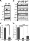
Daxx stabilization in 19S knockdown cells correlates with a defect in IE protein expression during HCMV infection. (A) HFs were transfected with Rpn1-specific or scrambled (Scr) control siRNAs for 72 h and then mock infected (M) or infected (V) at an MOI of 1. At 8 h postinfection, lysates were harvested and analyzed by immunoblotting. (B) HFs were transfected with the indicated siRNAs for 72 h and then infected at an MOI of 0.1 for 6 h. Lysates from three biological replicates were collected and analyzed by semiquantitative immunoblotting. IE1 protein expression was quantified and normalized to β-actin, and values are reported relative to the scrambled control. (C) HFs were transfected with the indicated siRNAs for 72 h and were infected and analyzed by immunoblotting as described for panel B. (D) Three biological replicates from the experiment shown in panel C were analyzed by semiquantitative immunoblotting and reported as described for panel B.
FIG 2
FIG 2
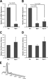
19S subunit depletion inhibits IE transcript accumulation during HCMV infection. (A) HFs transfected with scrambled (Scr) or Rpn1-targeting siRNAs for 72 h were infected at an MOI of 0.1 for 6 h. RNA and DNA were isolated from three biological replicates and analyzed by reverse transcription and real-time PCR. IE transcript levels (IE exon 3 primers) were normalized to both 16S mitochondrial rRNA transcripts and the number of HCMV genomes delivered per cell, and IE gene expression is reported relative to the scrambled control. (B) HFs were transfected with the indicated siRNAs and analyzed as described for panel A. (C) Lysates from the experiment shown in panel A were also analyzed with real-time PCR for genome delivery. The number of HCMV genomes (IE exon 3 primers) was normalized to cellular genomes (16S mitochondrial rRNA gene) and reported relative to scrambled controls. (D) Lysates from the experiment shown in panel B were also analyzed as described for panel C. (E) For 72 h, HFs were transfected with scrambled or Rpn1-specific siRNAs. Cells were then harvested, fixed, stained with propidium iodide, and analyzed by flow cytometry. Data were analyzed using FlowJo software.
FIG 3
FIG 3
Dismantling the intrinsic immune defense is not the only role the 19S RP plays in promoting IE gene expression. (A) Telomerase-immortalized human fibroblasts (tHF) and shDaxx knockdown cells were transfected with scrambled (Scr) or Rpn1-targeting siRNA. After 72 h, HFs were mock infected (M) or infected (V) with HCMV at an MOI of 1 for 10 h. Cell lysates were collected and analyzed by immunoblotting. (B) HFs were transfected with scrambled control (Scr) or Rpn1-specific siRNAs for 72 h and then were mock infected (M) or infected (V) with HCMV at an MOI of 0.5. At 20 h prior to infection, cells were treated with DMSO (D) or TSA (T). At the start of infection, the indicated scrambled control groups were also treated with lactacystin (L). Cell lysates were collected at 4 h postinfection and analyzed by immunoblotting. (C) HFs were treated as described for panel B, with the addition of HFs transfected with siRNAs targeting the chymotrypsin-like subunit of the 20S core (β5), and nucleic acids were isolated and analyzed by real-time PCR. IE transcript levels (exon 3) were normalized to the number of HCMV genomes (exon 3) delivered per cell (β-actin). IE gene expression of TSA-treated samples is reported relative to each sample's respective DMSO control. Three biological replicates (closed squares) are reported and averaged (closed square with dash).
FIG 4
FIG 4
The 19S RP is recruited to the MIE locus. (A) HFs were infected with HCMV at an MOI of 3 for 3 h. Lysates were collected, fixed, and then subjected to ChIP using the indicated antibodies. Isolated DNA was quantified using real-time PCR, and enrichment at the MIEP was normalized to IgG controls. Three biological replicates are represented. β7 versus IgG, P = 0.62. (B) Samples from the experiment shown in panel A were also analyzed for enrichment at exon 3 of IE1/2. β7 versus IgG, P = 0.46. (C) Samples from the experiment shown in panel A were evaluated for enrichment at the 5′ end of UL47. β7 versus IgG, P = 0.32. (D) Samples from the experiment shown in panel A were analyzed for association with the 3′ end of UL48. Rpn7 versus IgG, P = 0.05; β7 versus IgG, P < 0.01. (E) Aliquots from the experiment shown in panel A were analyzed by immunoblotting to examine the efficacy of the immunoprecipitation.
FIG 5
FIG 5
19S knockdown cells exhibit a defect in RNAPII elongation at the MIE locus. (A) Cartoon schematic of the MIE gene locus and the primer sets used during real-time analysis of RNAPII ChIP. Beginning nucleotides of exon 4 and exon 5 are numbered relative to the transcription start site (+1). (B) At 72 h after transfection with scrambled (Scr) or Rpn1 siRNA, HFs were infected with HCMV at an MOI of 1 for 3 h. Lysates were subjected to ChIP using the 8WG16 monoclonal antibody that recognizes the nonphosphorylated form of RNAPII. Enrichment of the indicated regions of the IE gene was analyzed by real-time PCR. Five biological replicates are represented. (C) Samples from the experiment shown in panel B were also analyzed for exon 5 occupancy with the H5 monoclonal antibody that recognizes the (elongating) Ser2-phosphorylated (Ser2P) form of RNAPII. Three biological replicates were completed.
FIG 6
FIG 6
20S knockdown inhibits RNAPII recruitment to the MIEP but not RNAPII occupancy at the 3′ end of the MIE locus. (A) A cartoon representing the IE gene and the primer sets used during real-time analysis of ChIP samples. Beginning nucleotides of exon 4 and exon 5 are numbered relative to the transcription start site (+1). (B) At 72 h after transfection with scrambled (Scr) or β5 siRNA, HFs were infected with HCMV at an MOI of 1 for 3 h. Lysates were subjected to ChIP using the 8WG16 monoclonal antibody that recognizes the nonphosphorylated form of RNAPII. Enrichment of the indicated regions of the IE gene was analyzed by real-time PCR. Five biological replicates are represented. Scr versus β5, P = 0.14 (exon 4) and P = 0.44 (exon 5). (C) Samples from the experiment shown in panel B were also analyzed for exon 5 occupancy using the H5 monoclonal antibody that recognizes the Ser2-phosphorylated (the elongating) form of RNAPII. Three biological replicates are shown. Scr versus β5, P = 0.32.
FIG 7
FIG 7
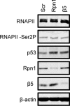
19S RP or 20S CP knockdown does not alter steady-state levels of RNAPII. HFs were transfected with the indicated siRNAs and analyzed by immunoblotting to examine steady-state levels of the indicated proteins.
FIG 8
FIG 8
The 19S RP promotes HCMV IE gene expression through proteolytic and nonproteolytic mechanisms. (A) At the start of HCMV infection, Daxx induces a repressive chromatin structure at the MIEP, prohibiting RNAPII recruitment. (B) To induce IE gene expression, tegument-delivered pp71 degrades Daxx using both the 19S RP and the 20S CP. (C) Once Daxx has been eliminated and the local chromatin structure is relaxed, RNAPII can access the MIEP. RNAPII is then recruited, and the 19S RP promotes the efficient elongation of transcription complexes.

Similar articles

Cited by

References

    1. Jung T, Catalgol B, Grune T. 2009. The proteasomal system. Mol. Aspects Med. 30:191–296. 10.1016/j.mam.2009.04.001 - DOI - PubMed
    1. Loureiro J, Ploegh HL. 2006. Antigen presentation and the ubiquitin-proteasome system in host-pathogen interactions. Adv. Immunol. 92:225–305. 10.1016/S0065-2776(06)92006-9 - DOI - PMC - PubMed
    1. Blanchette P, Branton PE. 2009. Manipulation of the ubiquitin-proteasome pathway by small DNA tumor viruses. Virology 384:317–323. 10.1016/j.virol.2008.10.005 - DOI - PubMed
    1. Choi AG, Wong J, Marchant D, Luo H. 2013. The ubiquitin-proteasome system in positive-strand RNA virus infection. Rev. Med. Virol. 23:85–96. 10.1002/rmv.1725 - DOI - PMC - PubMed
    1. Hwang J, Winkler L, Kalejta RF. 2011. Ubiquitin-independent proteasomal degradation during oncogenic viral infections. Biochim. Biophys. Acta 1816:147–157. 10.1016/j.bbcan.2011.05.005 - DOI - PMC - PubMed

Publication types

Substances

LinkOut - more resources