Review
. 2014 Jul 15;5(13):4603-50.
doi: 10.18632/oncotarget.2209.

Deregulation of the EGFR/PI3K/PTEN/Akt/mTORC1 pathway in breast cancer: possibilities for therapeutic intervention

Affiliations
Review

Deregulation of the EGFR/PI3K/PTEN/Akt/mTORC1 pathway in breast cancer: possibilities for therapeutic intervention

Nicole M Davis et al. Oncotarget. .

Abstract

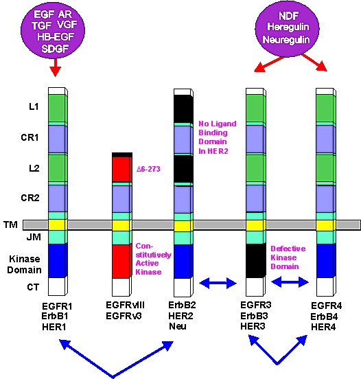
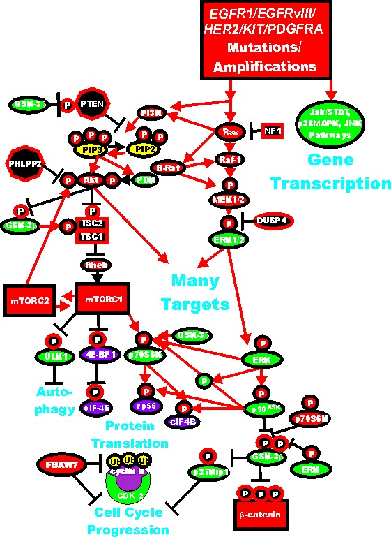
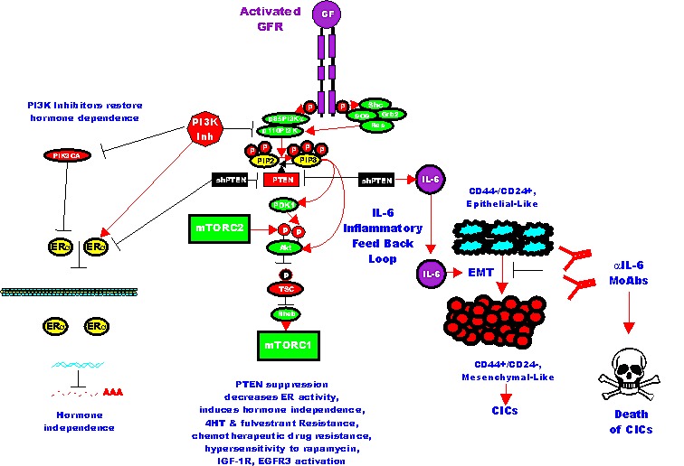
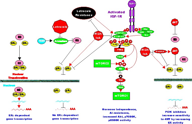
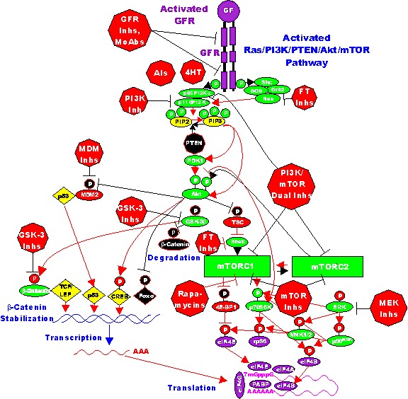
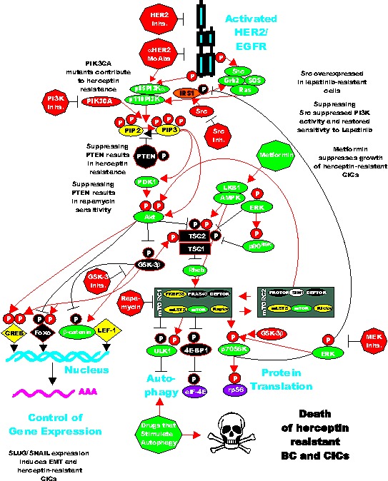
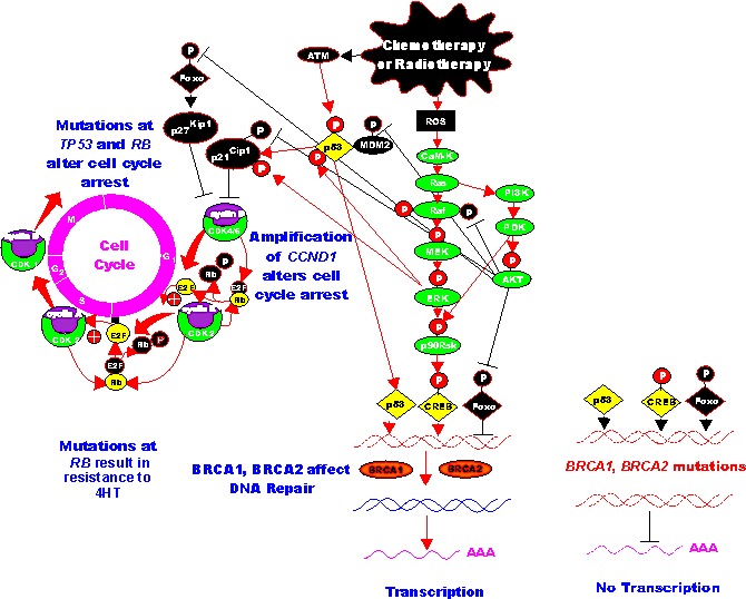
The EGFR/PI3K/PTEN/Akt/mTORC1/GSK-3 pathway plays prominent roles in malignant transformation, prevention of apoptosis, drug resistance and metastasis. The expression of this pathway is frequently altered in breast cancer due to mutations at or aberrant expression of: HER2, ERalpha, BRCA1, BRCA2, EGFR1, PIK3CA, PTEN, TP53, RB as well as other oncogenes and tumor suppressor genes. In some breast cancer cases, mutations at certain components of this pathway (e.g., PIK3CA) are associated with a better prognosis than breast cancers lacking these mutations. The expression of this pathway and upstream HER2 has been associated with breast cancer initiating cells (CICs) and in some cases resistance to treatment. The anti-diabetes drug metformin can suppress the growth of breast CICs and herceptin-resistant HER2+ cells. This review will discuss the importance of the EGFR/PI3K/PTEN/Akt/mTORC1/GSK-3 pathway primarily in breast cancer but will also include relevant examples from other cancer types. The targeting of this pathway will be discussed as well as clinical trials with novel small molecule inhibitors. The targeting of the hormone receptor, HER2 and EGFR1 in breast cancer will be reviewed in association with suppression of the EGFR/PI3K/PTEN/Akt/mTORC1/GSK-3 pathway.


Figures

Figure 1
Figure 1. Epidermal Growth Factor Receptor Family
Conserved domains of the four different EGFR family members are indicated by similar shading. L = ligand binding domain, CR = cysteine-rich domains. TM = transmembrane domain. CT = C-terminal domain which contains the phosphorylation sites. JM = juxta membrane domain. HER2 does not bind a ligand. The kinase domain in ERB3 is defective. Alternative names for each receptor are written underneath each receptor. Ligands which bind the receptors are indicated in purple circles above the receptors. Epidermal growth factor (EGF), vaccinia growth factor (VGF), amphiregulin (AR), heparin binding-EGF (HB-EGF), schwannoma-derived growth factor (SDGF), Neu differentiation factor (NDF), heregulin, neuregulin. Arrows between receptors indicate possible heterodimer formation between various EGFR family receptors.
Figure 2
Figure 2. Dysregulated Expression of Upstream Receptors and Kinases Can Result in Activation of the Ras/Raf/MEK/ERK and Ras/PI3K/PTEN/Akt/mTOR and Other Signaling Pathways and Contribute to Malignant Transformation
Sometimes dysregulated expression of growth factor receptors occurs by increased expression, genetic translocations or genomic amplifications which can lead to activation of the Ras/Raf/MEK/ERK, Ras/PI3K/PTEN/Akt/mTOR and other signaling pathways. Alternatively chromosomal translocations can occur in non-receptor kinases and other genes which result in activation of these pathways. Genes in the Ras/Raf/MEK/ERK and Ras/PI3K/PTEN/Akt/mTOR pathways that have activating mutations detected in human cancer and proliferative diseases are indicated in red ovals and squares. Tumor suppressor genes inactivated in certain cancer are indicated in black squares or octagons. Other key genes are indicated in green ovals. Red arrows indicate activating events in pathways. Blocked black arrows indicating inactivating events in pathways.
Figure 3
Figure 3. Effects of Targeting PI3K/PTEN/Akt/mTORC1 and IL-6 on Breast Cancer Hormonal Dependency, EMT and CICs
Mutations at PIK3CA can alter hormonal dependency. Silencing PI3K with PI3K inhibitors can restore hormonal dependency. Silencing PTEN can also result in hormonal-independenc, 4HT and fulvestrant resistance. Silencing PTEN can in some cases also lead in some cases to IL-6 production and an IL-6 inflammatory feed back loop which results in EMT and CIC formation. Silencing of IL-6 with monoclonal antibodies (MoAb) can prevent this loop and result in the death of the breast CICs.
Figure 4
Figure 4. Effects of PI3K/PTEN/Akt/mTORC1 on Aromatase Resistance and Sensitivity to Tamoxifen (4HT)
The aromatase inhibitor (AI) letrozole prevents the conversion of testosterone (TEST) into estrogen (ER) and hence there is no ERalpha mediated gene transcription. Letrozole alters the expression of the PI3K/PTEN/Akt/mTORC1 pathway. PI3K inhibitors will restore ERalpha mediated gene expression in Letrozole resistant cells and the cells revert to hormonal sensitivity. Likewise PI3K inhibitors will restore the sensitivity of 4HT-resistant cells to 4HT. Breast cells with mutations at PIK3CA may develop resistance to 4HT and PI3K inhibitors may restore sensitivity to 4HT.
Figure 5
Figure 5. Sites of Targeting the EGFR/PI3K/PTEN/Akt/mTORC Pathway with Small Molecule Membrane-Permeable Inhibitors and Monoclonal Antibodies (MoAbs)
The PI3K/PTEN/Akt/mTORC1 pathway is regulated by Ras (indicated in green ovals), as well as various upstream growth factor receptors (indicated in purple) and PTEN indicated in a black rectangle. Sites where various small molecule inhibitors suppress this pathway are indicated by red octagons. The downstream transcription factors regulated by this pathway are indicated in yellow diamond shaped outlines. The Ras/Raf/MEK/ERK pathway also interacts with key proteins involved in protein translation (indicated in green ovals). The two pathways aid in the assembly of the protein translation complex (indicated in purple ovals) responsible for the translation of “weak” mRNAs (indicated in a red line folding over on itself) important in the prevention of apoptosis. Other key proteins inactivated by PI3K/PTEN/Akt/mTORC1 pathway (e.g., TSC, PTEN, beta-catenin, Foxo and 4E-BP1 are indicated by black ovals, diamonds and rectangles. Red arrows indicate activating events in pathways. Blocked black arrows indicating inactivating events in pathways. GF = growth factor, GFR = growth factor receptor.
Figure 6
Figure 6. Sites of Resistance in the HER2/PI3K/PTEN/Akt/mTORC Pathway and Potential Sites for Intervention with Small Molecule Membrane-Permeable Inhibitors and Monoclonal Antibodies (MoAbs)
The HER2/EGFR receptor is indicated in a blue figure. The downstream PI3K/PTEN/Akt/mTORC1 pathway is regulated by Ras (indicated in green ovals), PTEN indicated in a black octagon, activated Src is indicated by a red oval, IRS1 is indicated by an orange oval. Sites where various small molecule inhibitors suppress this pathway are indicated by red octagons. Sites which stimulate proteins involved in autophagy are indicated by green octagons and ovals. The Serine/threonine-protein kinase ULK1 (ULK1) which is regulated by mTORC1 is indicated in a black oval. The downstream transcription factors regulated by this pathway are indicated in yellow diamond shaped outlines. The Ras/Raf/MEK/ERK pathway also interacts with key proteins involved in protein translation (indicated in green ovals). The two pathways aid in the assembly of the protein translation complex (indicated in purple ovals) responsible for the translation of “weak” mRNAs (indicated in a red line folding over on itself) important in the prevention of apoptosis. Other key proteins inactivated by PI3K/PTEN/Akt/mTORC1 pathway (e.g., TSC, PTEN, GSK-3beta, beta-catenin, Foxo and 4E-BP1 are indicated by black ovals, diamonds and rectangles. Red arrows indicate activating events in pathways. Blocked black arrows indicating inactivating events in pathways.
Figure 7
Figure 7. Induction of the Ras/Raf/MEK/ERK Pathway after Leukemia Therapy and Subsequent Effects on Cell Cycle Progression, Survival Pathways and Protein Translation
After chemotherapy or radiotherapy there can be activation of signaling pathways which can actually promote cell survival and may lead to therapy resistance. Chemotherapeutic drug treatment (shown in irregular black oval) frequently results in the induction of reactive oxygen species (ROS) (shown in black square). ROS can induce the calcium calmodulin kinase (CaM-K) cascade which can induce Ras which can subsequently activate both the Raf/MEK/ERK and PI3K/PTEN/Akt/mTOR cascades (most components of the two cascades which promote signaling are show in green ovals, transcription factors activated by events are shown in yellow diamonds, transcription factors inactivated by events shown in black diamonds). Induction of the Ras/Raf/MEK/ERK and PI3K/PTEN/Akt/mTOR pathways can result in the activation of many survival pathways, and regulate both cell cycle progression as well as protein translation. Some of the phosphorylation events mediated by Akt actually serve to inhibit the activities of key proteins such as the Foxo transcription factors and the murine double minute (MDM2) ubiquitin ligase (depicted in black ovals). MDM2 serves to regulate p53 protein stability by ubiquitination, however when it is phosphorylated by Akt it is inactivated. Moreover chemotherapeutic drugs and radiotherapy can induce the ataxia telangiectasia mutated (ATM) protein shown in a black oval, which can in turn phosphorylate and regulate p53. p53 can have complex positive and negative effects on cell growth (depicted in yellow diamond), it can regulate the expression of p21 cyclin dependent kinase inhibitory protein-1 (p21Cip1) which controls cell cycle progression. p53 can also control the transcription of genes such as Puma, Noxa and Bax which are involved in apoptosis (all of these molecules are shown in black ovals, as they tend to inhibit cell cycle progression or promote apoptosis). Both Akt and ERK can phosphorylate p21Cip1 which alters its activity and ability to inhibit cell growth (shown as black phosphorylation sites) and subsequently influence cell growth and therapy resistance. p27Kip1 can also be phosphorylated by both Akt and ERK, however the effects of these phosphorylation events are unclear. Akt phosphorylation of p27Kip1 may result in its cytoplasmic localization, while ERK phosphorylation of p27Kip1 may result in elevated levels of the protein. Hence phosphorylation of proteins by ERK and Akt can have dramatic effects on cell proliferation and contribute to the therapy resistance. Chemo- and radiotherapy also induce breaks in the DNA. In the presence of functional BRCA1 and BRCA2 (indicated in yellow ovals) these breaks may be repaired and normal gene transcription can occur. However when BRCA1 or BRCA2 are mutated, the repair of these genes may not occur and proper gene transcription might not occur. This figure serves as an introduction as to how activation of these pathways by chemotherapy and radiotherapy may contribute to therapeutic resistance.

Similar articles

Cited by

References

    1. Siegel R, Naishadham D, Jemal A. Cancer statistics, 2013. CA Cancer J Clin. 2013;63:11–30. - PubMed
    1. Weiss JR, Moysich KB, Swede H. Epidemiology of male breast cancer. Cancer Epidemiol Biomarkers Prev. 2005;14:20–26. - PubMed
    1. Deb S, Jene N, Investigators Kconfab, Fox SB. Genotypic and phenotypic analysis of familial male breast cancer shows under representation of the HER2 and basal subtypes in BRCA-associated carcinomas. BMC Cancer. 2012;12:510. - PMC - PubMed
    1. Miki Y, Swensen J, Shattuck-Eidens D, Futreal PA, Harshman K, Tavtigian S, Liu Q, Cochran C, Bennett LM, Ding W, Bell R, Rosenthal J, Hussey C, Tran T, McClure M, Fry C, et al. A strong candidate for the breast and ovarian cancer susceptibility gene BRCA1. Science. 1994;266:66–71. - PubMed
    1. Easton DF, Steele L, Fields P, Ormiston W, Averill D, Daly PA, McManus R, Neuhausen SL, Ford D, Wooster R, Cannon-Albright LA, Stratton MR, Goldgar DE. Cancer risks in two large breast cancer families linked to BRCA2 on chromosome 13q12-13. Am J Hum Genet. 1997;61:120–128. - PMC - PubMed

Publication types

MeSH terms

Substances