

Neonatal overexpression of estrogen receptor-α alters midbrain dopamine neuron development and reverses the effects of low maternal care in female offspring
- PMID: 25044746
- PMCID: PMC4284154
- DOI: 10.1002/dneu.22206
Neonatal overexpression of estrogen receptor-α alters midbrain dopamine neuron development and reverses the effects of low maternal care in female offspring
Abstract
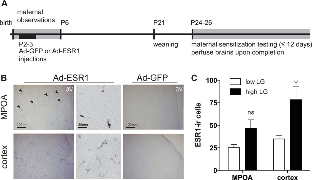
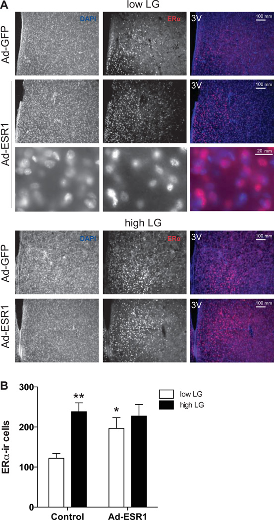
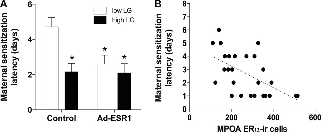
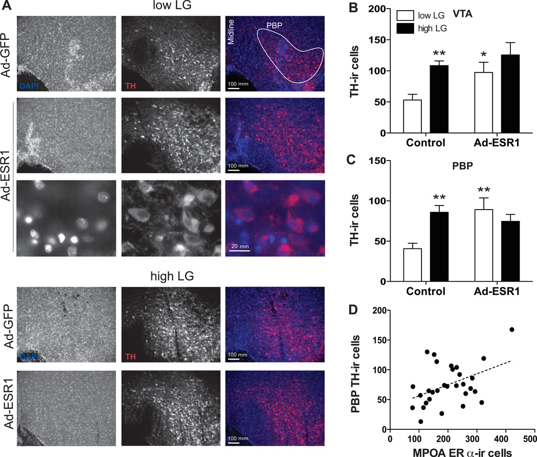
Maternal behavior is dependent on estrogen receptor-alpha (ERα; Esr1) and oxytocin receptor (OTR) signaling in the medial preoptic area (MPOA) of the hypothalamus, as well as dopamine signaling from the ventral tegmental area (VTA) to forebrain regions. Previous studies in rats indicate that low levels of maternal care, particularly licking/grooming (LG), lead to reduced levels of MPOA ERα and VTA dopamine neurons in female offspring and predict lower levels of postpartum maternal behavior by these offspring. The aim of this study was to determine the functional impact on maternal behavior of neonatal manipulation of ERα in females that had experienced low versus high levels of postnatal maternal LG. Adenovirus expressing ESR1 was targeted to the MPOA in female pups from low and high LG litters on postnatal day 2-3. Overexpression of ESR1 in low LG offspring elevated the level of ERα-immunoreactive cells in the MPOA and of tyrosine hydroxylase cells in the VTA to that observed in high LG females. Amongst juvenile female low LG offspring, ESR1 overexpression also decreased the latency to engage in maternal behavior toward donor pups. These results show that virally mediated expression of ESR1 in the neonatal rat hypothalamus results in lasting changes in ESR1 expression through the juvenile period, and can "rescue" hormone receptor levels and behavior of offspring reared by low LG dams, potentially mediated by downstream alterations within reward circuitry. Thus, the transmission of maternal behavior from one generation to the next can be augmented by neonatal ERα in the MPOA.
Keywords: dopamine; estrogen receptor-alpha; maternal care; medial preoptic area; ventral tegmental area.
© 2014 Wiley Periodicals, Inc.
Figures
Similar articles
-
Developmental timing of the effects of maternal care on gene expression and epigenetic regulation of hormone receptor levels in female rats.Endocrinology. 2013 Nov;154(11):4340-51. doi: 10.1210/en.2013-1595. Epub 2013 Sep 3. Endocrinology. 2013. PMID: 24002038 Free PMC article.
-
Natural variations in maternal care are associated with estrogen receptor alpha expression and estrogen sensitivity in the medial preoptic area.Endocrinology. 2003 Nov;144(11):4720-4. doi: 10.1210/en.2003-0564. Epub 2003 Jul 24. Endocrinology. 2003. PMID: 12959970
-
Effects of maternal care on the development of midbrain dopamine pathways and reward-directed behavior in female offspring.Eur J Neurosci. 2014 Mar;39(6):946-956. doi: 10.1111/ejn.12479. Epub 2014 Jan 21. Eur J Neurosci. 2014. PMID: 24446918
-
Adaptations in reward-related behaviors and mesolimbic dopamine function during motherhood and the postpartum period.Front Neuroendocrinol. 2020 Apr;57:100839. doi: 10.1016/j.yfrne.2020.100839. Epub 2020 Apr 16. Front Neuroendocrinol. 2020. PMID: 32305528 Free PMC article. Review.
-
Medial preoptic area interactions with dopamine neural systems in the control of the onset and maintenance of maternal behavior in rats.Front Neuroendocrinol. 2009 Jan;30(1):46-64. doi: 10.1016/j.yfrne.2008.10.002. Epub 2008 Nov 5. Front Neuroendocrinol. 2009. PMID: 19022278 Review.
Cited by
-
Prospects for sociogenomics in avian cooperative breeding and parental care.Curr Zool. 2020 Jun;66(3):293-306. doi: 10.1093/cz/zoz057. Epub 2019 Dec 4. Curr Zool. 2020. PMID: 32440290 Free PMC article.
-
The effects of early life stress on motivated behaviors: A role for gonadal hormones.Neurosci Biobehav Rev. 2020 Dec;119:86-100. doi: 10.1016/j.neubiorev.2020.09.014. Epub 2020 Oct 3. Neurosci Biobehav Rev. 2020. PMID: 33022296 Free PMC article. Review.
-
Overlapping representations of food and social stimuli in VTA dopamine neurons.bioRxiv [Preprint]. 2023 May 17:2023.05.17.541104. doi: 10.1101/2023.05.17.541104. bioRxiv. 2023. Update in: Neuron. 2023 Nov 15;111(22):3541-3553.e8. doi: 10.1016/j.neuron.2023.08.003. PMID: 37293057 Free PMC article. Updated. Preprint.
-
Naringenin stimulates aromatase expression and alleviates the clinical and histopathological findings of experimental autoimmune encephalomyelitis in C57bl6 mice.Histochem Cell Biol. 2023 Nov;160(5):477-490. doi: 10.1007/s00418-023-02217-1. Epub 2023 Jun 28. Histochem Cell Biol. 2023. PMID: 37378907
-
The Effects of Low-Dose Bisphenol A and Bisphenol F on Neural Differentiation of a Fetal Brain-Derived Neural Progenitor Cell Line.Front Endocrinol (Lausanne). 2018 Feb 9;9:24. doi: 10.3389/fendo.2018.00024. eCollection 2018. Front Endocrinol (Lausanne). 2018. PMID: 29479338 Free PMC article.
References
-
- Afonso VM, Grella SL, Chatterjee D, Fleming AS. Previous maternal experience affects accumbal dopaminergic responses to pup-stimuli. Brain Res. 2008;1198:115–123. - PubMed
-
- Afonso VM, King S, Chatterjee D, Fleming AS. Hormones that increase maternal responsiveness affect accumbal dopaminergic responses to pup- and food-stimuli in the female rat. Horm Behav. 2009;56:11–23. - PubMed
-
- Arling GL, Harlow HF. Effects of social deprivation on maternal behavior of rhesus monkeys. J Comp Physiol Psychol. 1967;64:371–377. - PubMed
-
- Benoit D, Parker KC. Stability and transmission of attachment across three generations. Child Dev. 1994;65:1444–1456. - PubMed
Publication types
MeSH terms
Substances
Grants and funding
LinkOut - more resources
Full Text Sources
Other Literature Sources
Miscellaneous
