

An autopsy case of infantile-onset vanishing white matter disease related to an EIF2B2 mutation (V85E) in a hemizygous region
- PMID: 25031760
- PMCID: PMC4097266
An autopsy case of infantile-onset vanishing white matter disease related to an EIF2B2 mutation (V85E) in a hemizygous region
Abstract
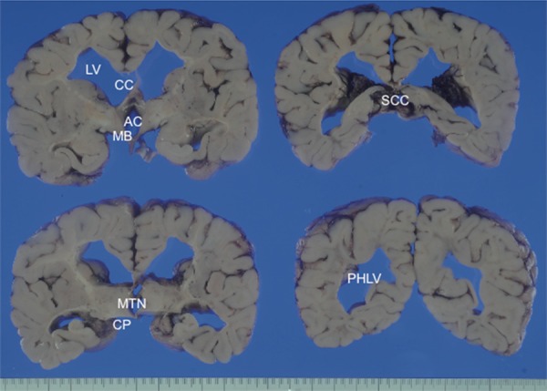
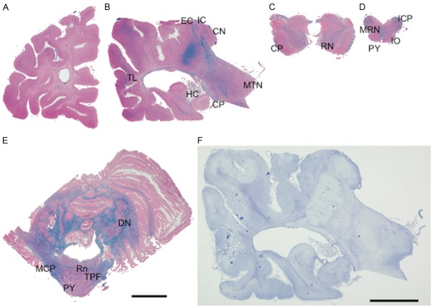
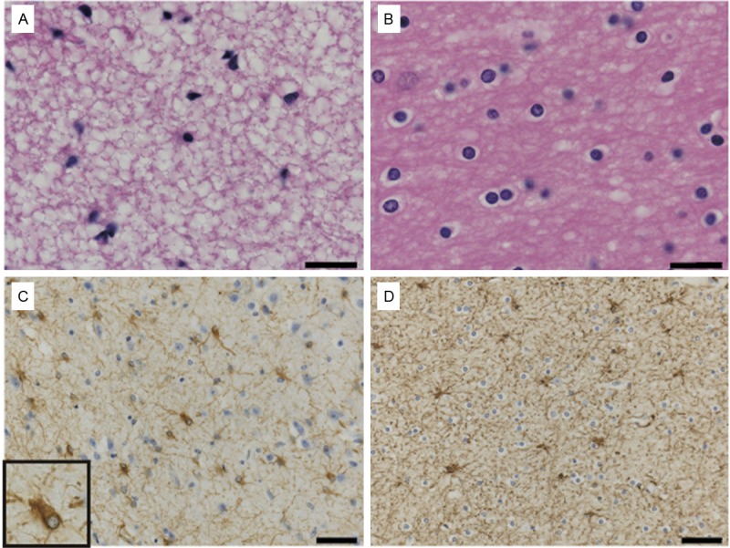
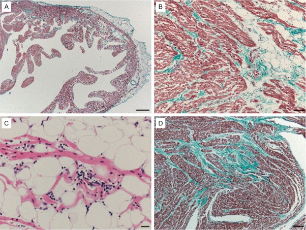
We report a rare autopsy case of early infantile-onset vanishing white matter disease, with a submicroscopic deletion of 14q24.3, which included EIF2B2 and a missense mutation of EIF2B2 (V85E) of the remaining allele. The patient was a 4-year-old boy, who was found to have suddenly died during sleep. Physical and mental development began to deteriorate after convulsions at 10 month of age, and did not recover to baseline measurements. At autopsy, the brain showed a marked decrease in volume of white matter, with no typical cystic rarefaction. Histopathologically, the affected white matter showed diffuse loss of myelin fibers, meager astrogliosis with dysmorphic astrocytes, and loss of oligodendrocytes. Proliferative and apoptotic markers were negative for oligodendrocytes in the severely affected area. These findings may be related to the severity of the disease, and might be a feature of the EIF2B2 mutation pattern of the patient. Additionally, unusual fatty infiltration of both ventricles of the heart was found. These findings were suspected as early pathology of arrhythmogenic right ventricular cardiomyopathy due to characteristic gene mutation in the present case. In the present case, the defect EIF2B2 caused by hemizygosity may be related to early onset of the disease and the unusual pathological changes with vulnerability of oligodendrocytes and astrocytes, as well as cardiac abnormalities and sudden unexpected death.
Keywords: EIF2B2 mutation; Vanishing white matter disease; arrhythmogenic right ventricular cardiomyopathy; hemizygosity; sudden unexpected death.
Figures
Similar articles
-
EIF2B2 gene mutation causing early onset vanishing white matter disease: a case report.Ital J Pediatr. 2022 Jul 27;48(1):128. doi: 10.1186/s13052-022-01325-3. Ital J Pediatr. 2022. PMID: 35897042 Free PMC article.
-
An unmasked mutation of EIF2B2 due to submicroscopic deletion of 14q24.3 in a patient with vanishing white matter disease.Am J Med Genet A. 2012 Jul;158A(7):1771-7. doi: 10.1002/ajmg.a.35431. Epub 2012 Jun 7. Am J Med Genet A. 2012. PMID: 22678813
-
Adult-onset leukoencephalopathies with vanishing white matter with novel missense mutations in EIF2B2, EIF2B3, and EIF2B5.Neurogenetics. 2011 Aug;12(3):259-61. doi: 10.1007/s10048-011-0284-7. Epub 2011 Apr 12. Neurogenetics. 2011. PMID: 21484434 No abstract available.
-
Vanishing white matter disease: an Italian case with A638G mutation in exon 5 of EIF2B2 gene, an unusual early onset and a long course.Neurol Sci. 2013 Jul;34(7):1235-8. doi: 10.1007/s10072-012-1129-3. Epub 2012 Jun 23. Neurol Sci. 2013. PMID: 22729508 Review.
-
Leukoencephalopathy with vanishing white matter: a review.J Neuropathol Exp Neurol. 2010 Oct;69(10):987-96. doi: 10.1097/NEN.0b013e3181f2eafa. J Neuropathol Exp Neurol. 2010. PMID: 20838246 Review.
Cited by
-
Astrocytes are central in the pathomechanisms of vanishing white matter.J Clin Invest. 2016 Apr 1;126(4):1512-24. doi: 10.1172/JCI83908. Epub 2016 Mar 14. J Clin Invest. 2016. PMID: 26974157 Free PMC article.
-
Modeling vanishing white matter disease with patient-derived induced pluripotent stem cells reveals astrocytic dysfunction.CNS Neurosci Ther. 2019 Jun;25(6):759-771. doi: 10.1111/cns.13107. Epub 2019 Feb 5. CNS Neurosci Ther. 2019. PMID: 30720246 Free PMC article.
-
EIF2B2 gene mutation causing early onset vanishing white matter disease: a case report.Ital J Pediatr. 2022 Jul 27;48(1):128. doi: 10.1186/s13052-022-01325-3. Ital J Pediatr. 2022. PMID: 35897042 Free PMC article.
-
eIF2B activator prevents neurological defects caused by a chronic integrated stress response.Elife. 2019 Jan 9;8:e42940. doi: 10.7554/eLife.42940. Elife. 2019. PMID: 30624206 Free PMC article.
References
-
- Van der Knaap MS, Pronk JC, Scheper GC. Vanishing white matter disease. Lancet Neurol. 2006;5:413–423. - PubMed
-
- Bugiani M, Boor I, Powers JM, Scheper GC, Van der Knaap MS. Leukoencephalopathy with vanishing white matter: A review. J Neuropathol Exp Neurol. 2010;69:987–996. - PubMed
-
- Shimada S, Miya K, Oda N, Watanabe Y, Kumada T, Sugawara M, Shimojima K, Yamamoto T. An unmasked mutation of EIF2B2 due to submicroscopic deletion of 14q24.3 in a patient with vanishing white matter disease. Am J Med Genet A. 2012;158A:1771–1777. - PubMed
-
- Hata Y, Mori H, Tanaka A, Fujita Y, Shimomura T, Tabata T, Kinoshita T, Yamaguchi Y, Ichida F, Kominato Y, Ikeda N, Nishida N. Identification and characterization of a novel genetic mutation with prolonged QT syndrome in an unexplained postoperative death. Int J Legal Med. 2014;128:105–115. - PubMed
-
- Schiffmann R, Fogli A, Van der Knaap MS, Boespflug-Tanguy O. GeneReviews™ [Internet] Seattle (WA): University of Washington, Seattle; 2003. Childhood Ataxia with Central Nervous System Hypomyelination/Vanishing White Matter; pp. 1993–2013. http://www.ncbi.nlm.nih.gov/books/NBK1258/ - PubMed
Publication types
MeSH terms
Substances
LinkOut - more resources
Full Text Sources