. 2014 Sep;88(18):10767-77.
doi: 10.1128/JVI.03296-13. Epub 2014 Jul 9.

Envelope glycoprotein binding to the integrin α4β7 is not a general property of most HIV-1 strains

Affiliations

Envelope glycoprotein binding to the integrin α4β7 is not a general property of most HIV-1 strains

Lautaro G Perez et al. J Virol. 2014 Sep.

Abstract

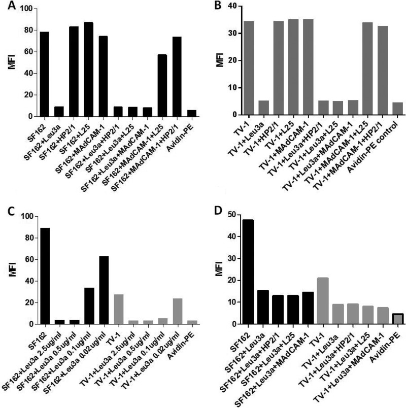
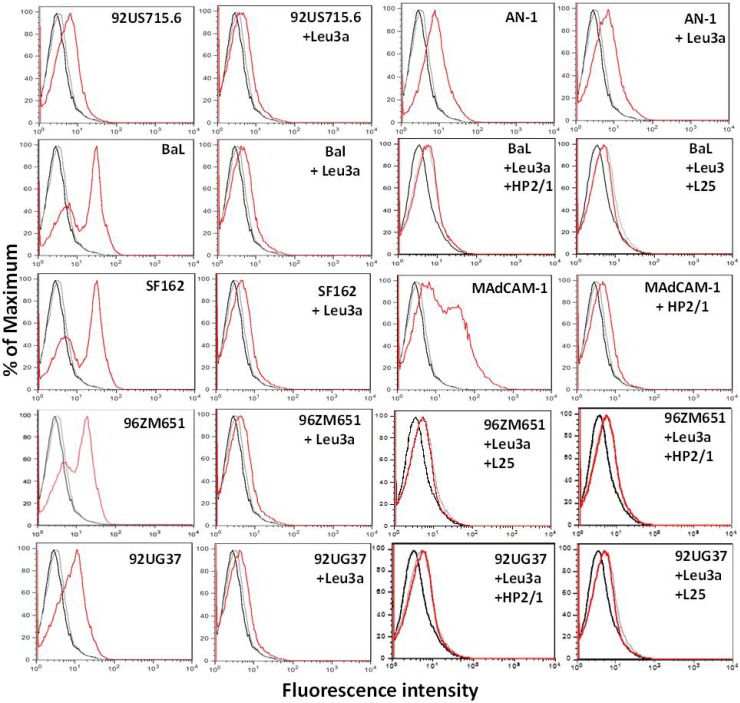
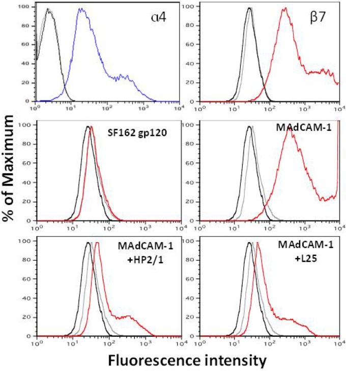
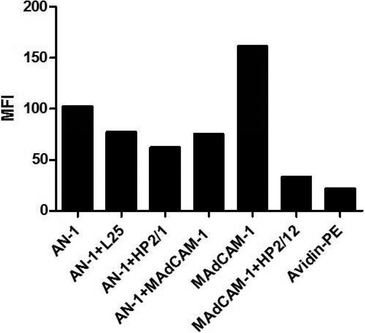
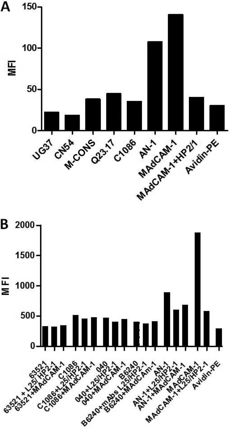
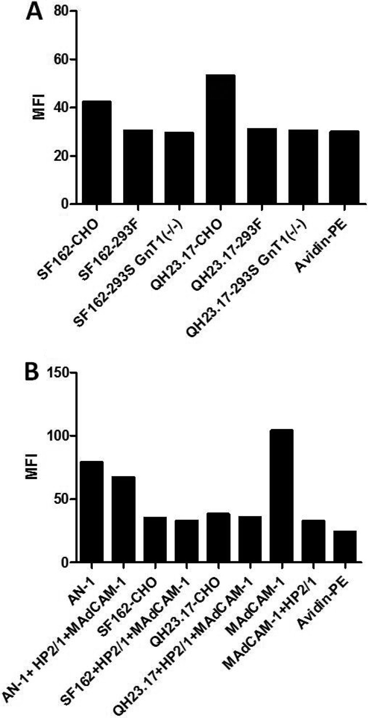
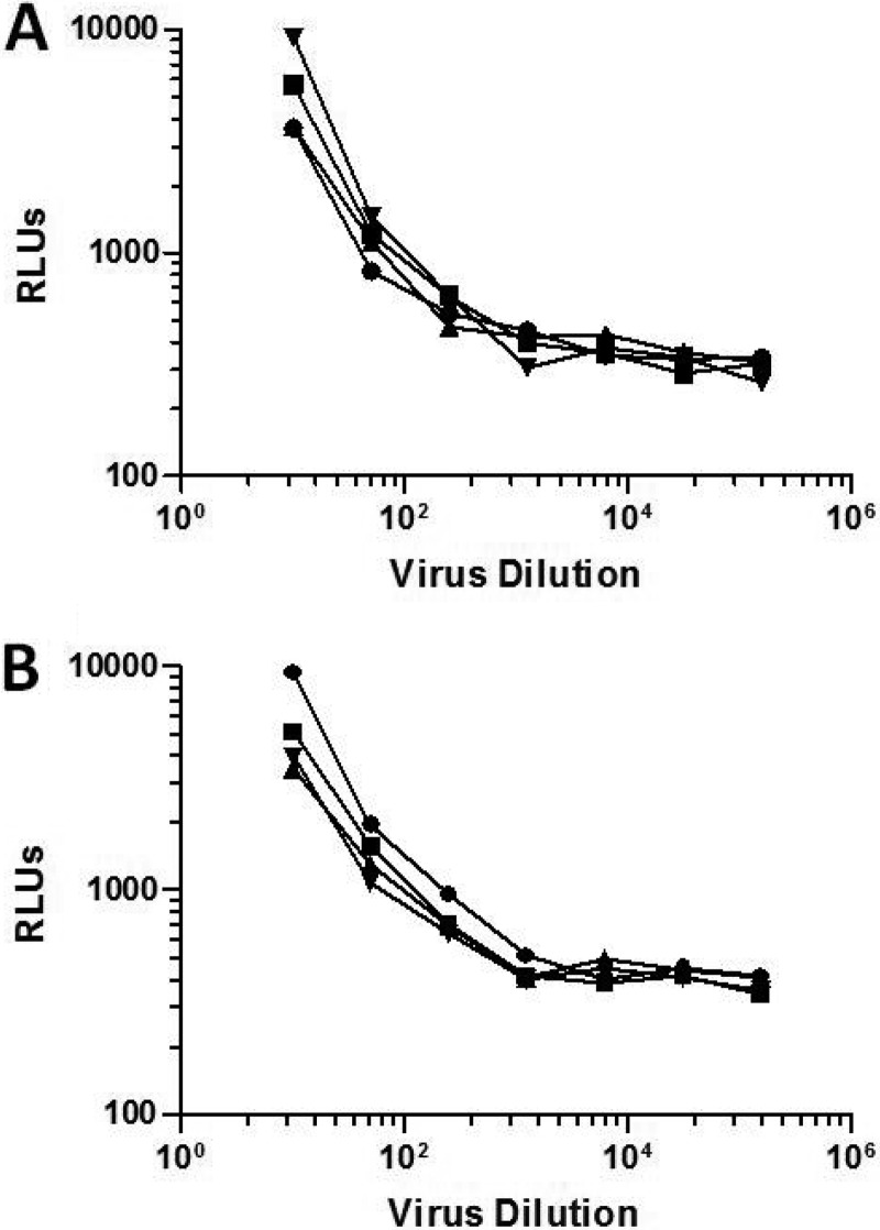
The HIV-1 surface glycoprotein gp120 has been reported to bind and signal through α4β7 by means of a tripeptide motif in the V2 loop that mimics structures present in the natural ligands for α4β7, suggesting that α4β7 may facilitate HIV-1 infection of CD4(+) T cells in the gut. Furthermore, immune correlates in the RV144 vaccine efficacy trial generated the hypothesis that V1V2 antibodies to an epitope near the putative α4β7 binding motif may play a role in protection against HIV-1 infection. In the interest of developing an assay to detect antibodies that block gp120 binding to α4β7, we used retinoic acid (RA)-activated human peripheral blood mononuclear cells (PBMCs) and transfected HEK293T (293T) cells expressing the integrin complex to study the α4β7 binding properties of 16 HIV-1 envelope glycoproteins. The natural ligand for α4β7, mucosal addressin cell adhesion molecule-1 (MAdCAM-1), bound efficiently to RA-activated PBMCs and transfected 293T cells, and this binding was blocked by antibodies to α4. gp120 from multiple HIV-1 subtypes bound to RA-activated PBMCs from three donors in a CD4-dependent manner, but little or no α4β7 binding was detected. Similarly, little or no binding to α4β7 on transfected 293T cells was detected with multiple gp120s and gp140s, including gp120s from transmitted/founder strains, or when gp120 was produced in CHO, 293T, and 293S/GnT1(-/-) cells. Finally, we found no evidence that infectious HIV-1 virions produced in either PBMCs or 293T cells could bind α4β7 on transfected 293T cells. Infectious HIV-1 virions and most gp120s/gp140s appear to be poor ligands for the α4β7 integrin complex under the conditions tested here.

Importance: Certain HIV-1 gp120 envelope glycoproteins have been shown to bind the gut-homing receptor α4β7, and it has been suggested that this binding facilitates mucosal transmission and virus replication in the gut mucosa. Additional evidence has generated the hypothesis that antibodies that bind near the putative α4β7 binding motif in the V2 loop of gp120, possibly disrupting gp120-α4β7 binding, may be important for HIV-1 vaccines. Our evidence indicates that infectious HIV-1 virions and many gp120s lack detectable α4β7 binding activity, suggesting that this homing receptor may play a limited role in direct HIV-1 infection of cells.


Figures

FIG 1
FIG 1
RA upregulation of α4β7 integrins on three donor human PBMCs and MAdCAM-1 binding. (A) Expression of α4β7 integrins on PBMCs from three donors (D1, D2, and D3) after RA treatment was analyzed by flow cytometry. Cells were cultured with and without RA for 7 days in medium containing IL-2 and PHA. Cells were stained with an APC-conjugated anti-α4 MAb or PE-conjugated anti-β7 MAb, washed, and fixed with formaldehyde before analysis with a BD FACSCalibur analyzer. The mean fluorescence intensity (MFI) obtained from the analysis of the data with FlowJo software is shown. α4 and β7, the anti-human integrin MAbs used. (B) Binding of MAdCAM-1, the natural ligand for the α4β7 integrin complex, was measured by flow cytometry using RA-treated PBMCs from donor 3 incubated with a biotinylated recombinant human MAdCAM-1 Fc chimera and PE-avidin at 4°C. In parallel assays, RA-treated PBMCs were preincubated with the anti-α4 MAbs HP2/1 and L25 prior to the addition of MAdCAM-1. Black bars, mean fluorescence intensity obtained by analysis with FlowJo software.
FIG 2
FIG 2
CD4-specific binding of gp120 to RA-treated human PBMCs. Biotinylated gp120 from HIV-1 isolates SF162 (subtype B) and TV-1 (subtype C) was incubated with RA-treated PBMCs at 4°C for 15 min in HEPES buffer containing Ca2+ and Mn2+. Bound gp120 was detected with PE-avidin and flow cytometry analysis. The binding of gp120 to RA-treated PBMCs preincubated with a saturating concentration (25 μg/ml) of the mouse anti-human CD4 MAb Leu3a was also measured in this assay. Assays were performed with PBMCs from three donors. Black curves in the histograms, unstained PBMCs; gray curves, PBMCs incubated with avidin-PE only; red curves, PBMCs incubated with biotinylated gp120 from the indicated HIV-1 isolate.
FIG 3
FIG 3
Lack of α4β7-specific binding of gp120 from two different HIV-1 isolates and effect of subsaturating concentrations of Leu3a. (A and B) Binding of biotinylated SF162gp120 (A) and TV-1gp120 (B) to RA-treated PBMCs from donor 3 with and without preincubation with either unlabeled MAdCAM-1, the anti-α4 MAbs HP2/1 and L25, or the anti-CD4 MAb Leu3a at a saturating concentration or combinations of these reagents. (C) Titration of Leu3a for suboptimal inhibition of gp120 binding to donor 3 RA-treated PBMCs. (D) Effect of subsaturating concentrations of Leu3a on the binding of SF162gp120 or TV-1gp120 to donor 3 RA-treated PBMCs in the presence and absence of either HP2/1, L25, or MAdCAM-1. A subsaturating concentration of 0.08 μg/ml of Leu3a was chosen for both gp120s. Binding was assessed by flow cytometry. The mean fluorescence intensity obtained for each reagent and combinations is shown. Black bars, SF162gp120; gray bars, TV-1gp120. The same avidin-PE control was used for both gp120s in panels C and D (gray bar with a black border).
FIG 4
FIG 4
CD4-dependent binding of gp120 from multiple HIV-1 subtypes to human PBMCs. Biotinylated gp120s from HIV-1 subtypes A, B, and C were tested for binding to RA-treated PBMCs by flow cytometry. In addition, PBMCs preincubated with a saturating concentration of Leu3a prior to the addition of biotinylated gp120 were tested in parallel. Biotinylated MAdCAM-1 was used as a positive control to confirm the presence of the α4β7 integrins on the PBMCs. Black curves in the histograms, unstained PBMCs; gray curves, PBMCs incubated with avidin-PE only; red curves, PBMCs incubated with gp120 from HIV-1 isolates 92US715.6 (subtype B), 92UG37 (subtype A), BaL (subtype B), AN-1 (deduced ancestral subtype), SF162 (subtype B), and 96ZM651 (subtype C), in which binding was detected with avidin-PE. The results also correspond to those for PBMCs incubated with biotinylated MAdCAM-1, in which binding was detected with avidin-PE.
FIG 5
FIG 5
Weak inhibition of AN-1gp120 binding to RA-treated PBMCs in the presence of anti-α4 MAbs and MAdCAM-1. Binding of biotinylated AN-1gp120 to RA-treated human PBMCs preincubated with either Leu3a, L25, HP2/1, or MAdCAM-1 was tested by flow cytometry. Binding of biotinylated MAdCAM-1 to the same PBMCs preincubated with HP2/1 was used as a control. Black bars, the mean fluorescence intensity obtained by analysis with FlowJo software for each reagent.
FIG 6
FIG 6
Binding of SF162gp120 and MAdCAM-1 to α4β7 expressed on 293T cells. 293T cells were transfected with expression vectors for the cDNA of the human α4 and β7 integrins. The cell surface expression levels of the integrins were detected by flow cytometry 48 h later (top; blue and red for α4 and β7, respectively). (Middle) Binding of biotinylated SF162gp120 and MAdCAM-1 to transfected cells (red plots); (bottom) specificity of binding determined by testing the ability of HP2/1 and L25 to block the binding of biotinylated MAdCAM-1. Black curves, untreated cells; gray curves, cells incubated with avidin-PE only.
FIG 7
FIG 7
Binding of AN-1gp120 to α4β7 expressed on 293T cells. Flow cytometry was used to test the binding of biotinylated AN-1gp120 to 293T cells transiently transfected with the human α4 and β7 cDNAs. In parallel, AN-1gp120 was tested for binding to α4β7-expressing cells preincubated with either L25, HP2/1, or MAdCAM-1. Biotinylated MAdCAM-1was used as a control to monitor α4β7 expression levels. Black bars, the mean fluorescence intensity obtained from the analysis of the data with FlowJo software.
FIG 8
FIG 8
Binding of gp140 and T/F gp120s to α4β7 expressed on 293T cells. (A) The gp140s of HIV-1 strains CN54 (subtype C), 92UG037 (UG37; subtype A), M-CONS (a consensus sequence of group M Env), the transmitted/founder subtype C strain C.1086, and Q23.17 (subtype A), all produced in CHO cells, were biotinylated and tested for binding to α4β7-expressing 293T cells by flow cytometry. The mean fluorescence intensity representing the binding of the different gp140s to the integrin-expressing cells, in which binding was detected by avidin-PE, is shown. (B) gp120s from the transmitted/founder subtype B isolates B6240D11 (B6240), B040D11 (040), and 63521D11 (63521) and from the subtype C isolate C.1086 were biotinylated and tested in parallel for binding to α4β7-expressing cells by flow cytometry. Black bars, the mean fluorescence intensity obtained by analysis of the data with FlowJo software. The basal background level of fluorescence determined for cells incubated only with avidin-PE is also shown.
FIG 9
FIG 9
The α4β7 reactivity of gp120s produced in three different cell lines. (A) Biotinylated SF162 and Q23.17 gp120s produced in CHO, 293F, and 293S/GnT1−/− cell lines were tested in parallel for binding to 293T cells transiently expressing α4β7 integrins. (B) The same gp120s described for panel A were produced in CHO cells and tested for binding to α4β7-expressing 293T cells in the presence and absence of a combination of anti-α4 MAb HP2/1 and MAdCAM-1. Binding of biotinylated MAdCAM-1 to α4β7-expressing 293T cells in the presence and absence of the anti-α4 MAb HP2/1 was used as a control. Black bars, the mean fluorescence intensity obtained by analysis with FlowJo software. The basal background level of fluorescence determined for cells incubated only with avidin-PE is also shown.
FIG 10
FIG 10
Capture of HIV-1 infectious virions by α4β7 expressed on 293T cells. Serial dilutions of the HIV-1 infectious molecular clones R2184c4.IMC.LucR (A) and WEAU3-3.IMC.LucR (B) were incubated with either α4β7-expressing 293T cells or mock-transfected 293T cells for 1 h at 37°C. Cells were washed and overlaid with TZM-bl cells. Activation of the Luc reporter gene in the TZM-bl cells was measured 48 h later using a Britelite luminescence reaction kit. Circles, virus grown in PBMCs and incubated with mock-transfected 293T cells; squares, virus grown in 293T cells and incubated with mock-transfected 293T cells; triangles, virus grown in PBMCs and incubated with α4β7-expressing 293T cells; inverted triangles, virus grown in 293T cells and incubated with α4β7-expressing 293T cells. RLU, relative light units.

Similar articles

Cited by

References

    1. Desgrosellier JS, Cheresh DA. 2010. Integrins in cancer: biological implications and therapeutic opportunities. Nat. Rev. Cancer 10:9–22. 10.1038/nrc2748 - DOI - PMC - PubMed
    1. Hynes RO. 2002. Integrins: bidirectional, allosteric signaling machines. Cell 110:673–687. 10.1016/S0092-8674(02)00971-6 - DOI - PubMed
    1. DeNucci CC, Pagán AJ, Mitchell JS, Shimizu Y. 2010. Control of α4β7 integrin expression and CD4 T cell homing by the β1 integrin subunit. J. Immunol. 184:2458–2467. 10.4049/jimmunol.0902407 - DOI - PMC - PubMed
    1. Berlin C, Bargatze RF, Campbell JJ, von Andrian UH, Szabo MC, Hasslen SR, Nelson RD, Berg EL, Erlandsen SL, Butcher EC. 1995. α4 integrins mediate lymphocyte attachment and rolling under physiologic flow. Cell 80:413–422. 10.1016/0092-8674(95)90491-3 - DOI - PubMed
    1. Butcher EC, Picker LJ. 1996. Lymphocyte homing and homeostasis. Science 272:60–67. 10.1126/science.272.5258.60 - DOI - PubMed

Publication types

MeSH terms

Substances