. 2014 Sep;35(29):8427-38.
doi: 10.1016/j.biomaterials.2014.06.021. Epub 2014 Jun 28.

Branched-linear and agglomerate protein polymers as vaccine platforms

Affiliations

Branched-linear and agglomerate protein polymers as vaccine platforms

Leyi Wang et al. Biomaterials. 2014 Sep.

Abstract

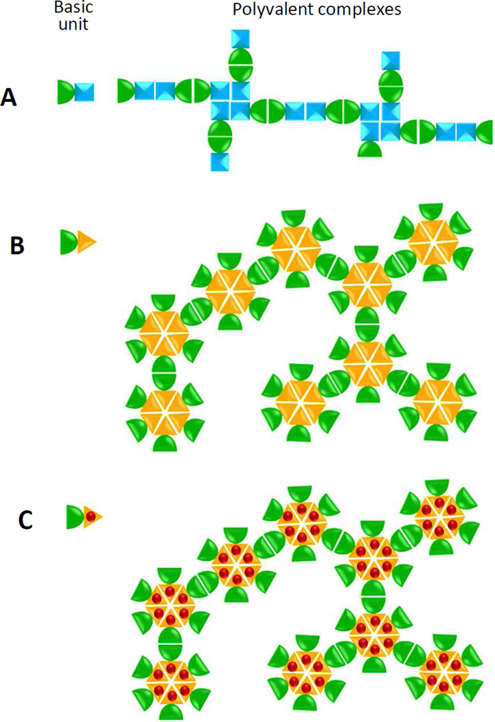
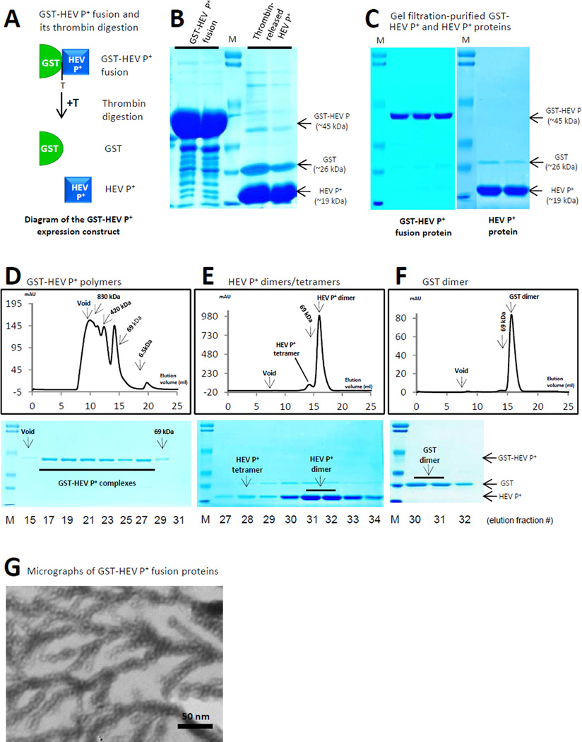
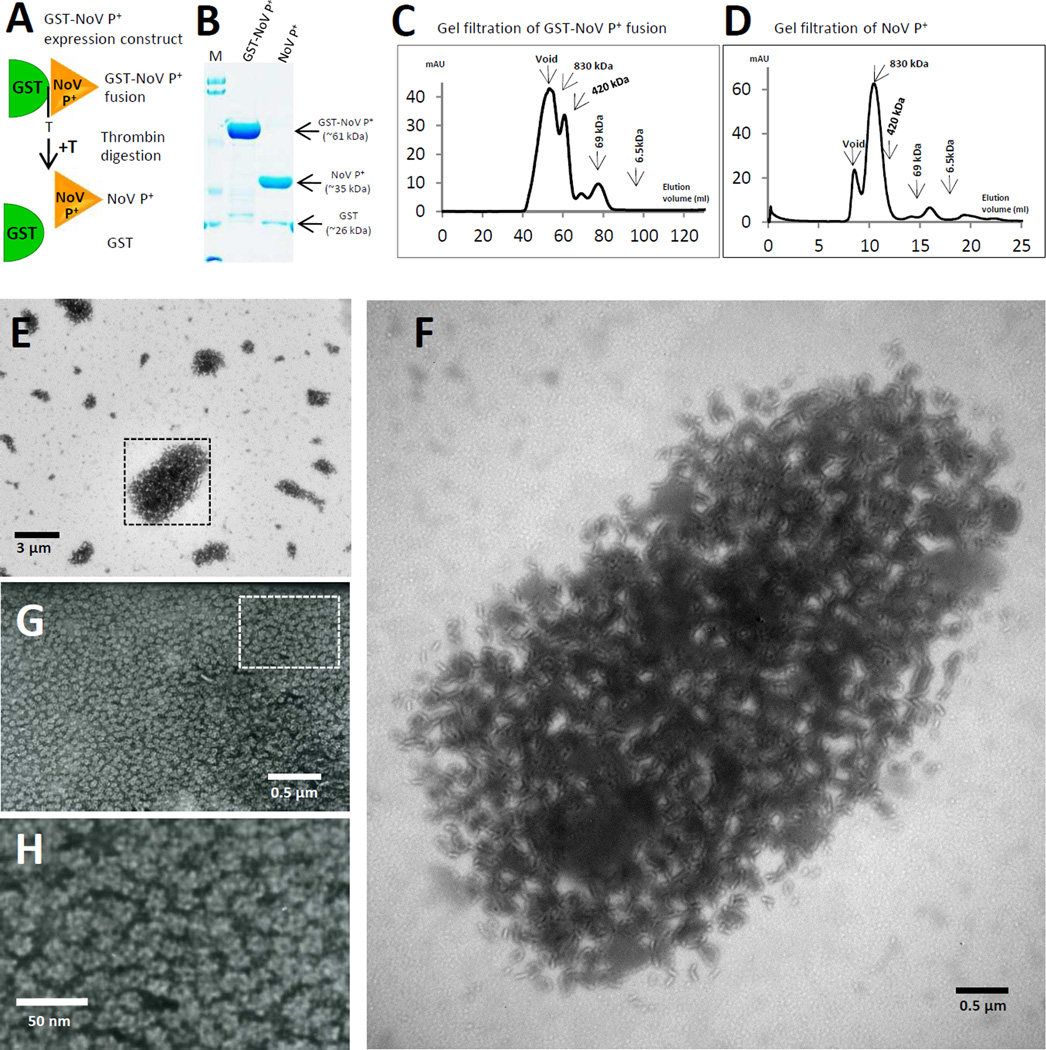
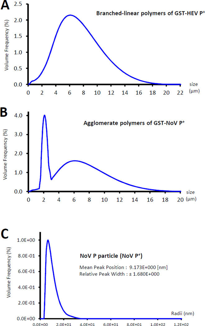
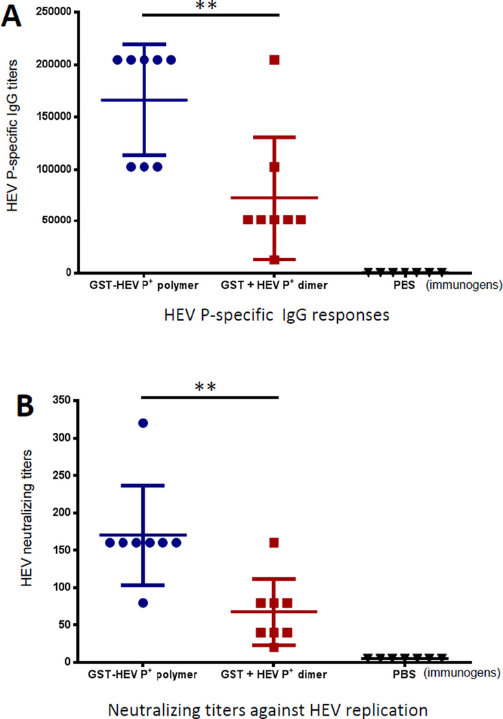
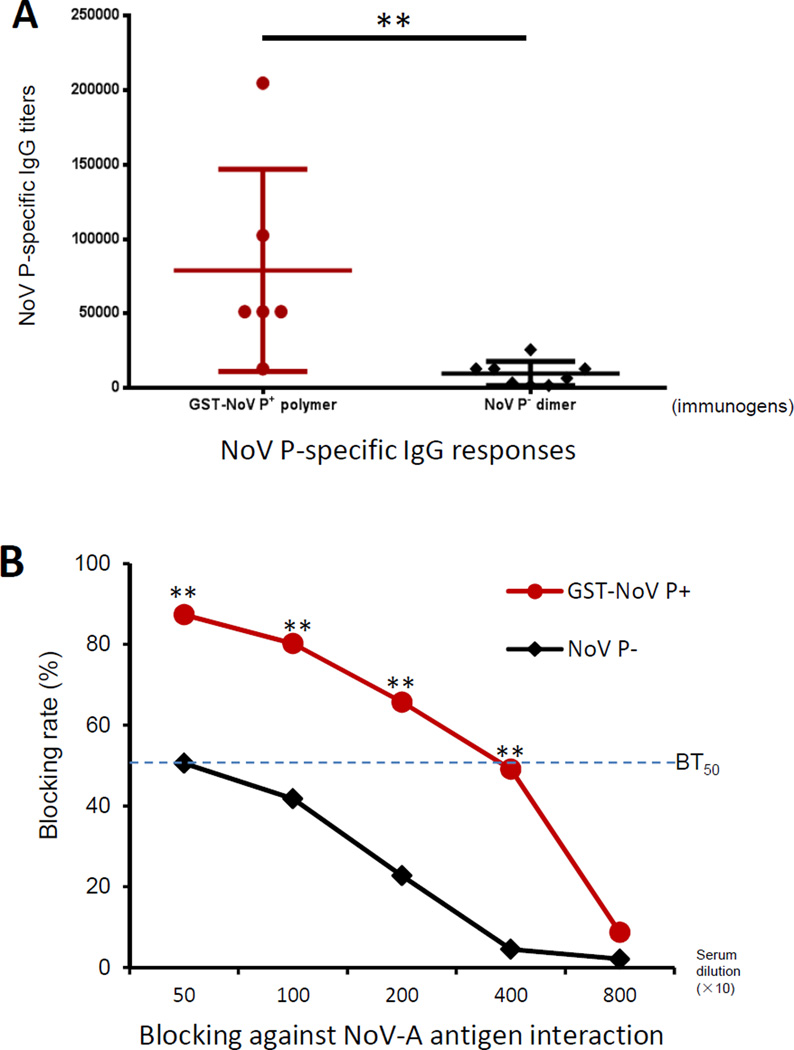
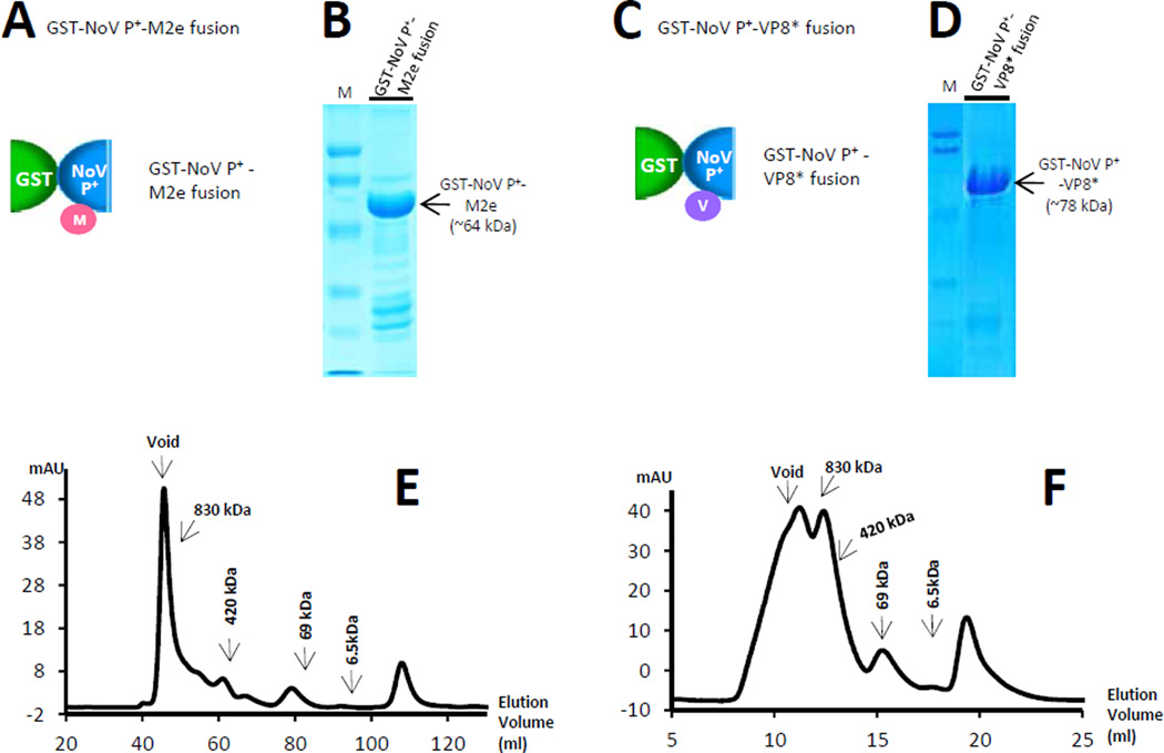
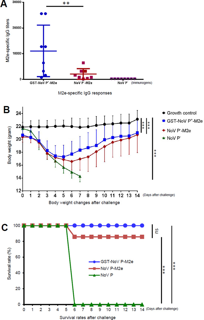
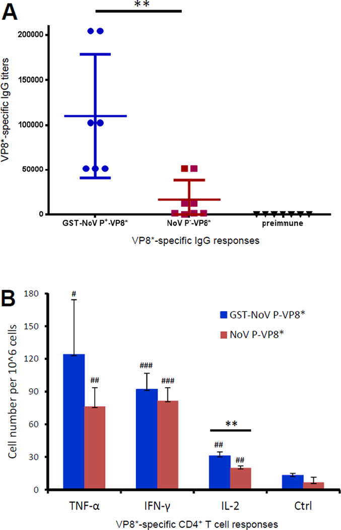
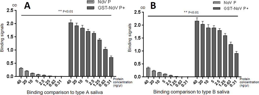
Many viral structural proteins and their truncated domains share a common feature of homotypic interaction forming dimers, trimers, and/or oligomers with various valences. We reported previously a simple strategy for construction of linear and network polymers through the dimerization feature of viral proteins for vaccine development. In this study, technologies were developed to produce more sophisticated polyvalent complexes through both the dimerization and oligomerization natures of viral antigens. As proof of concept, branched-linear and agglomerate polymers were made via fusions of the dimeric glutathione-s-transferase (GST) with either a tetrameric hepatitis E virus (HEV) protruding protein or a 24-meric norovirus (NoV) protruding protein. Furthermore, a monomeric antigen, either the M2e epitope of influenza A virus or the VP8* antigen of rotavirus, was inserted and displayed by the polymer platform. All resulting polymers were easily produced in Escherichia coli at high yields. Immunization of mice showed that the polymer vaccines induced significantly higher specific humoral and T cell responses than those induced by the dimeric antigens. Additional evidence in supporting use of polymer vaccines included the significantly higher neutralization activity and protective immunity of the polymer vaccines against the corresponding viruses than those of the dimer vaccines. Thus, our technology for production of polymers containing different viral antigens offers a strategy for vaccine development against infectious pathogens and their associated diseases.

Keywords: Flu vaccine; Hepatitis E virus vaccine; Norovirus; Norovirus vaccine; Vaccine development; Vaccine platform.


Figures

Figure 1
Figure 1
Schematic illustration of the principles of branched-linear and agglomerate polymer formations and the application of the agglomerate complex as a platform for antigen display. (A) Branched-linear polymer formation. Fusion of a dimeric protein (left, green half oval) with a dimeric/tetrameric protein (left, cyan square) forms in a branched-linear polymer (right). The linear portion of the polymer is formed by homotypic dimerizations of the two proteins, respectively, (ovals and double squares), while the branchings are formed by tetramerization of the tetrameric protein (tetra-squares). (B) Agglomerate polymer formation. Fusion of a dimeric protein (left, green half oval) with an oligomeric protein (left, yellow triangle) forms an agglomerate polymer (right) through intermolecular dimerization (green ovals) and oligomerization (yellow hexagon) of the two protein components. In both (A) and (B) only small portions of the large branched-linear (A) and agglomerate (B) polymers are shown for clarity, while the actual polymers are much more complex. The oligomers in (B) are shown in a cross-sectional view, while they may be spherical in the actual agglomerate polymers. (C) A monomeric antigen (red ball) can be inserted into the surface of a component of the fusion protein unit (left). Through formation of the agglomerate polymers, the inserted antigen becomes polyvalent in the polymer (right).
Figure 2
Figure 2
Production and characterization of branched-linear polymers of GST-HEV P+. (A) Schematic illustration of GST-HEV P+ fusion and its thrombin digestion (+T) into free GST (green half oval) and HEV P+ (blue square) proteins. The thrombin digestion site (T) between the GST and the HEV P+ protein is indicated. (B) The affinity column-purified GST-HEV P+ fusion protein (left) and the thrombin released HEV P+ protein (right) were analyzed on a SDS PAGE gel. (C) The gel filtration-purified GST-HEV P+ (left) and HEV P+ (right) proteins are analyzed on SDS PAGE gels. Positions of the GST-HEV P+ fusion protein (~45 kDa), GST (~26 kDa), and HEV P+ (~19 kDa) proteins are indicated. M represents the pre-stained protein markers (Bio-Rad, low range), with bands from top to bottom representing 113, 92, 52, 34, 25, and 18 kDa, respectively. (D) to (F) Elution curves of gel filtration chromatography of the GST-HEV P+ (D), HEV P+ (E), and GST (F) proteins through the size-exclusion column (Superdex 200, 10/300 GL, GE Healthcare Life Sciences). The gel filtration column was calibrated by the Gel Filtration Calibration Kit (GE Healthcare Life Sciences) and the purified recombinant P particles, small P particles, and P dimers of NoV (VA387). The elution positions of blue Dextran 2000 (~2000 kDa, void), P particles (~830 kDa), small P particles (420 kDa), P dimers (~69 kDa), and/or aprotinins (~6.5kDa) were indicated. The protein peaks were analyzed by SDS-PAGE shown below the corresponding elution curves of gel filtrations. Fraction #15 represents the beginning of the void volume (>800 kDa) (D), fractions #28 and #31/32 represent the peaks of the HEV P+ tetramers (~76 kDa) and dimers (~38 kDa) (E), respectively, while fractions #30/31, represent the peak of GST dimer (~56 kDa). (G) An electron micrograph of negatively stained GST-HEV P+ protein revealed branched-linear polymers.
Figure 3
Figure 3
Production and Characterization of agglomerate polymers of GST-NoV P+. (A) Schematic illustration of the GST-NoV P+ fusion protein and its thrombin digestion (+T) into GST and NoV P+ protein. The thrombin digestion site (T) between the GST (green half oval) and the NoV P+ (yellow triangle) is indicated. (B) The affinity column-purified GST-NoV P+ and the thrombin released NoV P+ proteins were analyzed on a SDS PAGE. Positions of the GST-NoV P+ fusion (~61 kDa), NoV P+ (~35 kDa) and GST (~26kDa) proteins are indicated. M represents the pre-stained protein markers (Bio-Rad, low range), with bands from top to bottom representing 113, 92, 52, 34, 25, and 18 kDa, respectively. (C) and (D) The elution curves of gel filtration chromatography of the GST-NoV P+ (C) and NoV P+ (D) proteins through the size exclusion column (Superdex 200, 10/300 GL, GE Healthcare Life Sciences). The gel filtration columns were calibrated by the Gel Filtration Calibration Kit (GE Healthcare Life Sciences) and the purified recombinant P particles, small P particles, and P dimers of NoV. The elution positions of blue Dextran 2000 (~2000 kDa, void), P particles (~830 kDa), small P particles (420 kDa), P dimers (~69 kDa), and aprotinin (~6.5kDa) were indicated. (E) to (H) Electron micrographs of negatively stained GST-NoV P+ (E and F) and NoV P+ (G and H) proteins. The GST-NoV P+ protein forms large agglomerate polymers with variable sizes (E), while the NoV P+ forms the 24-meric P particles measuring ~20 nm in diameter (G and H). A large agglomerate polymer in (E) is enlarged in (F). The rectangular labeled region in (G) is enlarged in (H) to show the morphologies of the 24mer P particles.
Figure 4
Figure 4
Size distributions of the branched-linear and agglomerate polymers. (A and B) The size distributions of the two polymers were measured by light scatting using a high definition digital particle size analyzer (Saturn DigiSizer 5200, Micromeritics), in which the size distribution curves of the branched-linear (A) and agglomerate (B) polymers based on volume frequency are shown. The branched-linear polymers of GST-HEV P+ showed a mean size of ~5.8 µm. The agglomerate polymers of GST-NoV P+ exhibited a major sharp peak at ~2.2 µm, while the remaining complexes showed a normal distribution curve with a peak at ~6.5 µm. (C) The previously characterized P particles (NoV P+, ~20 nm) were measured by the DSL Compact goniometer system (ALV, Langen, Germany) as control.
Figure 5
Figure 5
The branched-linear polymers increased immunogenicity of HEV P antigen. (A) HEV P specific IgG responses after immunization. Mice were immunized with equal molar amount of GST-HEV P+ polymers and a mixture of free GST and HEV P+ proteins (GST + HEV P+, 1:1 in mole amount) intranasally three times without an adjuvant in two-week intervals (n = 8 mice/group). Phosphate buffer saline (PBS) was given as negative controls (n = 7 mice/group). (B) The resulting mouse antisera blocked HEV replication. The neutralizing titers of the resulting antisera against HEV (Kernow P6 strain, genotype 3) infection in HepG2/C3A cells were determined. ** (P < 0.01) indicates a statistically very significant difference.
Figure 6
Figure 6
The agglomerate polymers increased immune responses of NoV P antigen. (A) NoV P specific IgG responses after immunization. Mice (n=6–8/group) were immunized with equal molar amounts of GST-NoV P+ polymers and NoV P dimers intranasally three times without an adjuvant in two-week intervals. NoV P-specific antibody IgG titers of the resulting antisera were determined by EIAs. (B) The resulting mouse antisera blocked NoV-ligand interaction. The mouse antisera were examined for their blocking activity against interaction of NoV P particle with A antigen. The 50% blocking titers (BT50s) of both antisera are indicated by a flashed line. ** (P < 0.01) indicates statistically very significant differences between data pairs of sera at different given dilutions.
Figure 7
Figure 7
A monomeric antigen can be displayed by the agglomerate polymers. (A and C) schematic illustration of a monomeric antigen (red or purple circle) being attached to the surface of the NoV P+ component of the GST-NoV P+ fusion protein. M indicates the M2e epitope of influenza A virus (A), while V indicates the VP8* antigen of rotavirus (C). (B and D), affinity column-purified GST-NoV P+-M2e (B) and GST-NoV P+-VP8* (D) fusion proteins were analyzed on SDS PAGE gels. (E and F) Elusion curves of gel filtration chromatography of GST-NoV P+-M2e (E) and GST-NoV P+-VP8* (F) fusion proteins indicated the formation of large complexes. The gel filtration column was calibrated by the Gel Filtration Calibration Kit (GE Healthcare Life Sciences) and the purified recombinant P particles, small P particles, and P dimers of NoV (VA387). The elution positions of blue Dextran 2000 (~2000 kDa, void), P particles (~830 kDa), small P particles (420 kDa), P dimers (~69 kDa), and aprotinin (~6.5kDa) were indicated.
Figure 8
Figure 8
The agglomerate polymer-displayed M2e epitope exhibited improved immunogenicity (A) and protective immunity (B and C). (A) M2e-specific antibody (IgG) responses of polymer- and dimer-displayed M2e epitope. Mice (n=8 mice/group) were immunized with equal molar amounts of GST-NoV P+-M2e polymers, NoV P-M2e dimers and NoV P dimers intranasally three times without an adjuvant in two-week intervals. The M2e-specific IgG titers of the resulting antisera were determined by EIAs. (B and C) Body weight changes (B) and survival rates (C) of vaccinated and various control mice after challenge with influenza A virus (IAV). The mice were challenged with lethal dose of mouse-adapted IAV (strain PR8, H1N1) and monitored for body weight changes (B) and survival rates (C) for 14 days. In (B) “Growth control” mice were not vaccinated and were not challenged. Each data point was a mean value of four to eight mice. The standard deviations are indicated in one direction for clarity of the figure. In (C) “GST-NoV P-M2e”, “NoV-P-M2e” and “NoV P” groups were vaccinated with GST-NoV P+-M2e, NoV-P-M2e and NoV P, respectively, followed by a challenge with IAV. ** (P < 0.01) indicates a statistically very significant difference, while *** (P < 0.001) indicates a statistically extremely significant difference.
Figure 9
Figure 9
The polymer-presented VP8* antigen showed improved humoral (A) and T cell cytokine (B) responses. (A) Antibody (IgG) responses of polymer- and dimer-displayed VP8* antigen. Mice (n=7/group) were immunized with equal molar amounts of GST-NoV P+-VP8* polymers or NoV P-VP8* dimers intranasally three times without an adjuvant in two-week intervals. The VP8*-specific IgG titers were determined by EIAs. (B) CD4+ T cell cytokine responses of polymer- and dimer-displayed VP8* antigen. Mice (n=3/group) were immunized as in (A). After stimulation of the splenocytes with a VP8*-specific CD4+ T cell epitope, the resulting CD4+ T cell cytokines IL-2, IFN-γ, and TNF-α were measured by intracellular cytokine staining. Cytokine-producing T cells from the polymer- (GST-NoV P-VP8*) and dimer- (NoV P-VP8*) immunized mice were shown in blue and red, respectively. Those from unimmunized mice were negative controls (Ctrl). Data were presented as cytokine-producing T cell numbers per 106 cells. The statistical significances between the immunized groups and unimmunized controls (Ctrl) are shown by # symbols (# P<0.05, ## P<0.01, ### P<0.001), while those between the polymer- and dimer-immunized groups (blue and red) are shown by ** (P<0.01).
Figure 10
Figure 10
The agglomerate polymers increased binding activities of NoV P domain to HBGA ligands. The free NoV P dimers bound weakly, while the agglomerate polymers of GST-NoV P+ bound strongly to type A (A) and B (B) saliva samples that were defined as type A and B nonsecretor. Y-axes indicate the binding signal intensities in optical density (OD), while X-axes indicate protein concentrations that were used for the binding assays.

Similar articles

Cited by

References

    1. Tan M, Jiang X. Subviral particle as vaccine and vaccine platform. Curr Opin Virol. 2014;6C:24–33. - PMC - PubMed
    1. McAleer WJ, Buynak EB, Maigetter RZ, Wampler DE, Miller WJ, Hilleman MR. Human hepatitis B vaccine from recombinant yeast. Nature. 1984;307:178–180. - PubMed
    1. Andre FE, Safary A. Summary of clinical findings on Engerix-B, a genetically engineered yeast derived hepatitis B vaccine. Postgrad Med J. 1987;63(Suppl 2):169–177. - PubMed
    1. Kirnbauer R, Booy F, Cheng N, Lowy DR, Schiller JT. Papillomavirus L1 major capsid protein self-assembles into virus-like particles that are highly immunogenic. Proc Natl Acad Sci U S A. 1992;89:12180–12184. - PMC - PubMed
    1. Jagu S, Kwak K, Garcea RL, Roden RB. Vaccination with multimeric L2 fusion protein and L1 VLP or capsomeres to broaden protection against HPV infection. Vaccine. 2010;28:4478–4486. - PMC - PubMed

Publication types

MeSH terms