

The kinetochore
- PMID: 24984773
- PMCID: PMC4067989
- DOI: 10.1101/cshperspect.a015826
The kinetochore
Abstract
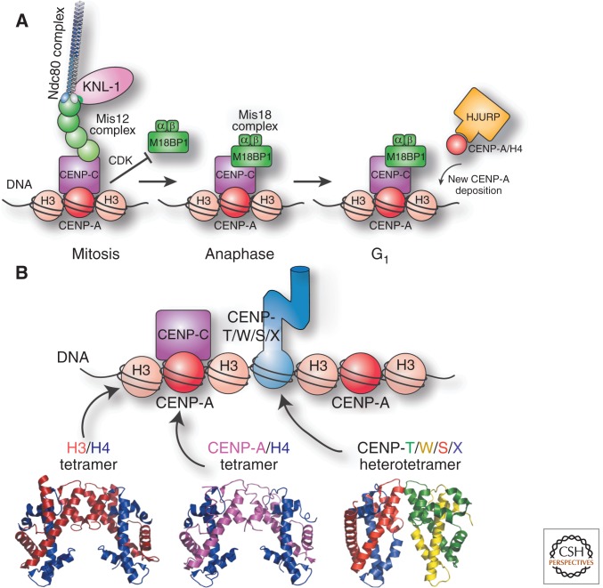
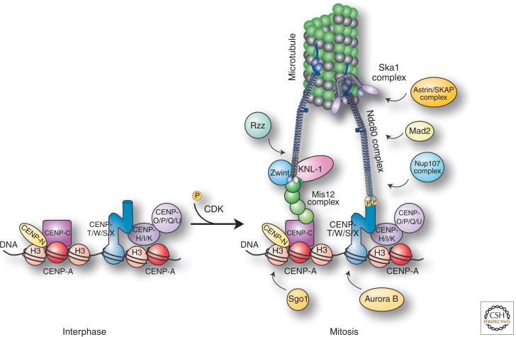
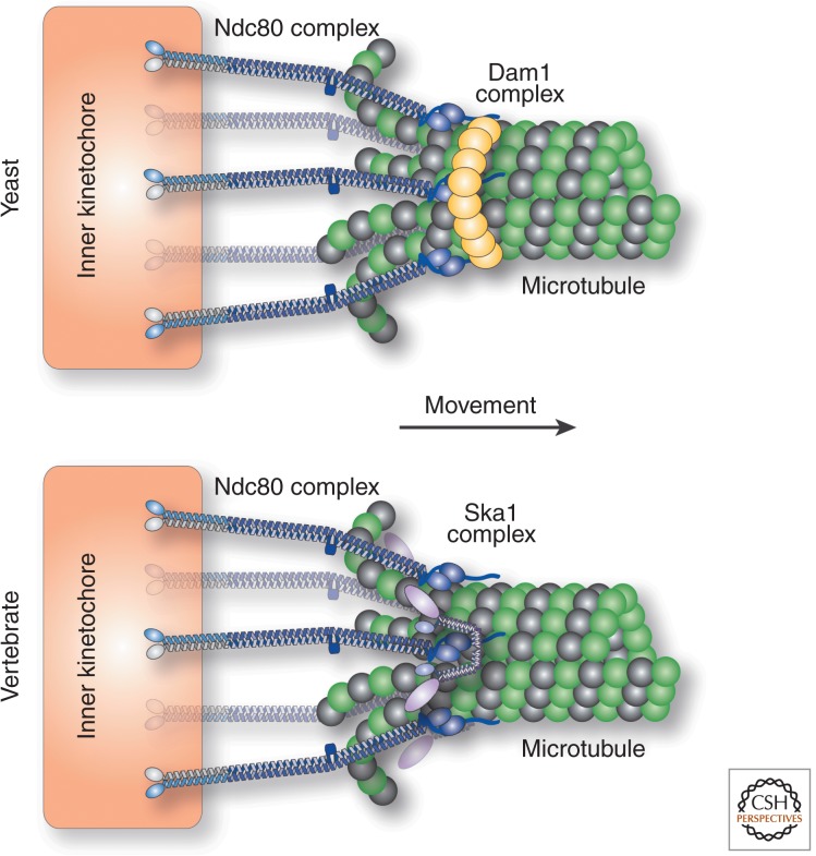
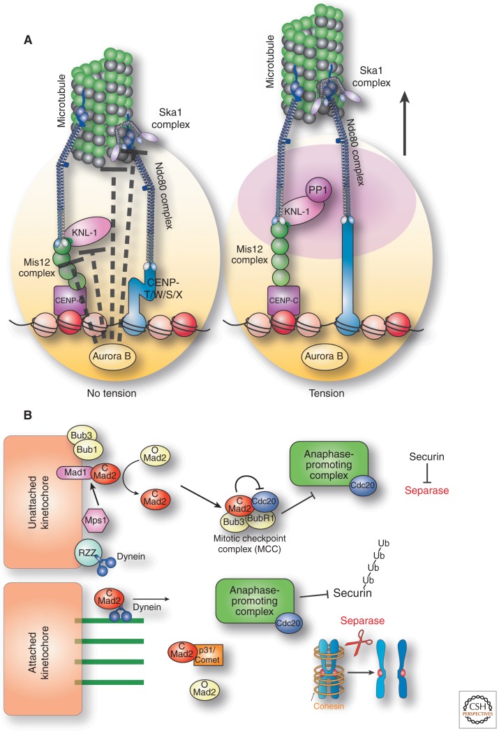
A critical requirement for mitosis is the distribution of genetic material to the two daughter cells. The central player in this process is the macromolecular kinetochore structure, which binds to both chromosomal DNA and spindle microtubule polymers to direct chromosome alignment and segregation. This review will discuss the key kinetochore activities required for mitotic chromosome segregation, including the recognition of a specific site on each chromosome, kinetochore assembly and the formation of kinetochore-microtubule connections, the generation of force to drive chromosome segregation, and the regulation of kinetochore function to ensure that chromosome segregation occurs with high fidelity.
Copyright © 2014 Cold Spring Harbor Laboratory Press; all rights reserved.
Figures

Similar articles
-
The dynamic kinetochore-microtubule interface.J Cell Sci. 2004 Nov 1;117(Pt 23):5461-77. doi: 10.1242/jcs.01536. J Cell Sci. 2004. PMID: 15509863 Review.
-
CENP-E kinesin interacts with SKAP protein to orchestrate accurate chromosome segregation in mitosis.J Biol Chem. 2012 Jan 6;287(2):1500-9. doi: 10.1074/jbc.M111.277194. Epub 2011 Nov 22. J Biol Chem. 2012. PMID: 22110139 Free PMC article.
-
The kinetochore-microtubule interface at a glance.J Cell Sci. 2018 Aug 16;131(16):jcs214577. doi: 10.1242/jcs.214577. J Cell Sci. 2018. PMID: 30115751 Free PMC article. Review.
-
Mitotic Protein CSPP1 Interacts with CENP-H Protein to Coordinate Accurate Chromosome Oscillation in Mitosis.J Biol Chem. 2015 Nov 6;290(45):27053-27066. doi: 10.1074/jbc.M115.658534. Epub 2015 Sep 16. J Biol Chem. 2015. PMID: 26378239 Free PMC article.
-
Septin 7 interacts with centromere-associated protein E and is required for its kinetochore localization.J Biol Chem. 2008 Jul 4;283(27):18916-25. doi: 10.1074/jbc.M710591200. Epub 2008 May 6. J Biol Chem. 2008. PMID: 18460473 Free PMC article.
Cited by
-
Meiotic Nuclear Architecture in Distinct Mole Vole Hybrids with Robertsonian Translocations: Chromosome Chains, Stretched Centromeres, and Distorted Recombination.Int J Mol Sci. 2020 Oct 15;21(20):7630. doi: 10.3390/ijms21207630. Int J Mol Sci. 2020. PMID: 33076404 Free PMC article.
-
Synchronization of human retinal pigment epithelial-1 cells in mitosis.J Cell Sci. 2020 Sep 17;133(18):jcs247940. doi: 10.1242/jcs.247940. J Cell Sci. 2020. PMID: 32878943 Free PMC article.
-
Both tails and the centromere targeting domain of CENP-A are required for centromere establishment.J Cell Biol. 2015 Mar 2;208(5):521-31. doi: 10.1083/jcb.201412011. Epub 2015 Feb 23. J Cell Biol. 2015. PMID: 25713413 Free PMC article.
-
Quiescent Cells Actively Replenish CENP-A Nucleosomes to Maintain Centromere Identity and Proliferative Potential.Dev Cell. 2019 Oct 7;51(1):35-48.e7. doi: 10.1016/j.devcel.2019.07.016. Epub 2019 Aug 15. Dev Cell. 2019. PMID: 31422918 Free PMC article.
-
Sumoylation regulates protein dynamics during meiotic chromosome segregation in C. elegans oocytes.J Cell Sci. 2019 Jul 18;132(14):jcs232330. doi: 10.1242/jcs.232330. J Cell Sci. 2019. PMID: 31243051 Free PMC article.
References
Publication types
MeSH terms
Substances
Grants and funding
LinkOut - more resources
Full Text Sources
Other Literature Sources