. 2014 Jun 30;15(6):R82.
doi: 10.1186/gb-2014-15-5-r82.

Insulator function and topological domain border strength scale with architectural protein occupancy

Insulator function and topological domain border strength scale with architectural protein occupancy

Kevin Van Bortle et al. Genome Biol. .

Abstract

Background: Chromosome conformation capture studies suggest that eukaryotic genomes are organized into structures called topologically associating domains. The borders of these domains are highly enriched for architectural proteins with characterized roles in insulator function. However, a majority of architectural protein binding sites localize within topological domains, suggesting sites associated with domain borders represent a functionally different subclass of these regulatory elements. How topologically associating domains are established and what differentiates border-associated from non-border architectural protein binding sites remain unanswered questions.

Results: By mapping the genome-wide target sites for several Drosophila architectural proteins, including previously uncharacterized profiles for TFIIIC and SMC-containing condensin complexes, we uncover an extensive pattern of colocalization in which architectural proteins establish dense clusters at the borders of topological domains. Reporter-based enhancer-blocking insulator activity as well as endogenous domain border strength scale with the occupancy level of architectural protein binding sites, suggesting co-binding by architectural proteins underlies the functional potential of these loci. Analyses in mouse and human stem cells suggest that clustering of architectural proteins is a general feature of genome organization, and conserved architectural protein binding sites may underlie the tissue-invariant nature of topologically associating domains observed in mammals.

Conclusions: We identify a spectrum of architectural protein occupancy that scales with the topological structure of chromosomes and the regulatory potential of these elements. Whereas high occupancy architectural protein binding sites associate with robust partitioning of topologically associating domains and robust insulator function, low occupancy sites appear reserved for gene-specific regulation within topological domains.


Figures

Figure 1
Figure 1
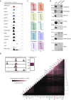
Genome-wide mapping of dTFIIIC220 in D. melanogaster. (a) Example ChIP-seq profile shown for dTFIIIC220 (red) over a tRNA cluster on Drosophila chromosome 2R, co-bound by TFIIIB subunits TRF1 and BRF. (b,c) Tag density enrichment profiles for dTFIIIC220 over all annotated tDNAs (B) and over sites previously identified as bound by TFIIIB complex subunits TRF1 and BRF (C) confirms the expected genome-wide localization patterns for Drosophila (overlap significance P < 0.00001, permutation test). RPM, reads per million. (d) Consensus sequences identified de novo by MEME-ChIP reveals evolutionarily conserved Drosophila B box and A box elements present in dTFIIIC220-bound tRNA genes. (e) Central motif enrichment (CentriMo) plot for B box and A box sequences with respect to dTFIIIC220 ChIP-seq peaks at tRNA genes. (f) Overlap between dTFIIIC220 peaks, independently identified in two biological replicates at a false discovery rate of 5%, with annotated tRNA genes obtained from Flybase (P < 0.00001, permutation test). Non-overlapping sites indicate thousands of ETC loci in D. melanogaster, of which 348 contain the B box binding motif (14.5%, P < 0.00001, permutation test).
Figure 2
Figure 2
SMC-containing cohesin and condensin complexes localize to a subset of tDNAs and ETC loci. (a) Number of overlapping peaks identified by ChIP-seq against α-kleisen subunits Rad21 (cohesin), Barren (condensin I), and CAP-H2 (condensin II) in Kc167 cells (P < 0.00001 for overlap between Rad21 with CAP-H2 or Barren, permutation test). (b) Heatmap representation of ChIP-seq read intensities of SMC-containing complexes and TFIIIC subunit dTFIIIC220, anchored across all dTFIIIC220 peaks (top), plus or minus 5 kb. Heatmap representation (bottom) of overlap frequencies between dTFIIIC220 peaks and those of SMC-containing complexes (overlap significance for dTFIIIC220 with each factor P < 0.00001, permutation test). (c) Read intensity plots for Rad21, Barren, and CAP-H2 at TFIIIC-bound tDNAs (left) and ETC loci (right) plus or minus 5 kb. Tag density is represented as rank-order normalized reads per million (RPM) across all three ChIP-seq experiments. (d,e) Example genomics viewer profiles of overlapping dTFIIIC220 sites at tRNA genes and ETC loci. (f) Heatmap representation shown for DNase-seq and ChIP-seq read intensities at 1,311 active enhancers previously defined by STARR-seq, and marked by active enhancer characteristics in the Kc167 cell line, including DNase I hypersensitivity, H3K4me1 and H3K27ac. (g) Percentage of enhancers bound by dTFIIIC220 and SMC-containing complexes.
Figure 3
Figure 3
Drosophila TFIIIC clusters with CTCF at sites combinatorially bound by architectural proteins, cohesin, and condensin II. (a) Example genomics viewer profile of a combinatorially bound APBS, co-bound by dTFIIIC220, SMC-containing cohesin and condensin complexes, dCTCF, BEAF-32, Su(Hw), CP190, Mod(mdg4), DREF, Chromator, L(3)mbt, and marked by strong DHS. (b) Heatmap representation of co-factor co-localization at 3,728 genomic loci combinatorially bound by architectural proteins. Overlap frequency is the fraction of combinatorially bound loci bound by each individual factor. Inset: sites were identified as genomic fragments having four or more proteins in Kc167 cells using MACS called summits ±200 bp for factors dCTCF, BEAF-32, Su(Hw), CP190, Mod(mdg4), Zw5, DREF, Chromator, and L(3)mbt, and mapped independently of TFIIIC and SMC complexes; size distribution (bp) of combinatorially bound loci. P < 0.00001 for overlap between combinatorially bound loci with dTFIIIC220, Rad21, and CAP-H2, permutation test. Overlap frequency matrix hierarchically clustered (absolute centered, single linkage). (c) Heatmaps depict ChIP-seq tag densities for each Drosophila architectural protein as a function of distance, ±5 kb, from ETC loci. (d) Western blot analysis of control preimmune and α-dTFIIIC220 immunoaffinity purifications detect interactions between dTFIIIC220 and CP190, Mod(mdg4), and BEAF-32.
Figure 4
Figure 4
High occupancy APBSs delineate TADs and associate with robust enhancer-blocking activity. (a) Heatmap representing Hi-C interaction frequencies at single fragment resolution for a 1 Mb region across Drosophila chromosome 3 L in Kc167 cells. White lines demarcate previously defined TAD boundaries [1]. A high occupancy APBS (left) is present at a single fragment topological domain border strongly separating two TADs (white arrowhead). Colorbar represents (log2) interaction frequencies observed between restriction fragments, ranging from low (blue) to high (red). (b) Percentage of TADs defined in Kc167 cells delineated by a high, medium, or low occupancy APBSs ± one restriction cut site (TAD borders n = 1,110, high occupancy APBSs n = 1,638, P < 0.00001, permutation test). (c) Topological border strength defined by the ratio of intra- versus inter-TAD interaction frequencies scales with the occupancy (number of bound proteins) at APBSs. (d) Architectural protein occupancy and DNase I hypersensitivity at DNA fragments previously tested for enhancer-blocking activity in transgenic reporter assays [13,51,52]. Sequences shown to possess robust activity (red) correlate with both the highest occupancy and DNase I activity, whereas sites incapable of insulator activity are marked by low occupancy (P < 0.01, Wilcoxon rank sum test, two-sided). RPM, reads per million. (e) Quantification of topological domain border strengths at sequences tested for insulator function within their endogenous context. Robust insulator sequences are characterized by significantly greater topological border strength than non-enhancer-blocking sequences (P < 0.05, Wilcoxon rank sum test, two-sided). (f) Tag density plots of rank-order normalized DNase-seq profiles throughout embryonic stages of development at APBSs [53], and at transcription factor binding sites shown to function as developmental enhancers during early embryogenesis. The progressive loss of DNase accessibility at highly bound transcription factor binding sites (right) contrasts with the combinatorially bound APBSs (left), which are marked by strong DNase I hypersensitivity throughout each stage of development.
Figure 5
Figure 5
Clustering of architectural proteins is a conserved feature of genome organization. (a) Heatmap depicting overlap enrichment between architectural proteins mapped by ChIP-seq in mESCs. Red to blue squares represent depletion (red) or enrichment (blue), determined as the log2 (observed/expected) frequency of overlap when compared to randomized, simulated data. (b) Example genomics viewer profile (left) of a high occupancy APBSs in mESCs, bound by CTCF, TFIIIC (-220, -110, and -90), Rad21, condensin II (CAP-D3 and CAP-H2), and PRDM5, and marked by strong DHS. Hi-C interaction matrix (right) illustrates the corresponding TAD separation observed in vivo (TAD boundary defined by black arrowhead). (c) Sites combinatorially bound by CTCF and other factors (CTCF plus three or more proteins) are significantly enriched at TAD borders in mESCs. P values (*P < 0.05, ** P < 0.01, *** P < 0.001) were calculated using permutation tests. (d) Relationship between protein occupancy, defined by the presence of CTCF, Rad21, PRDM5, TFIIIC (any or all subunits -220, -110, -90) and condensin II (CAP-H2 and/or CAP-D3), and topological domain border strength in mESCs. (e) Parallel analysis of topological domain border strength in human IMR90 fibroblasts as a function of protein occupancy at CTCF binding sites. Co-binding determined by cross-comparison of ChIP-seq datasets for transcription factors and DNA binding proteins in human K562 cells. (f) Relationship between cell-type specificity of CTCF binding sites and localization to TAD borders. CTCF ubiquity determined by cross-comparison of 62 CTCF ChIP-seq datasets across 31 human cell lines. The x-axis represents CTCF sites grouped into eight bins (approximately 15,000 sites each) of increasing ubiquity ranging from cell type-specific to constitutive. For a list of human cell lines, ubiquity scores and exact number of CTCF binding sites in each bin, see Materials and methods and Additional file 8.
Figure 6
Figure 6
Combinatorial binding of architectural proteins shapes topological domain structure. Model illustrating the relationship between protein occupancy at APBSs and observed heterogeneity in TAD border strengths. We uncover a spectrum of architectural protein co-localization, ranging from low (blue) to high (red), which scales with the strength of TAD border formation. We propose that differences in TAD border strength reflect the role of architectural proteins in mediating long-range interactions. Interaction frequencies and/or interaction stability are greatest at high occupancy APBSs (red), whereas fewer or less stable interactions at intermediate APBSs (green) allows for inter-TAD interactions, resulting in comparatively weaker TAD borders observed by Hi-C.

Comment in

Similar articles

Cited by

References

    1. Hou C, Li L, Qin Z, Corces V. Gene density, transcription and insulators contribute to partitioning the Drosophila genome into physical domains. Mol Cell. 2012;48:471–484. - PMC - PubMed
    1. Sexton T, Yaffe E, Kenigsberg E, Bantignies F, Leblanc B, Hoichman M, Parrinello H, Tanay A, Cavalli G. Three-dimensional folding and functional organization principles of the Drosophila genome. Cell. 2012;148:458–472. - PubMed
    1. Dixon JR, Selvaraj S, Yue F, Kim A, Li Y, Shen Y, Hu M, Liu JS, Ren B. Topological domains in mammalian genomes identified by analysis of chromatin interactions. Nature. 2012;485:376–380. - PMC - PubMed
    1. Phillips-Cremins JE, Sauria ME, Sanyal A, Gerasimova TI, Lajoie BR, Bell JS, Ong CT, Hookway TA, Guo C, Sun Y, Bland MJ, Wagstaff W, Dalton S, McDevitt TC, Sen R, Dekker J, Taylor J, Corces VG. Architectural protein subclasses shape 3D organization of genomes during lineage commitment. Cell. 2013;153:1281–1295. - PMC - PubMed
    1. Seitan VC, Faure AJ, Zhan Y, McCord RP, Lajoie BR, Ing-Simmons E, Lenhard B, Giorgetti L, Heard E, Fisher AG, Flicek P, Dekker J, Merkenschlager M. Cohesin-based chromatin interactions enable regulated gene expression within preexisting architectural compartments. Genome Res. 2013;23:2066–2077. - PMC - PubMed

Publication types

MeSH terms