

Mortalin, apoptosis, and neurodegeneration
- PMID: 24970131
- PMCID: PMC4030873
- DOI: 10.3390/biom2010143
Mortalin, apoptosis, and neurodegeneration
Abstract
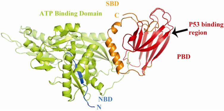
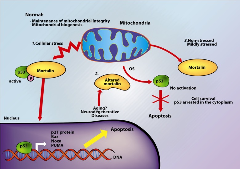
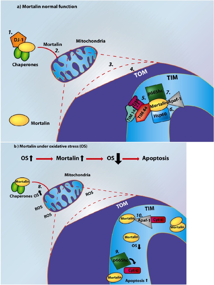
Mortalin is a highly conserved heat-shock chaperone usually found in multiple subcellular locations. It has several binding partners and has been implicated in various functions ranging from stress response, control of cell proliferation, and inhibition/prevention of apoptosis. The activity of this protein involves different structural and functional mechanisms, and minor alterations in its expression level may lead to serious biological consequences, including neurodegeneration. In this article we review the most current data associated with mortalin's binding partners and how these protein-protein interactions may be implicated in apoptosis and neurodegeneration. A complete understanding of the molecular pathways in which mortalin is involved is important for the development of therapeutic strategies for cancer and neurodegenerative diseases.
Figures

Similar articles
-
Mortalin: Protein partners, biological impacts, pathological roles, and therapeutic opportunities.Front Cell Dev Biol. 2023 Feb 2;11:1028519. doi: 10.3389/fcell.2023.1028519. eCollection 2023. Front Cell Dev Biol. 2023. PMID: 36819105 Free PMC article. Review.
-
Structural studies of UBXN2A and mortalin interaction and the putative role of silenced UBXN2A in preventing response to chemotherapy.Cell Stress Chaperones. 2016 Mar;21(2):313-26. doi: 10.1007/s12192-015-0661-5. Epub 2015 Dec 4. Cell Stress Chaperones. 2016. PMID: 26634371 Free PMC article.
-
Abrogating the Interaction Between p53 and Mortalin (Grp75/HSPA9/mtHsp70) for Cancer Therapy: The Story so far.Front Cell Dev Biol. 2022 Apr 14;10:879632. doi: 10.3389/fcell.2022.879632. eCollection 2022. Front Cell Dev Biol. 2022. PMID: 35493098 Free PMC article. Review.
-
Crystal structure of the nucleotide-binding domain of mortalin, the mitochondrial Hsp70 chaperone.Protein Sci. 2014 Jun;23(6):833-42. doi: 10.1002/pro.2466. Epub 2014 Apr 17. Protein Sci. 2014. PMID: 24687350 Free PMC article.
-
On the brotherhood of the mitochondrial chaperones mortalin and heat shock protein 60.Cell Stress Chaperones. 2006 Summer;11(2):116-28. doi: 10.1379/csc-144r.1. Cell Stress Chaperones. 2006. PMID: 16817317 Free PMC article. Review.
Cited by
-
A Review of Proteins Associated With Neuroprotection and Regeneration in Alzheimer's Disease.Cureus. 2022 Oct 18;14(10):e30412. doi: 10.7759/cureus.30412. eCollection 2022 Oct. Cureus. 2022. PMID: 36407141 Free PMC article. Review.
-
Hydrogen peroxide initiates oxidative stress and proteomic alterations in meningothelial cells.Sci Rep. 2022 Aug 25;12(1):14519. doi: 10.1038/s41598-022-18548-3. Sci Rep. 2022. PMID: 36008468 Free PMC article.
-
Characterization and prognostic significance of mortalin, Bcl-2 and Bax in intrahepatic cholangiocarcinoma.Oncol Lett. 2018 Feb;15(2):2161-2168. doi: 10.3892/ol.2017.7570. Epub 2017 Dec 8. Oncol Lett. 2018. PMID: 29434920 Free PMC article.
-
Identification and validation of aging-related genes in atrial fibrillation.PLoS One. 2023 Nov 13;18(11):e0294282. doi: 10.1371/journal.pone.0294282. eCollection 2023. PLoS One. 2023. PMID: 37956134 Free PMC article.
-
Markers of acute toxicity of DDT exposure in pancreatic beta-cells determined by a proteomic approach.PLoS One. 2020 Oct 26;15(10):e0229430. doi: 10.1371/journal.pone.0229430. eCollection 2020. PLoS One. 2020. PMID: 33104727 Free PMC article.
References
-
- Wadhwa R., Kaul S.C., Ikawa Y., Sugimoto Y. Identification of a novel member of mouse hsp70 family. Its association with cellular mortal phenotype. J. Biol. Chem. 1993;268:6615–6621. - PubMed
-
- Bhattacharyya T., Karnezis A.N., Murphy S.P., Hoang T., Freeman B.C., Phillips B., Morimoto R.I. Cloning and subcellular localization of human mitochondrial hsp70. J. Biol. Chem. 1995;270:1705–1710. - PubMed
-
- Webster T.J., Naylor D.J., Hartman D.J., Hoj P.B., Hoogenraad N.J. cDNA cloning and efficient mitochondrial import of pre-mtHSP70 from rat liver. DNA Cell Biol. 1994;13:1213–1220. - PubMed
LinkOut - more resources
Full Text Sources
Other Literature Sources
