. 2014 Sep;34(9):2012-22.
doi: 10.1161/ATVBAHA.114.303399. Epub 2014 Jun 26.

Monocytes are recruited from venules during arteriogenesis in the murine spinotrapezius ligation model

Affiliations

Monocytes are recruited from venules during arteriogenesis in the murine spinotrapezius ligation model

Anthony C Bruce et al. Arterioscler Thromb Vasc Biol. 2014 Sep.

Abstract

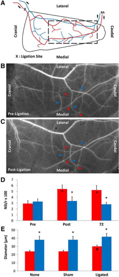
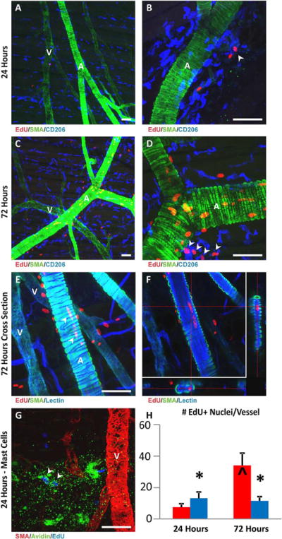
Objective: Chronic arterial occlusion results in arteriogenesis of collateral blood vessels. This process has been shown to be dependent on the recruitment of growth-promoting macrophages to remodeling collaterals. However, the potential role of venules in monocyte recruitment during microvascular arteriogenesis is not well demonstrated. First, we aim to document that arteriogenesis occurs in the mouse spinotrapezius ligation model. Then, we investigate the temporal and spatial distribution, as well as proliferation, of monocytes/macrophages recruited to collateral arterioles in response to elevated fluid shear stress.

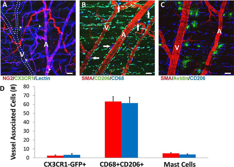
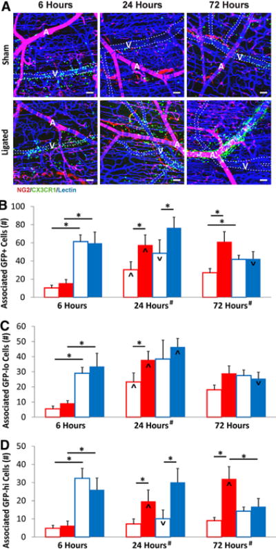
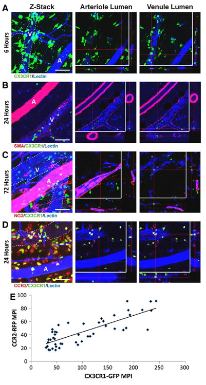
Approach and results: Laser speckle flowmetry confirmed a postligation increase in blood velocity within collateral arterioles but not within venules. After 72 hours post ligation, collateral arteriole diameters were increased, proliferating cells were identified in vessel walls of shear-activated collaterals, and perivascular CD206(+) macrophages demonstrated proliferation. A 5-ethynyl-2'-deoxyuridine assay identified proliferation. CD68(+)CD206(+) cells around collaterals were increased 96%, whereas CX3CR1((+/GFP)) cells were increased 126% in ligated versus sham groups after 72 hours. CX3CR1((+/GFP)) cells were predominately venule associated at 6 hours after ligation; and CX3CR1((+/GFP hi)) cells shifted from venule to arteriole associated between 6 and 72 hours after surgery exclusively in ligated muscle. We report accumulation and extravasation of adhered CX3CR1((+/GFP)) cells in and from venules, but not from arterioles, after ligation.

Conclusions: Our results demonstrate that arteriogenesis occurs in the murine spinotrapezius ligation model and implicate postcapillary venules as the site of tissue entry for circulating monocytes. Local proliferation of macrophages is also documented. These data open up questions about the role of arteriole-venule communication during monocyte recruitment.

Keywords: arterioles; blood flow velocity; macrophages; microvasculature; monocytes.


Figures

Figure 1
Figure 1. Blood velocity and vessel diameter increase in collateral arterioles 72 hours post feeder arteriole ligation
A. Representative cartoon of a spinotrapezius ligation. Red lines indicate arteriole network; blue lines indicate venules. Arrows indicate flow direction, “RA” indicates the redundant feeding arteriole, and the dashed box represents the region analyzed for blood velocity changes and collateral arteriole remodeling. B. Pre-ligation laser speckle index map of murine spinotrapezius blood velocity in measured collateral arterioles (red arrows) and draining venules (blue arrows). Increased intensity of white signal indicates increased blood velocity. C. Post-ligation speckle index map of the same spinotrapezius presenting an increase in laser speckle intensity of collateral arterioles but not draining venules following feeder arteriole ligation. D. Normalized Speckle Intensity per radius (NSI/r) was similar between collateral arterioles (red bars) and venules (blue bars) pre-ligation. Collateral arterioles, but not venules, experienced an 86% increase in NSI/r following feeder arteriole ligation. This difference was maintained after 72 hours. n=11 arterioles and 9 venules total from 3 mice. E. Dilated diameters of collateral arterioles (red bars) in ligated muscle were significantly increased from sham dilated diameters at 72 hours post-surgery and from the unoperated group. No difference was detected for venules (blue bars). n=24 fields of view (FOVs) from 6 mice per group. * indicates p<0.05 between vessel types (arteriole vs venule); ^ indicates p<0.05 increase from pre-ligation vessel group. # indicates p<0.05 increase in dilated arteriolar diameter compared to both unoperated and contralateral sham.
Figure 2
Figure 2. Macrophages and mast cells reside in the un-operated spinotrapezius
A. Example FOV from un-operated muscle showing CX3CR1(+/GFP) cells (arrowheads) in an NG2-DsRed hemizygote with DsRed expression evident in arteriolar smooth muscle, pericytes, and glial cells. CX3CR1(+/GFP) cell counts were very low in un-operated muscles with no difference in numbers surrounding arterioles compared to venules. “A” designates arteriole; “V” designates venule; white dashed line indicates venule outline. B. Example FOV from un-operated wild type muscle showing resident CD68+(blue)CD206+(green) macrophages and CD68+ mast cells exhibiting granules (arrows). Equal numbers of macrophages were associated with both vessel types. C. Staining with conjugated Avidin confirmed presence and positioning of mast cells. All scale bars indicate 50 μm. D. No significant differences were detected between microvessel types for associated resident cell populations. Red bars indicate arteriole; blue bars indicate venule. The number of vessel associated cells were counted along a single arteriole or venule stretched across a 640 micron FOV. n=12 FOVs from 3 mice per group.
Figure 3
Figure 3. Macrophage and mast cell recruitment is increased in ligated versus sham muscles
A. CD68+(blue)CD206+(green) macrophages and CD68+ mast cells (arrows) surrounding SMA+(red) microvasculature in sham and ligated muscles at 72 hours post-ligation surgery. “A” designates arteriole; “V” designates venule; arrowheads indicate mast cells associated with microvessels. All scale bars indicate 50 μm. B. Vessel-associated CD68+CD206+ macrophages were not increased significantly from un-operated for either collateral arterioles (red bars) or venules (blue bars) in sham muscles after 72 hours. In contrast, CD68+CD206+ macrophages associated with both arterioles and venules in ligated muscles were significantly increased compared to sham surgery. C. Arteriole-associated mast cells were significantly increased in the ligated group compared to muscles within the sham group. Also, significantly more mast cells were associated with arterioles (red bars) compared to venules (blue bars) in the ligated group. * indicates p<0.05 between vessel types within surgery group. ^ designates p<0.05 increase between surgery treatments for the indicated group. n=12 FOVs from 3 mice per group.
Figure 4
Figure 4. Differential recruitment of CX3CR1(+/GFP) cells is evident in ligated versus sham surgery muscles
A. CX3CR1(+/GFP) (green) cell recruitment around isolectin (blue) stained vasculature (with arterioles exhibiting NG2-DSRED) in ligated and sham surgery muscles for each time point. “A” designates arteriole; “V” designates venule; white dashed line indicates venule outline. All scale bars indicate 50 μm. B-D. Red indicates arteriole; blue indicates venule. Outlined bars indicate sham surgery; solid bars indicate ligation surgery. * indicates p<0.05 between indicated groups; ^ indicates p<0.05 increase from the previous time point; ∨ indicates p<0.05 decrease from the previous time point; # indicates that upon combining arteriole and venule cell counts, p<0.05 between surgery treatments within the indicated time. n=12 FOVs from 3 mice per group. For comparison to unoperated values, see data presented in Figure 2D. The number of vessel associated cells were counted along a single arteriole or venule stretched across a 640 micron FOV. B. CX3CR1(+/GFP) cells were primarily associated with venules 6 hours post-ligation in both surgery treatments. Associations with arterioles increased significantly by 24 hours within both surgery treatments, and numbers associated with arterioles at 24 hours were significantly higher in the ligated group versus the sham. Significantly more GFP+ cells were present in ligated muscles versus sham at both 24 and 72 hours post-surgery. GFP+ cells associated with venules decreased significantly in the sham group at 24 hours and the ligated group at 72 hours. C. CX3CR1(+/GFP lo) cells were primarily associated with venules at 6 hours post-surgery in both treatment groups. Associations with arterioles increased significantly by 24 hours in both treatments, and numbers associated with arterioles at 24 hours were significantly higher in the ligated group versus the sham. Significantly more of these cells were present in ligated muscles versus sham at 24 hours. Cell counts associated with venules increased significantly by 24 hours in the ligated group before decreasing significantly by 72 hours. This change in venule-associated numbers did not occur in the sham group. Note that no significant differences existed between any groups within the 72 hour time point. D. CX3CR1(+/GFP hi) cells were primarily associated with venules at 6 hours post-surgery in both surgery treatments. Cell counts increased significantly between 6 and 24 hours in the ligated group only. At 24 hours, significantly more cells were associated with arterioles in the ligated group versus the sham group. Cell numbers associated with venules decreased by 24 hours in the sham group, but not in the ligated group. CX3CR1(+/GFP hi) cells associated with arterioles increased between 24 and 72 hours in the ligated group but not the sham. These cells were preferentially associated with arterioles at 72 hours in only the ligated group. Significantly more of these cells were present in ligated muscles versus sham at both 24 and 72 hours.
Figure 5
Figure 5. Monocytes accumulate within venules but not arterioles post-ligation
A-D. Left panels are volume renders of Z-stacks taken through the full thickness of arteriole/venule pairs in ligated murine spinotrapezius. Center panels display FOVs focused on the arteriolar lumen. Right panels display FOV focused on the venular lumen. “A” designates arteriole; “V” designates venule; white dashed line indicates venule outline. Endothelium was labeled with isolectin (blue). CX3CR1(+/GFP) (green) and CCR2(+/RFP) (red) cells visible within the two dimensional areas of the arterioles in some volume renders were confirmed to not be within the arteriolar lumina. All scale bars indicate 50 μm. A. CX3CR1(+/GFP) cells extravasating from a post-capillary venule next to a collateral arteriole at 6 hours post-ligation. B. Accumulation of CX3CR1(+/GFP) and isolectin+ leukocytes within a venular lumen adjacent to a collateral arteriole at 24 hours post-ligation in muscle stained for alpha-smooth muscle actin (red). Note the accumulation of adhered leukocytes in venular lumina and lack of this accumulation in arteriolar lumina. C. CX3CR1(+/GFP) cell recruitment at 72 hours post-ligation in an NG2-DsRed hemizygote with DsRed expression evident in arteriolar smooth muscle, pericytes, and glial cells. D. Recruitment at 24 hours post-ligation of CCR2(+/RFP) and CX3CR1(+/GFP) leukocytes. Note that most recruited CX3CR1(+/GFP) cells also exhibited expression of CCR2-RFP. E. Recruited CX3CR1(+/GFP) cells exhibit a positive correlation between CX3CR1-GFP and CCR2-RFP. Mean pixel intensities (MPI) of CX3CR1-GFP and CCR2-RFP correlate strongly within individual cells present at 24 hours post-ligation. Correlation coefficient=0.81; p=2.1X10⁻¹². n=50 CX3CR1(+/GFP) cells from 2 mice.
Figure 6
Figure 6. EdU proliferation assays indicate that collateral walls remodel and that local proliferation of macrophages is occurring
A-G. “A” designates arteriole; “V” designates venule. All scale bars indicate 50 μm. A. EdU+ nuclei were primarily associated with the perivascular space of venules at 24 hours post-ligation, and were not yet evident in walls of collateral arterioles. B. EdU+ nuclei were few at 24 hours post-ligation. Some EdU+ nuclei were in cells expressing macrophage markers. Arrowhead indicates EdU+ nucleus in a CD206+ cell. C. EdU+ cells were increased in the perivascular space of collateral arterioles and were evident in the walls of collateral arterioles in ligated muscle after 72 hours. D. Macrophages with EdU+ nuclei were more prevalent in the perivascular spaces of collateral arterioles after 72 hours. In addition, EdU+ nuclei were now prevalent in collateral vessel walls, especially in the intima. Arrowheads indicate EdU+ nuclei in CD206+ cells. E. EdU+ nuclei in endothelial cells (indicated by arrowheads) were recognizable by their distinct elongated shape. F. Examination of collateral cross-sections confirmed EdU+ nuclei to be within endothelium. G. EdU+ mast cells were rare but present at time points examined. H. Bars represent total EdU+ nuclei present within microvessels and in their perivascular spaces in 200X FOVs. Red indicates arteriole; blue indicates venule. EdU+ nuceli were commonly seen in perivascular positions at 24 hours post-ligation with significantly more being associated with venules. At 72 hours post-ligation, increased numbers in the perivascular spaces of collateral arterioles, in addition to the appearance of EdU+ nuclei within the walls of arterioles, resulted in significantly more EdU+ nuclei being associated with collateral arterioles. * indicates p<0.05 between vessel types. ^ indicates significant increase from previous time point for vessel type. n=12 FOVs from 4 mice per group. Scale bars are 50 μm.

Similar articles

Cited by

References

    1. Arras M, Ito WD, Scholz D, Winkler B, Schaper J, Schaper W. Monocyte activation in angiogenesis and collateral growth in the rabbit hindlimb. J Clin Invest. 1998 Jan 1;101:40–50. - PMC - PubMed
    1. Ziegelhoeffer T, Fernandez B, Kostin S, Heil M, Voswinckel R, Helisch A, Schaper W. Bone marrow-derived cells do not incorporate into the adult growing vasculature. Circ Res. 2004 Feb 6;94:230–238. - PubMed
    1. Fung E, Helisch A. Macrophages in collateral arteriogenesis. Front Physiol. 2012;3:353. - PMC - PubMed
    1. Meisner JK, Price RJ. Spatial and temporal coordination of bone marrow-derived cell activity during arteriogenesis: Regulation of the endogenous response and therapeutic implications. Microcirculation. 2010 Nov;17:583–599. - PMC - PubMed
    1. Heil M, Ziegelhoeffer T, Pipp F, Kostin S, Martin S, Clauss M, Schaper W. Blood monocyte concentration is critical for enhancement of collateral artery growth. Am J Physiol Heart Circ Physiol. 2002 Dec;283:H2411–9. - PubMed

Publication types

MeSH terms

LinkOut - more resources