. 2014 Jun 26;10(6):e1004165.
doi: 10.1371/journal.ppat.1004165. eCollection 2014 Jun.

Growth factor and Th2 cytokine signaling pathways converge at STAT6 to promote arginase expression in progressive experimental visceral leishmaniasis

Affiliations

Growth factor and Th2 cytokine signaling pathways converge at STAT6 to promote arginase expression in progressive experimental visceral leishmaniasis

E Yaneth Osorio et al. PLoS Pathog. .

Abstract

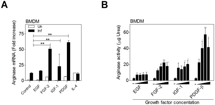
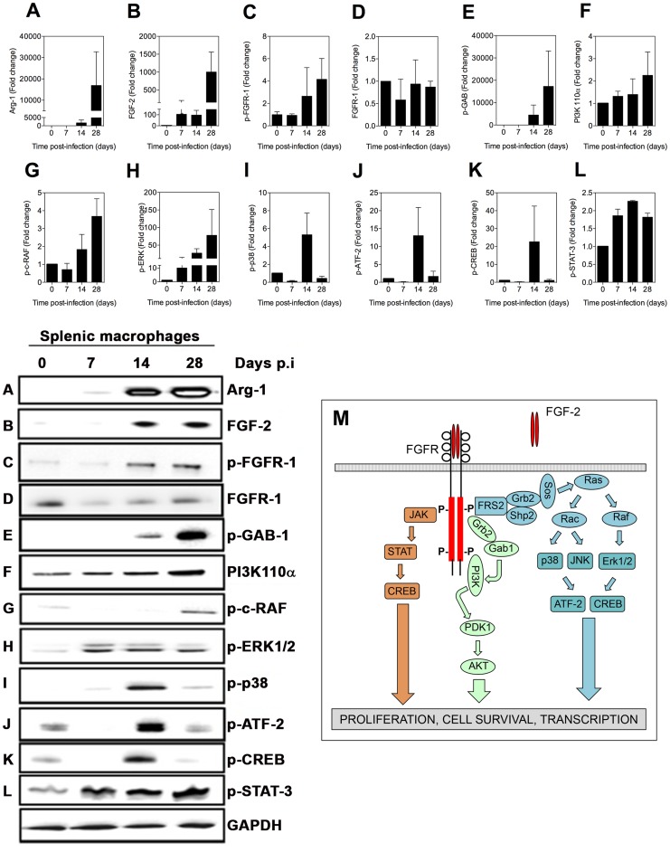
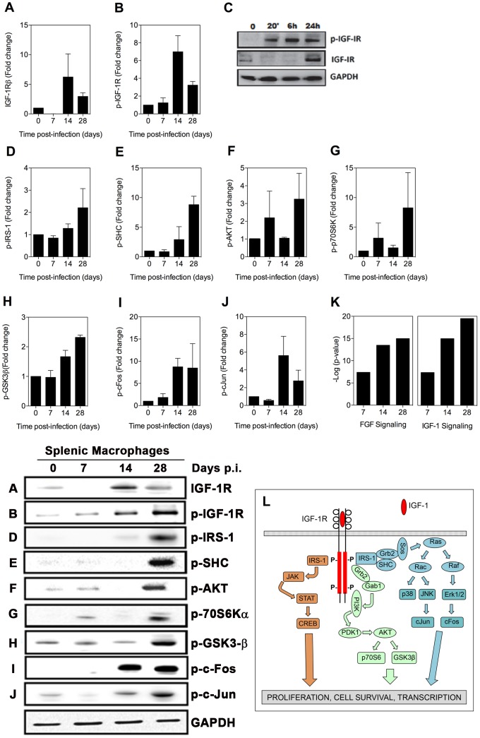
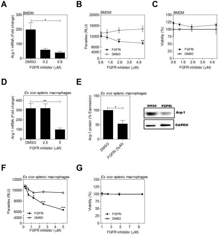
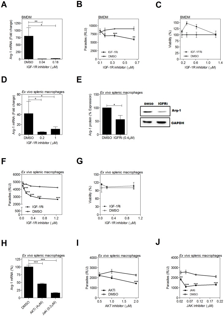
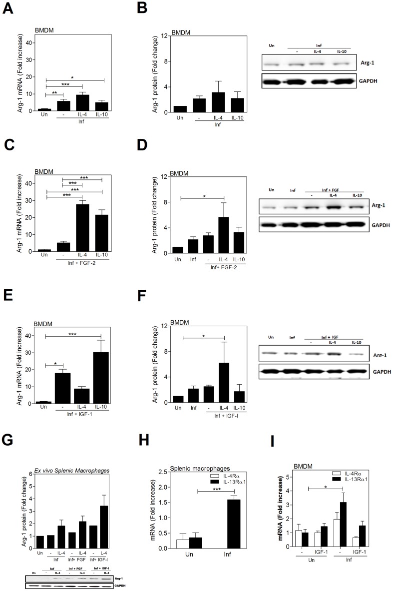
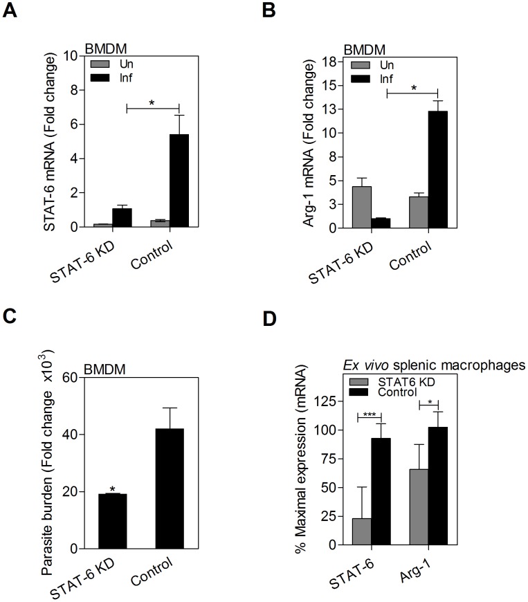
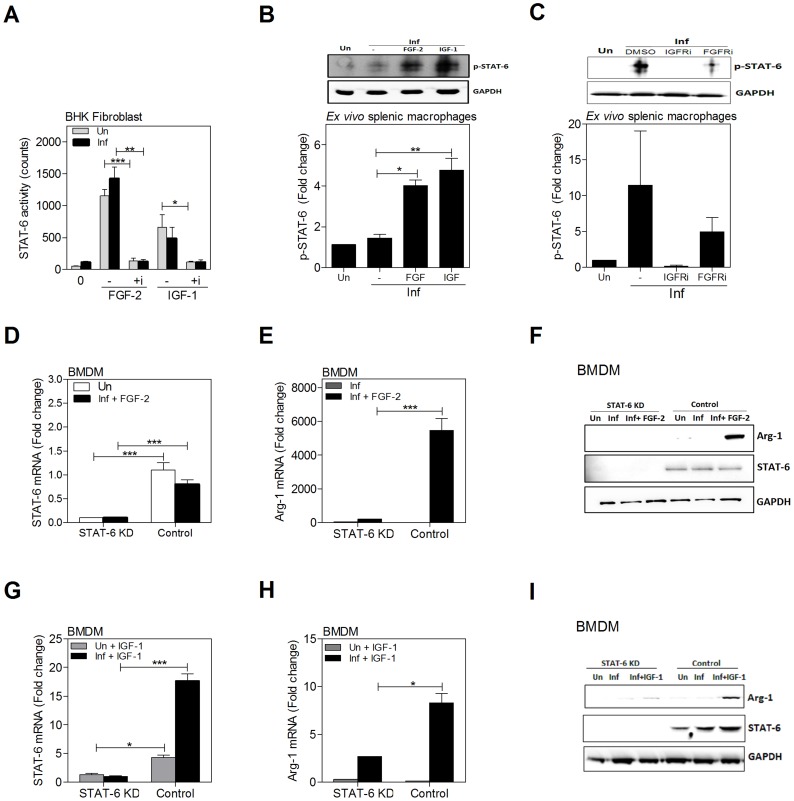
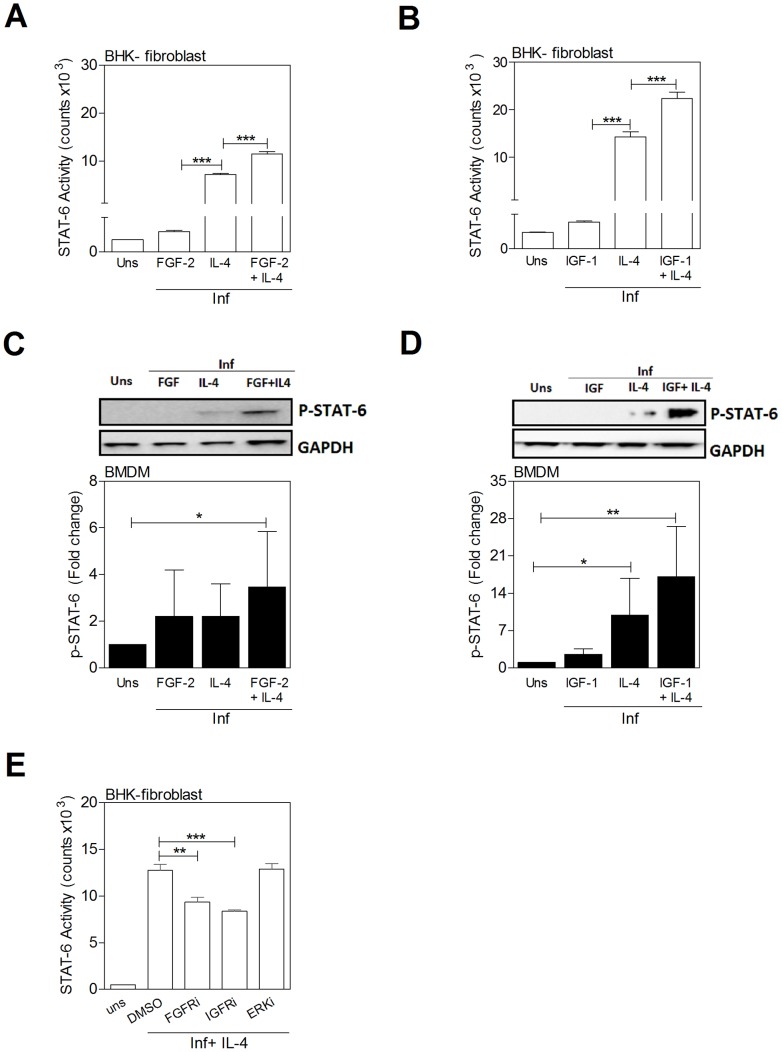
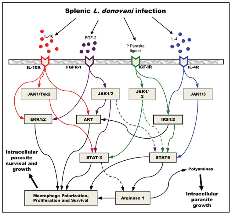
Host arginase 1 (arg1) expression is a significant contributor to the pathogenesis of progressive visceral leishmaniasis (VL), a neglected tropical disease caused by the intracellular protozoan Leishmania donovani. Previously we found that parasite-induced arg1 expression in macrophages was dependent on STAT6 activation. Arg1 expression was amplified by, but did not require, IL-4, and required de novo synthesis of unknown protein(s). To further explore the mechanisms involved in arg1 regulation in VL, we screened a panel of kinase inhibitors and found that inhibitors of growth factor signaling reduced arg1 expression in splenic macrophages from hamsters with VL. Analysis of growth factors and their signaling pathways revealed that the Fibroblast Growth Factor Receptor 1 (FGFR-1) and Insulin-like Growth Factor 1 Receptor (IGF-1R) and a number of downstream signaling proteins were activated in splenic macrophages isolated from hamsters infected with L. donovani. Recombinant FGF-2 and IGF-1 increased the expression of arg1 in L. donovani infected hamster macrophages, and this induction was augmented by IL-4. Inhibition of FGFR-1 and IGF-1R decreased arg1 expression and restricted L. donovani replication in both in vitro and ex vivo models of infection. Inhibition of the downstream signaling molecules JAK and AKT also reduced the expression of arg1 in infected macrophages. STAT6 was activated in infected macrophages exposed to either FGF-2 or IGF-1, and STAT6 was critical to the FGFR-1- and IGF-1R-mediated expression of arg1. The converse was also true as inhibition of FGFR-1 and IGF-1R reduced the activation of STAT6 in infected macrophages. Collectively, these data indicate that the FGFR/IGF-1R and IL-4 signaling pathways converge at STAT6 to promote pathologic arg1 expression and intracellular parasite survival in VL. Targeted interruption of these pathological processes offers an approach to restrain this relentlessly progressive disease.


Conflict of interest statement

The authors have declared that no competing interests exist.

Figures

Figure 1
Figure 1. Growth factors upregulate arginase 1 in macrophages.
A) Induction of arg1 mRNA expression in macrophages exposed to recombinant growth factors. Uninfected and L. donovani infected hamster BMDMs were stimulated with EGF (100 ng/mL), FGF-2 (20 ng/mL), IGF-1 (100 ng/mL), PDGF (100 ng/mL), or IL-4 (25 IU/mL) for 24 hrs and the expression of arg1 mRNA determined by qRT-PCR. Shown is the mean and standard error of the mean (SEM; error bars) of 4 replicates from a single experiment that is representative of 2 independent experiments. B) Dose-dependent induction of arginase activity (urea production) in hamster BMDMs infected with L. donovani and exposed to 2-fold increasing concentrations of growth factors for 48 h. The concentration of the growth factors was: EGF: 12.5–100 ng/mL; FGF-2: 6.25–50 ng/mL; IGF-1: 50–400 ng/mL; and PDGF: 25–100 ng/mL. Shown is the mean and SEM of 2 replicates per dose that is representative of 4 independent experiments. *p<0.05; **p<0.01; ***p<0.001.
Figure 2
Figure 2. Activation of signaling proteins in the FGFR canonical pathway in splenic macrophages from hamsters with VL.
(A–L) Splenic macrophages were isolated by adherence from the spleens of uninfected hamsters (time 0) or hamsters infected for 7, 14, and 28 days and whole cell lysates probed with antibodies directed against arg1 (panel A, representative blot A) or members of the FGF signaling pathway (panels and representative blots B–L). Bars represent the fold change with reference to control cells of uninfected hamsters calculated by densitometry analysis of immunoblot bands from samples pooled from 1–4 hamsters per determination from 2–3 independent experiments. M) Simplified schematic of the canonical FGF signaling pathway for reference.
Figure 3
Figure 3. Activation of signaling proteins in the IGF-1R canonical pathway in splenic macrophages from hamsters with VL.
(A–B, D–J) Immunoblot analysis of expression of proteins in the IGF-1R canonical signaling pathway in splenic macrophages was performed as described in Fig. 2. C) Detection of phospho-IGFR by immunoblot in BMDMs uninfected (Un) or infected in vitro with L. donovani for 20 min to 24 hrs. Shown is an immunoblot from a single experiment. K) Network analysis showing the activation of the FGF and IGF-1 canonical signaling pathways generated by comparing the fold change of 32 signaling proteins in splenic macrophages from uninfected and infected (7, 14, and 28 days) hamsters using Ingenuity Pathway Analysis software (Ingenuity Systems). The –log of the p value (vertical axis) represents the probability that the association of the data set in that pathway is due to chance. L) Simplified schematic of the canonical IGF-1 signaling pathway for reference.
Figure 4
Figure 4. Inhibition of FGFR signaling decreases arg1 expression and parasite burden in L. donovani infected macrophages.
(A–C) Hamster BMDMs were treated with a FGFR-1 inhibitor (PD 166866) or an equivalent concentration of vehicle control (DMSO) and infected in vitro with L. donovani for 24 or 48 hrs. A) arg1 mRNA expression determined by qRT-PCR at 24 hrs post-treatment. B) Intracellular parasite burden determined by luminometry from luciferase-transfected L. donovani at 48 hrs post-treatment. C) Viability of BMDMs determined by luminometry (Cell titer Glo) at 48 hrs post-treatment. D–G) Splenic macrophages from L. donovani infected hamsters (21–28 days p.i.) were isolated by adherence and cultured ex vivo with an inhibitor of FGFR-1 (PD 166866) or an equivalent concentration of vehicle control (DMSO) for 24 or 48 hrs. D) arg1 mRNA expression determined by qRT-PCR at 24 hrs post-treatment. E) arg1 protein expression determined at 48 hrs post-treatment. Bars represent the percent of expression with reference to control (DMSO treated) cells calculated by densitometry analysis of immunoblot bands from 3 independent experiments. A representative immunoblot is also shown. F) Intracellular parasite burden determined by luminometry from luciferase-transfected L. donovani at 48 hrs post-treatment. G) Viability of splenic macrophages determined by luminometry (Cell titer Glo) at 48 hrs post-treatment. Shown is the mean and SEM of from a single experiment that was representative of 2–4 independent experiments. *p<0.05; **p<0.01.
Figure 5
Figure 5. Inhibition of IGF-1R signaling decreases arg1 expression and parasite burden in L. donovani infected macrophages.
(A–C) Hamster BMDMs were treated with an IGF-1R inhibitor (Picropodophyllin, PPP) or an equivalent concentration of vehicle (DMSO) and infected in vitro with L. donovani for 24 or 48 hrs. (A) arg1 mRNA expression determined by qRT-PCR at 24 hrs post-treatment. B) Intracellular parasite burden determined by luminometry from luciferase-transfected L. donovani at 48 hrs post-treatment. C) Viability of BMDMs determined by luminometry (Cell titer Glo) at 48 hrs post-treatment. D–J) Splenic macrophages from L. donovani infected hamsters (21–28 days p.i.) were isolated by adherence and treated with an IGF-1R inhibitor, AKT inhibitor, or JAK inhibitor, or an equivalent concentration of vehicle control (DMSO) for 24 of 48 hrs. D) arg1 mRNA expression determined by qRT-PCR at 24 hrs post-treatment. E) arg1 protein expression determined at 48 hrs post-treatment. Bars represent the percent of expression with reference to control (DMSO treated) cells calculated by densitometry analysis of immunoblot bands from 3 independent experiments. A representative immunoblot is also shown. F) Intracellular parasite burden determined by luminometry from luciferase-transfected L. donovani at 48 hrs post-treatment. G) Viability of splenic macrophages determined by luminometry (Cell titer Glo) at 48 hrs post-treatment. H) arg1 mRNA expression determined by qRT-PCR at 24 hrs post-treatment with AKT inhibitor (AKTi; CAS# 612847-09-3, Calbiochem), JAK inhibitor (JAKi; CAS# 457081-03-07) or DMSO control. I–J) Intracellular parasite burden determined by luminometry from luciferase-transfected L. donovani at 48 hrs post-treatment with AKT inhibitor (I) or JAK inhibitor (J), compared to DMSO treated controls. In each of the panels the mean and SEM from a single experiment that was representative of 2–3 independent experiments is shown. *p<0.05; ***p<0.001.
Figure 6
Figure 6. IL-4 enhances growth factor-induced arg1 in L. donovani infected macrophages.
Infected hamster BMDM were exposed or not to hamster IL-4 (25 IU/mL), recombinant human IL-10 (100 ng/mL), recombinant human FGF-2 (20 ng/mL) and/or recombinant human IGF-1 (100 ng/mL) for 24 or 48 hrs. A, C, E) Arg1 mRNA expression determined by qRT-PCR at 24 hrs post-treatment. Shown is the mean and SEM of 6 replicates from a single experiment that was representative of 2 independent experiments. B, D, F) Arg1 protein expression determined in L. donovani infected BMDM exposed to IL-4, IL-10, and growth factors, alone or in combination, for 48 hrs. The membranes were stripped and stained with antibody against GAPDH to confirm equivalent protein loading. Bars represent the fold change with reference to control cells of uninfected hamsters calculated by densitometry analysis of immunoblot bands from 3 independent experiments. Also shown is a representative individual immunoblot. G) Arg1 protein expression determined in splenic macrophages from uninfected and L. donovani infected hamsters exposed ex vivo to IL-4 and growth factors, alone or in combination, for 48 hrs. The membranes were stripped and stained with antibody against GAPDH to confirm equivalent protein loading. Bars represent the fold change with reference to control cells of uninfected hamsters calculated by densitometry analysis of immunoblot bands from 3 independent experiments. Also shown is a representative individual immunoblot. H) Expression of IL-13Rα1 and IL-4Rα mRNA in splenic macrophages from uninfected (0) or 18-day infected hamsters determined by qRT-PCR. I) Expression of IL-13Rα1 and IL-4Rα mRNA in BMDM from uninfected (Un) and L. donovani infected (Inf) BMDMs (24 hrs p.i.) stimulated or not with IGF-1 or FGF-2. Shown is mean and SEM of the fold increase of receptor expression over uninfected, unstimulated controls from a single experiment representative of 2 independent experiments. *p<0.05; **p<0.01; ***p<0.001.
Figure 7
Figure 7. Parasite-induced arg1 expression in macrophages is dependent on STAT6.
Expression of A) STAT6 mRNA and B) arg1 mRNA in BMDMs that were uninfected (Un) or infected in vitro with L. donovani (Inf) for 24 h after transfection with STAT6-specific knockdown siRNA (STAT6 KD) or scrambled siRNA (Control). Shown is the mean and SEM of the fold-change in mRNA compared to unstimulated controls as determined by qRT-PCR in 6 replicates from 2 independent experiments. C) Parasite burden at 24 h post-infection of STAT6 KD BMDMs or control. Shown is the mean and SEM of the parasite burden with reference to control (uninfected) cells in 4 replicates determined by qRT-PCR. D) STAT6 and arg1 mRNA expression in splenic macrophages from L. donovani infected hamsters 48 hrs after ex vivo transfection with STAT6-specific siRNA (STAT6 KD) or scrambled siRNA (Control). Data are shown as the mean and SEM of the percent of maximal mRNA expression in the control samples. *p<0.05; ***p<0.001.
Figure 8
Figure 8. Growth factors and cytokines converge at STAT6 to induce arg1 expression in L. donovani infected macrophages.
A) Growth factors activate STAT6. Hamster BHK fibroblasts transfected with a STAT6 luciferase reporter were uninfected (Un) or infected (Inf) with L. donovani and stimulated for 24 h with FGF-2 (20 ng/mL) or IGF-1 (100 ng/mL) in the absence or presence (+i) of 250 nM of an inhibitor of FGFR-1 (PD166866) or IGF-1R (PPP). Shown is the mean and SEM of luciferase activity from 3 replicates from a single experiment that was representative of 2 independent experiments. B) phospho-STAT6 expression in BMDMs stimulated with IGF-1 and FGF-2. BMDMs were uninfected (Un) or infected in vitro with L. donovani (Inf) and stimulated or not with FGF-2 (20 ng/mL) or IGF-1 (100 ng/mL) for 20 min. STAT6 protein was immunoprecipitated in cell lysates and phosphorylated STAT6 determined by immunoblot. GAPDH was used to confirm that equivalent amounts of protein were subjected to the immunoprecipitation. Bars represent the mean and SEM of fold change with reference to the uninfected controls calculated by densitometry analysis of immunoblot bands from 6 independent experiments. Also shown is a representative individual immunoblot. C) Blockade of STAT6 activation by IGF-1R and FGFR inhibitors. Splenic macrophages from uninfected hamsters were pre-treated with IGF-1R inhibitor (100 nM PPP) or FGFR inhibitor (300 nM PD166866) and infected in vitro with L. donovani for 20 min in absence of the inhibitor. STAT6 protein was immunoprecipitated in cell lysates and the level of phosphorylated STAT6 determined by immunoblot. Data shown is from 3 independent experiments, with a representative individual immunoblot, as described for Fig. 8B. D) Expression of STAT6 mRNA in BMDMs that were uninfected-unstimulated (Un), infected (Inf), or infected and stimulated with FGF-2 after transfection with STAT6-specific siRNA (STAT6 KD) or scrambled siRNA (Control). Shown is the mean and SEM of the fold-increase in STAT6 mRNA with reference to uninfected control as determined by qRT-PCR in 4–10 replicates from a single experiment that was representative of 3 independent experiments. E) Abrogation of arg1 mRNA expression by knockdown of STAT6 in BMDMs infected in vitro with L. donovani and stimulated with FGF-2. BMDMs were transfected with the siRNA as described above and then infected and stimulated with FGF-2 (20 ng/mL) for 24 hrs. The data are shown as the mean and SEM of the fold-increase in arg1 mRNA relative to negative (uninfected) control cells from a single experiment that was representative of 3 independent experiments. F) Immunoblot showing efficiency of siRNAi-mediated knockdown of STAT6 protein in hamster BMDMs and the requirement of STAT6 in the FGF-2-induced arg1 expression in L. donovani infected cells. Following transfection with the STAT6-specific (STAT6 KD) or control siRNAi the BMDMs were uninfected (Un), infected with L. donovani (Inf), or infected and treated with FGF-2 (20 ng/mL) for 48 hrs. G) Expression of STAT6 mRNA in BMDMs that were uninfected and stimulated with IGF-1 (200 ng/mL) or infected (Inf) and stimulated with IGF-1 after transfection with STAT6-specific siRNA (STAT6 KD) or scrambled siRNA (Control). Shown is the mean and SEM of the fold-increase in STAT6 mRNA compared to IGF-1-treated STAT6 KD cells as determined by qRT-PCR in 3 replicates from a single experiment that was representative of 2 independent experiments. H) Abrogation of arg1 mRNA expression by knockdown of STAT6 in BMDMs infected in vitro with L. donovani and stimulated with IGF-1. BMDMs were transfected with the siRNA as described above and then infected and stimulated with IGF-1 (200 ng/mL) for 24 hrs. The data are shown as the mean and SEM of the fold-increase in arg1 mRNA as determined by qRT-PCR relative to nonstimulated cells from a single experiment that was representative of 2 independent experiments. I) Immunoblot showing efficiency of siRNAi-mediated knockdown of STAT6 protein in hamster BMDMs and the partial requirement of STAT6 in the IGF-1-induced arg1 expression in L. donovani infected cells. Experiment was designed and data presented as described for Fig. 8F. IGF-1 was used at 200 ng/mL. Shown is an immunoblot from a single experiment that was representative of 2 independent experiments. *p<0.05; **p<0.01; ***p<0.001.
Figure 9
Figure 9. IL-4 and growth factors amplify STAT6 activation.
A, B) STAT6 activation measured in a luciferase reporter assay in BHK cells stimulated for 24 h with growth factors and a sub-maximal concentration of IL-4. A) IL-4 (3 IU/mL) and/or FGF-2 (20 ng/mL) and B) IL-4 (6 IU/mL) and/or IGF-1 (100 ng/mL). Shown is the mean and SEM of STAT6 activity determined by luminometry from a single experiment that was representative of 3 independent experiments. C, D) Immunoblots of cell lysates (not immunoprecipitation as shown in Fig. 8B) showing phospho-STAT6 in BMDMs stimulated for 20 min with L. donovani promastigotes and C) IL-4 (8 IU/mL) and/or FGF-2 (20 ng/mL) or D) IL-4 (20 IU/mL) and/or IGF-1 (100 ng/mL). Bars represent the mean and SEM of fold change with reference to the uninfected, unstimulated (Uns) controls calculated by densitometry analysis of immunoblot bands from 3 independent experiments. Also shown is a representative individual immunoblot. E) IL-4-mediated activation of STAT6 in infected BHK fibroblasts is reduced by inhibition of FGFR and IGF-1R but not ERK. The cells were exposed to the control (DMSO) or FGFR inhibitor (FGFRi; PD166866; 10 µM), IGFR inhibitor (IGFRi; PPP; 5 µM), or ERK inhibitor (ERKi; PD98059; 5 µM) for 1 hr and then stimulated for another 24 hrs with IL-4 (25 IU/mL) in the presence or absence of inhibitor. Shown is the mean and SEM of STAT6 activity determined by luminometry from a single experiment that was representative of 2 independent experiments.
Figure 10
Figure 10. Working model for convergent signaling of growth factors and cytokines in the induction of arg1 in VL.
L. donovani infection induces the production of IL-4, IL-10 and FGF-2 in the spleen. FGFR is activated, as is IGF-IR by a yet to be identified host or parasite ligand. JAK kinases are phosphorylated through the activated cytokine or growth factor receptors, which lead to IRS-1/2, AKT, ERK, and STAT activation. Translocated STAT6, and possibly STAT3 lead to the transcriptional activation of arginase, which generates polyamines from arginine and leads to parasite growth. These transcription factors also contribute to the polarization macrophages toward an M2 phenotype, which is more permissive to L. donovani survival and growth. The collective effect of AKT, ERK, and STAT3 activation, and the generation of polyamines, are likely to lead to growth, proliferation and survival of arginase expressing cells, but this needs experimental confirmation in VL. Solid lines indicate known mechanistic interactions; dashed lines represent suppositional interactions. Only key shared signaling proteins are included in the model.

Similar articles

Cited by

References

    1. Alvar J, Velez ID, Bern C, Herrero M, Desjeux P, et al. (2012) Leishmaniasis worldwide and global estimates of its incidence. PLoS One 7: e35671. - PMC - PubMed
    1. Osorio EY, Zhao W, Espitia C, Saldarriaga O, Hawel L, et al. (2012) Progressive visceral leishmaniasis is driven by dominant parasite-induced STAT6 activation and STAT6-dependent host arginase 1 expression. PLoS Pathog 8: e1002417. - PMC - PubMed
    1. Green SJ, Nacy CA, Meltzer MS (1991) Cytokine-induced synthesis of nitrogen oxides in macrophages: a protective host response to Leishmania and other intracellular pathogens. J Leukoc Biol 50: 93–103. - PubMed
    1. Liew FY, Li Y, Moss D, Parkinson C, Rogers MV, et al. (1991) Resistance to Leishmania major infection correlates with the induction of nitric oxide synthase in murine macrophages. Eur J Immunol 21: 3009–3014. - PubMed
    1. Gordon S, Martinez FO (2010) Alternative activation of macrophages: mechanism and functions. Immunity 32: 593–604. - PubMed

MeSH terms