Crosstalk between BRCA-Fanconi anemia and mismatch repair pathways prevents MSH2-dependent aberrant DNA damage responses
- PMID: 24966277
- PMCID: PMC4194102
- DOI: 10.15252/embj.201387530
Crosstalk between BRCA-Fanconi anemia and mismatch repair pathways prevents MSH2-dependent aberrant DNA damage responses
Abstract
Several proteins in the BRCA-Fanconi anemia (FA) pathway, such as FANCJ, BRCA1, and FANCD2, interact with mismatch repair (MMR) pathway factors, but the significance of this link remains unknown. Unlike the BRCA-FA pathway, the MMR pathway is not essential for cells to survive toxic DNA interstrand crosslinks (ICLs), although MMR proteins bind ICLs and other DNA structures that form at stalled replication forks. We hypothesized that MMR proteins corrupt ICL repair in cells that lack crosstalk between BRCA-FA and MMR pathways. Here, we show that ICL sensitivity of cells lacking the interaction between FANCJ and the MMR protein MLH1 is suppressed by depletion of the upstream mismatch recognition factor MSH2. MSH2 depletion suppresses an aberrant DNA damage response, restores cell cycle progression, and promotes ICL resistance through a Rad18-dependent mechanism. MSH2 depletion also suppresses ICL sensitivity in cells deficient for BRCA1 or FANCD2, but not FANCA. Rescue by Msh2 loss was confirmed in Fancd2-null primary mouse cells. Thus, we propose that regulation of MSH2-dependent DNA damage response underlies the importance of interactions between BRCA-FA and MMR pathways.
Keywords: FANCJ; Fanconi anemia; MLH1; mismatch repair; replication stress.
© 2014 The Authors. Published under the terms of the CC BY 4.0 license.
Figures

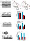
Immunoblot analysis of FANCJ, MLH1, and MSH2 expressions in U2OS cells treated with indicated siRNAs. β-actin was used as a loading control.
Graph shows the percentage of viable cells 5 days after indicated dose of MMC.
Immunoblot analysis of FANCJ and MSH2 expressions in human MSH2-null (HEC59) and MSH2-proficeint (HEC59+Chr2) cell lines treated with indicated siRNA reagents to Con or FANCJ (a or b).
Graph shows the percentage of viable cells 5 days after 500 nM MMC.
Immunoblot analysis of FANCJ and DNA-PKcs in DNA-PKcs-deficient (M059J) and DNA-PKcs-proficient (M059K) cells treated with siRNA reagents to Con or FANCJ (a or b).
Graph shows the percentage of viable cells 5 days after 250 nM MMC.
Immunoblot analysis showing 53BP1 or FANCJ expression in U2OS cells stably expressing shRNA vectors to either control or 53BP1 (a or b) that were also transfected with siRNAs to Con or FANCJ.
Graph shows the percentage of viable cells 5 days after 250 nM MMC.


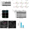
Immunoblot analysis of FANCJ and MSH2 expressions in the Fanconi anemia (FA)-J cell lines expressing indicated shRNAs.
Representative cell cycle profiles based on PI staining of DNA content for the indicated FA-J cell lines untreated (U) or at the indicated times following 0.25 μg/ml melphalan treatment.
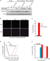
MSH2 depletion reduces DNA-PKcs Ser2056 phosphorylation after mitomycin C (MMC) treatment. Green fluorescent protein (GFP) expression indicates shRNA vector-infected FANCJK141/142A FA-J cells. Representative immunofluorescence images are shown.
Immunoblot analysis with indicated antibodies.
Genomic instability is suppressed by MSH2 depletion after 250 nM MMC for 16 h. Representative metaphases show examples of (a) broken and (b) quad-radial chromosomes that were suppressed by MSH2 depletion.
Graph shows number of breaks and radials quantified from 50 metaphases.


Similar articles
-
What is wrong with Fanconi anemia cells?Cell Cycle. 2014;13(24):3823-7. doi: 10.4161/15384101.2014.980633. Cell Cycle. 2014. PMID: 25486020 Free PMC article. Review.
-
Functional and physical interaction between the mismatch repair and FA-BRCA pathways.Hum Mol Genet. 2011 Nov 15;20(22):4395-410. doi: 10.1093/hmg/ddr366. Epub 2011 Aug 24. Hum Mol Genet. 2011. PMID: 21865299 Free PMC article.
-
The FANCJ/MutLalpha interaction is required for correction of the cross-link response in FA-J cells.EMBO J. 2007 Jul 11;26(13):3238-49. doi: 10.1038/sj.emboj.7601754. Epub 2007 Jun 21. EMBO J. 2007. PMID: 17581638 Free PMC article.
-
Assessing the link between BACH1/FANCJ and MLH1 in DNA crosslink repair.Environ Mol Mutagen. 2010 Jul;51(6):500-7. doi: 10.1002/em.20568. Environ Mol Mutagen. 2010. PMID: 20658644 Review.
-
Differential operation of MLH1/MSH2 and FANCD2 crosstalk in chemotolerant bladder carcinoma: a clinical and therapeutic intervening study.Mol Cell Biochem. 2023 Jul;478(7):1599-1610. doi: 10.1007/s11010-022-04616-9. Epub 2022 Nov 24. Mol Cell Biochem. 2023. Retraction in: Mol Cell Biochem. 2024 Jul;479(7):1865. doi: 10.1007/s11010-024-05021-0 PMID: 36434146 Retracted.
Cited by
-
Holding All the Cards-How Fanconi Anemia Proteins Deal with Replication Stress and Preserve Genomic Stability.Genes (Basel). 2019 Feb 22;10(2):170. doi: 10.3390/genes10020170. Genes (Basel). 2019. PMID: 30813363 Free PMC article. Review.
-
What is wrong with Fanconi anemia cells?Cell Cycle. 2014;13(24):3823-7. doi: 10.4161/15384101.2014.980633. Cell Cycle. 2014. PMID: 25486020 Free PMC article. Review.
-
Prime Editing and DNA Repair System: Balancing Efficiency with Safety.Cells. 2024 May 17;13(10):858. doi: 10.3390/cells13100858. Cells. 2024. PMID: 38786078 Free PMC article. Review.
-
Type-I Interferon Signaling in Fanconi Anemia.Front Cell Infect Microbiol. 2022 Feb 7;12:820273. doi: 10.3389/fcimb.2022.820273. eCollection 2022. Front Cell Infect Microbiol. 2022. PMID: 35198459 Free PMC article. Review.
-
FAN1 controls mismatch repair complex assembly via MLH1 retention to stabilize CAG repeat expansion in Huntington's disease.Cell Rep. 2021 Aug 31;36(9):109649. doi: 10.1016/j.celrep.2021.109649. Cell Rep. 2021. PMID: 34469738 Free PMC article.
References
-
- Adamo A, Collis SJ, Adelman CA, Silva N, Horejsi Z, Ward JD, Martinez-Perez E, Boulton SJ, La Volpe A. Preventing nonhomologous end joining suppresses DNA repair defects of Fanconi anemia. Mol Cell. 2010;39:25–35. - PubMed
-
- Alani E, Lee S, Kane MF, Griffith J, Kolodner RD. Saccharomyces cerevisiae MSH2, a mispaired base recognition protein, also recognizes Holliday junctions in DNA. J Mol Biol. 1997;265:289–301. - PubMed
-
- Alt A, Lammens K, Chiocchini C, Lammens A, Pieck JC, Kuch D, Hopfner KP, Carell T. Bypass of DNA lesions generated during anticancer treatment with cisplatin by DNA polymerase eta. Science. 2007;318:967–970. - PubMed
-
- Anderson CW, Dunn JJ, Freimuth PI, Galloway AM, Allalunis-Turner MJ. Frameshift mutation in PRKDC, the gene for DNA-PKcs, in the DNA repair-defective, human, glioma-derived cell line M059J. Radiat Res. 2001;156:2–9. - PubMed
Publication types
MeSH terms
Substances
Grants and funding
LinkOut - more resources
Full Text Sources
Other Literature Sources
Molecular Biology Databases
Research Materials
Miscellaneous

