. 2014 Aug 15;127(Pt 16):3425-33.
doi: 10.1242/jcs.149674. Epub 2014 Jun 24.

PKA-regulated VASP phosphorylation promotes extrusion of transformed cells from the epithelium

Affiliations

PKA-regulated VASP phosphorylation promotes extrusion of transformed cells from the epithelium

Katarzyna A Anton et al. J Cell Sci. .

Abstract

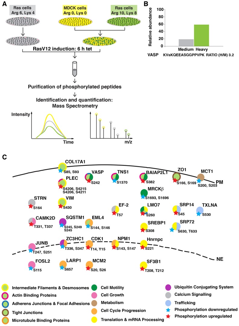
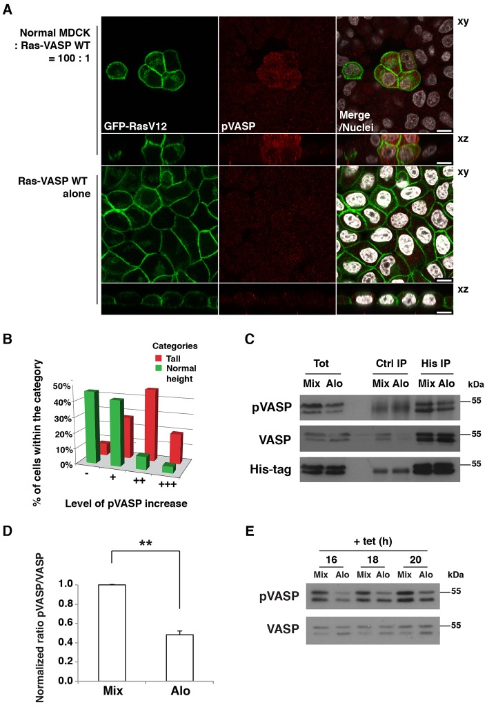
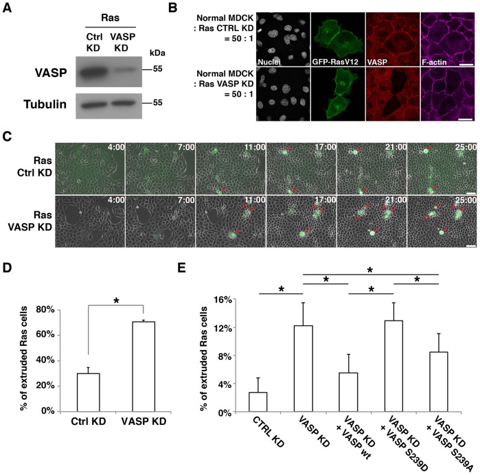
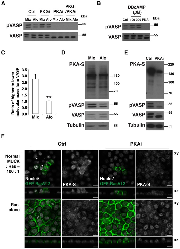
At the early stages of carcinogenesis, transformation occurs in single cells within tissues. In an epithelial monolayer, such mutated cells are recognized by their normal neighbors and are often apically extruded. The apical extrusion requires cytoskeletal reorganization and changes in cell shape, but the molecular switches involved in the regulation of these processes are poorly understood. Here, using stable isotope labeling by amino acids in cell culture (SILAC)-based quantitative mass spectrometry, we have identified proteins that are modulated in transformed cells upon their interaction with normal cells. Phosphorylation of VASP at serine 239 is specifically upregulated in Ras(V12)-transformed cells when they are surrounded by normal cells. VASP phosphorylation is required for the cell shape changes and apical extrusion of Ras-transformed cells. Furthermore, PKA is activated in Ras-transformed cells that are surrounded by normal cells, leading to VASP phosphorylation. These results indicate that the PKA-VASP pathway is a crucial regulator of tumor cell extrusion from the epithelium, and they shed light on the events occurring at the early stage of carcinogenesis.

Keywords: Apical extrusion; PKA; Ras-transformed; VASP.


Figures

Fig. 1.
Fig. 1.
Experimental outline of the SILAC screening. (A) MDCK pTR-GFP-RasV12 cells were labeled with medium (Arg 6, Lys 4) or heavy (Arg 10, Lys 8) arginine and lysine, and normal MDCK cells were labeled with light (Arg 0, Lys 0) arginine and lysine. Cells were plated either as Ras cells alone or as a 1∶1 mixed culture of heavy-labeled Ras∶MDCK cells. After 2 h, cells were incubated with tetracycline for 6 h to induce RasV12 expression. Phosphopeptides were isolated and analyzed by mass spectrometry. (B) Relative abundance of the VASP peptide from each experimental condition. The small ‘s’ represents the detected phosphorylation site. (C) Overview of proteins in which phosphorylation was identified as being upregulated (red star) or downregulated (blue star) in Ras cells upon co-incubation with normal cells. The key known functions of the selected proteins based on Gene NCBI, UniProt and PhosphoSitePlus databases are color-coded. Abbreviations of protein names are in agreement with those listed in the Gene NCBI database. PM, plasma membrane; NE, nuclear envelope.
Fig. 2.
Fig. 2.
Phosphorylation of VASP at S239 is increased in RasV12-transformed cells interacting with normal cells. (A) Immunofluorescence of phosphorylated S239 of VASP in MDCK pTR-GFP-RasV12-VASP-WT cells co-cultured with normal cells (upper panels) or cultured alone (lower panels) on collagen gels. Cells were stained with antibody against phosphorylated (p)S239-VASP (red) and Hoechst 33342 (white). Scale bars: 10 µm. (B) Correlation between the levels of pS239 VASP and cell height of Ras-VASP-WT cells surrounded by normal cells. Cells were divided into two categories according to their cell shape; tall and normal height (52% and 48% of all quantified cells, respectively). Within each category, cells were classified according to their pS239 VASP level as ‘−’ (no visible increase), ‘+’ (slight increase), ‘++’ (considerable increase) and ‘+++’ (strong increase). (C) Immunoprecipitation of VASP. Ras-VASP-WT cells and normal cells were co-cultured (Mix) or cultured alone and the respective cell lysates were mixed (Alo). Western blotting was performed with the indicated antibodies. Tot, total cell lysate; Ctrl, control. (D) Quantification of the ratio of pVASP∶VASP in the total cell lysates. Values are expressed as a ratio relative to that of Mix. Data show the mean±s.d. (three independent experiments); **P<0.005. (E) Time course of pVASP induction. Cells were cultured as described in C, and RasV12 expression was induced for the indicated times. Cell lysates were examined by western blotting. After plating, the cells were cultured for 2 h (A–D) (in the same way as for the SILAC screening) or 20–24 h (E) prior to tetracycline addition.
Fig. 3.
Fig. 3.
Phosphorylation of VASP at S239 promotes apical extrusion. (A) Knockdown of VASP in MDCK pTR-GFP-RasV12 cells. Ras cells were transfected with control siRNA (Ctrl KD) or VASP siRNA (VASP KD). Cell lysates were examined by western blotting with the indicated antibodies. (B) Immunofluorescence of VASP-knockdown Ras cells. Ras cells transfected with control siRNA or VASP siRNA were co-cultured with normal cells on glass. After induction of GFP–RasV12 expression with tetracycline for 8 h, cells were stained with anti-VASP antibody (red) and Alexa-Fluor-647–phalloidin (purple). Scale bars: 25 µm. (C) Time-lapse observation of apical extrusion of Ras cells. Ras cells were transfected with control siRNA or VASP siRNA and co-cultured with normal cells on collagen gels. Images were extracted from the representative time-lapse movies, and the indicated time reflects the duration of the tetracycline treatment. Red arrows indicate extruded Ras cells. Scale bars: 50 µm. (D) Quantification of the apical extrusion of Ras cells from time-lapse analyses (25 h). Data show the mean±s.d. (two independent experiments; n = 174 ctrl KD cells, n = 163 VASP KD cells). (E) The effect of expression of VASP proteins in VASP-knockdown RasV12-transformed cells on apical extrusion. Ras cells were transfected with siRNAs, followed by transfection with the wild-type (wt) VASP, VASP S239D or VASP S239A construct. Note that exogenously expressed human VASP is resistant to the siRNA that targets canine VASP. The transfected Ras cells were co-cultured with normal cells, followed by tetracycline treatment for 15 h. Data show the mean±s.d. (four independent experiments; n = 390 Ctrl KD cells, n = 388 VASP KD cells, n = 373 VASP KD+VASP WT cells, n = 343 VASP KD+VASP S239D cells, n = 324 VASP KD+VASP S239A cells); *P<0.05.
Fig. 4.
Fig. 4.
PKA is a crucial regulator of the phosphorylation of VASP and the cell shape of Ras-transformed cells. (A) The effect of PKA inhibitor treatment on pS239 VASP in mixed cultures of normal and Ras cells. Ras-VASP-WT cells and normal cells were co-cultured (Mix) or cultured alone followed by mixing of the cell lysates (Alo). Cells were incubated with tetracycline for 16 h in the presence of the PKA inhibitor H89 (PKAi) and/or the PKG inhibitor Rp-8-Br-PET-cGMPS (PKGi). Cell lysates were examined by western blotting with the indicated antibodies. Ctrl, control. (B) The effect of PKA inhibitor or activator on pS239 VASP in Ras cells. Ras cells were cultured for 4 h with the PKA activator dibutyryl-cAMP (DBcAMP) at the indicated concentrations or with the PKA inhibitor H89. Cell lysates were examined by western blotting with the indicated antibodies. (C) Quantification of the effect of co-culturing Ras and normal cells on the phosphorylation of S157 VASP. After a 20-h incubation of cells with tetracycline, cell lysates were analyzed by western blotting using an anti-VASP antibody. Data show the mean±s.d. (three independent experiments); **P<0.005. (D,E) Analyses of phosphorylated substrates of PKA in normal cells and Ras cells that were co-cultured (Mix) or cultured alone followed by mixing of the cell lysates (Alo). Cells were incubated with tetracycline for 20 h. In E, during the tetracycline treatment, cells were incubated in the absence (Ctrl) or presence (PKAi) of the PKA inhibitor H89. Cell lysates were examined by western blotting with the indicated antibodies. PKA-S, antibody against phosphorylated substrates of PKA. (F) Immunofluorescence analyses of phosphorylated substrates of PKA. Ras cells were co-cultured with normal cells (upper panels) or cultured alone (lower panels) on collagen gels. After a 20-h incubation with tetracycline in the absence or presence of H89 (PKAi), cells were stained with antibody against phosphorylated substrates of PKA and with Hoechst 33342 (white). Scale bars: 10 µm.

Similar articles

Cited by

References

    1. Barzik M., Kotova T. I., Higgs H. N., Hazelwood L., Hanein D., Gertler F. B., Schafer D. A. (2005). Ena/VASP proteins enhance actin polymerization in the presence of barbed end capping proteins. J. Biol. Chem. 280, 28653–28662 10.1074/jbc.M503957200 - DOI - PMC - PubMed
    1. Bear J. E., Svitkina T. M., Krause M., Schafer D. A., Loureiro J. J., Strasser G. A., Maly I. V., Chaga O. Y., Cooper J. A., Borisy G. G. et al.(2002). Antagonism between Ena/VASP proteins and actin filament capping regulates fibroblast motility. Cell 109, 509–521 10.1016/S0092-8674(02)00731-6 - DOI - PubMed
    1. Becker E. M., Schmidt P., Schramm M., Schröder H., Walter U., Hoenicka M., Gerzer R., Stasch J. P. (2000). The vasodilator-stimulated phosphoprotein (VASP): target of YC-1 and nitric oxide effects in human and rat platelets. J. Cardiovasc. Pharmacol. 35, 390–397 10.1097/00005344-200003000-00007 - DOI - PubMed
    1. Benz P. M., Blume C., Seifert S., Wilhelm S., Waschke J., Schuh K., Gertler F., Münzel T., Renné T. (2009). Differential VASP phosphorylation controls remodeling of the actin cytoskeleton. J. Cell Sci. 122, 3954–3965 10.1242/jcs.044537 - DOI - PMC - PubMed
    1. Blume C., Benz P. M., Walter U., Ha J., Kemp B. E., Renné T. (2007). AMP-activated protein kinase impairs endothelial actin cytoskeleton assembly by phosphorylating vasodilator-stimulated phosphoprotein. J. Biol. Chem. 282, 4601–4612 10.1074/jbc.M608866200 - DOI - PubMed

Publication types

MeSH terms

Substances

LinkOut - more resources