. 2014 Jul;42(13):8416-32.
doi: 10.1093/nar/gku529. Epub 2014 Jun 23.

ATM regulates NF-κB-dependent immediate-early genes via RelA Ser 276 phosphorylation coupled to CDK9 promoter recruitment

Affiliations

ATM regulates NF-κB-dependent immediate-early genes via RelA Ser 276 phosphorylation coupled to CDK9 promoter recruitment

Ling Fang et al. Nucleic Acids Res. 2014 Jul.

Abstract

Ataxia-telangiectasia mutated (ATM), a member of the phosphatidylinositol 3 kinase-like kinase family, is a master regulator of the double strand DNA break-repair pathway after genotoxic stress. Here, we found ATM serves as an essential regulator of TNF-induced NF-kB pathway. We observed that TNF exposure of cells rapidly induced DNA double strand breaks and activates ATM. TNF-induced ROS promote nuclear IKKγ association with ubiquitin and its complex formation with ATM for nuclear export. Activated cytoplasmic ATM is involved in the selective recruitment of the E3-ubiquitin ligase β-TrCP to phospho-IκBα proteosomal degradation. Importantly, ATM binds and activates the catalytic subunit of protein kinase A (PKAc), ribosmal S6 kinase that controls RelA Ser 276 phosphorylation. In ATM knockdown cells, TNF-induced RelA Ser 276 phosphorylation is significantly decreased. We further observed decreased binding and recruitment of the transcriptional elongation complex containing cyclin dependent kinase-9 (CDK9; a kinase necessary for triggering transcriptional elongation) to promoters of NF-κB-dependent immediate-early cytokine genes, in ATM knockdown cells. We conclude that ATM is a nuclear damage-response signal modulator of TNF-induced NF-κB activation that plays a key scaffolding role in IκBα degradation and RelA Ser 276 phosphorylation. Our study provides a mechanistic explanation of decreased innate immune response associated with A-T mutation.


Figures

Figure 1.
Figure 1.
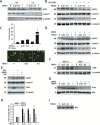
TNF-induced ATM activation and nuclear export. (A) A549 cells were treated with TNF (30 ng/ml) for the indicated time. Equal amounts of nuclear extract (NE) and cytoplasmic extract (CE) were analyzed by WBs to detect the level of ATM in both NE and CE. Lamin B and β-tubulin were also detected as internal control for NE and CE, respectively. (B) A549 cells were treated with TNF (30 ng/ml) for the indicated time with or without the pretreatment of KU-55933 (10 μM, 1 h). Equal amounts of NE and CE were analyzed by WBs to detect the level of pATM and ATM, respectively. Lamin B and β-tubulin were also detected as internal control for NE and CE, respectively. (C) Top panel. A549 cells were treated with TNF (30 ng/ml) or VP-16 (10 μM) for the indicated time and then assayed by Neutral Comet assay. 100 cells from each time interval were quantitated. Bottom panel. Representative images of comet moments. (D) A549 cells were treated with TNF (30 ng/ml) for the indicated time with or without the pretreatment of DMSO (2% in vol, 0.5 h). Equal amounts of NE and CE were analyzed by WBs to detect the level of pATM and ATM, respectively. Lamin B and β-tubulin were also detected as internal control for NE and CE, respectively. (E) A549 cells were treated with TNF (30 ng/ml) for 1 h with or without DMSO (2% in vol, 0.5 h) or NAC (15 mM, 1 h) pretreatment. Equal amounts of NE and CE were extracted and analyzed by WBs to detect the level of pATM and ATM, respectively. Lamin B and β-tubulin were also detected as internal control for NE and CE, respectively. (F) IKKγ+/+ and IKKγ−/− MEFs were treated with TNF (30 ng/ml) for the indicated time. Equal amounts of CE were analyzed by WB to detect the level of ATM. β-tubulin were detected as internal control. (G) A549 cells were treated with TNF (30 ng/ml) for the indicated time with or without the pretreatment of DMSO (2% in vol, 0.5 h). Equal amount of NE were immunoprecipitated by anti-IKKγ Ab. Interacting ATM were measured by WBs. (H) A549 cells were treated with TNF (30 ng/ml) for the indicated time with or without the pretreatment of DMSO (2% in vol, 0.5 h). Equal amount of NE were immunoprecipitated by Ubiquitin Ab and subjected to SID-SRM-MS analysis of IKKγ protein level. All of the values are presented as the ratios of native to SIS peptides. (I) A549 cells were treated with TNF (30 ng/ml) for the indicated times, Equal amount of NE were immunoprecipitated by anti-IKKγ Ab and assayed by western blot using anti-IKKγ Ab. * Significantly different from TNF (0 h)-treated samples, P < 0.05;** Significantly different from TNF (0 h)-treated samples, P < 0.01; Significantly different from ATM+/+ samples, P < 0.05; †† Significantly different from ATM+/+ samples, P < 0.01.
Figure 2.
Figure 2.
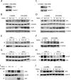
ATM is essential for IκBα degradation by recruiting β-TrCP. (A) A549 cells were transfected with ATM shRNA. 72 h later, equal amounts of NE were analyzed by WB to detect ATM. Lamin B were detected as internal control. (B) Control shRNA and ATM shRNA knockdown A549 cells were treated with TNF (30 ng/ml) for the indicated time. Equal amounts of CE were analyzed by WBs to detect the level of totalIκBα and pIκBα. β-tubulin were detected as internal control. (C) HeLa cells were transfected with ATM shRNA. 72 h later, equal amounts of NE were analyzed by WB to detect ATM. Lamin B were detected as internal control. (D) Control shRNA and ATM shRNA knockdown HeLa cells were treated with TNF (30 ng/ml) for the indicated time. Equal amounts of CE were analyzed by WBs to detect the level of total IκBα and pIκBα. β-tubulin were detected as internal control. (E) ATM+/+ and ATM−/− MEFs were treated with TNF (30 ng/ml) for the indicated time. Equal amount of CE were immunoprecipitated by anti-IκBα Ab. pIκBα levek were measured by WBs. (F) Control shRNA and ATM shRNA transfected A549 cells were TNF treated (30 ng/ml) for the indicated times. Equal amounts of WCE were analyzed by Western blot using anti-pIKKβ Ab. β-tubulin was used as an internal control. (G) ATM+/+ and ATM−/− MEFs were treated with TNF (30 ng/ml) for the indicated time. Equal amount of CE were were analyzed by WB to detect β-TrCP. β-tubulin were detected as internal control. (H) ATM+/+ and ATM−/− MEFs were pre-treated with MG132 prior to treatment with TNF (30 ng/ml) for the indicated time. Equal amount of CE were immunoprecipitated by anti-IκBα Ab. Interacting β-TrCP were measured by WBs. (I) A549 cells were treated with TNF (30 ng/ml) or VP16 (10 μM) for the indicated time. Equal amount of CE were immunoprecipitated by anti-β-TrCP Ab (lane 1–4), anti-p53 Ab (lane 5) or rabbit IgG (lane 6). Interacting ATM were measured by WBs. (J) Control shRNA and ATM shRNA-transfected A549 cells were treated with TNF (30 ng/ml) for the indicated times. Equal amounts of NE were analyzed by western to detect the level of RelA. Lamin B was detected as internal control.
Figure 3.
Figure 3.
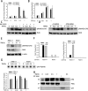
ATM is required for TNF-induced RelA Ser 276 phosphorylation through PKAc pathway. (A) A549 cells were treated with TNF (30 ng/ml) for the indicated time with or without the pretreatment of KU-55933 (10 μM, 1 h). Equal amounts of WCE were immunoprecipitated by pan anti-RelA Ab and subjected for SID-SRM-MS analysis using a phospho-Ser 276 RelA proteotypic peptide. All of the values are presented as the ratios of native to SIS peptides. (B) A549 cells were treated with TNF (30 ng/ml) for the indicated time with or without the pretreatment of KU-55933 (10 μM, 1 h). Equal amounts of WCE were immunoprecipitated by pan anti-RelA Ab and subjected for SID-SRM-MS analysis using a phospho-Ser 536 RelA proteotypic peptide. All of the values are presented as the ratios of native to SIS peptides. (C) A549 cells were treated with TNF (30 ng/ml) for the indicated time with or without the pretreatment of KU-55933 (10 μM, 1 h). Equal amounts of WCE were analyzed by WBs to detect the level of phospho-Ser 276 RelA. RelA were detected as internal control. (D) Control shRNA and ATM shRNA knockdown A549 cells were treated with TNF (30 ng/ml) for the indicated time. Equal amounts of WCE were analyzed by WBs to detect the level of phospho-Ser 276 RelA. RelA were detected as internal control. (E) RelA+/+ and RelA−/− MEFs were treated with TNF (30 ng/ml) for 1 h. Equal amounts of WCE were assayed by western blot using anti-RelA and anti-phospho-Ser 276 RelA Abs. β-actin was measured as internal control (F) Equal amounts of WCE from TNF treated RelA+/+ and RelA−/− MEFs were immunoprecipitated by anti-RelA Ab and then applied for SID-SRM-MS analysis to determine the RelA and phospho-Ser 276 RelA abundance. All of the values are presented as the ratios of native to SIS peptides. (G) Top panel. ATM+/+ and ATM−/− MEFs were treated with TNF (30 ng/ml) for the indicated time. WCE were collected and PKAc activity was measured with PepTag nonradioactive assay reagents as described in the ‘Materials and Methods’ section. Bottom panel. Quantitation of the top panel. (H) A549 cells were treated with TNF (30 ng/ml) or VP16 (10 μM) for the indicated time. Equal amount of CE were immunoprecipitated by PKAc Ab (lane1–4), p53 Ab (lane 5) or rabbit preimmune serum (lane 6). Interacting ATM were measured by WBs.* Significantly different from TNF (0 h)-treated samples, P < 0.05;** Significantly different from TNF (0 h)-treated samples, P < 0.01; Significantly different from ATM+/+ samples, P < 0.05; †† Significantly different from ATM+/+ samples, P < 0.01.
Figure 4.
Figure 4.
ATM in NF-κB-dependent immediate-early cytokine gene expression. (A). A549 cells were treated with TNF (30 ng/ml) for the indicated time with or without the pretreatment of KU-55933 (10 μM, 1 h). Total RNA was extracted. The mRNA levels of Gro-β, IκBα and IL-8 were measured. The results are expressed as fold change as compared with untreated cells after normalizing to internal controls, cyclophilin. Data represent the mean and STD of three independent experiments. (B). Control shRNA and ATM shRNA knockdown A549 cells were treated with TNF (30 ng/ml) for the indicated time. The experiment was performed as described for panel A. (C). Control shRNA and ATM shRNA knockdown HeLa cells were treated with TNF (30 ng/ml) for the indicated time. The experiment was performed as described for panel A. * Significantly different from TNF (0 h)-treated samples, P < 0.05;** Significantly different from TNF (0 h)-treated samples, P < 0.01; Significantly different from ATM+/+ samples, P < 0.05; †† Significantly different from ATM+/+ samples, P < 0.01.
Figure 5.
Figure 5.
ATM in recruitment of RelA and co-activators to immediate-early gene promoters. (A) Control shRNA and ATM shRNA knockdown A549 cells were treated with TNF (30 ng/ml) for the indicated time. The corresponding chromatin was immunoprecipitated with anti-RelA, CDK9 and pPol II Abs. IgG was the negative control. q-RT-PCR was performed using the Gro-β 5′ primer set, and the fold change was calculated compared with unstimulated samples. (B) The experiment was performed as described for panel A. q-RT-PCR was performed using the IL-8 5′ primer set, and the fold change was calculated compared with unstimulated samples. (C) The experiment was performed as described for panel A. q-RT-PCR was performed using the IκBα 5′ primer set, and the fold change was calculated compared with unstimulated samples. * Significantly different from TNF (0 h)-treated samples, P < 0.05;** Significantly different from TNF (0 h)-treated samples, P < 0.01; Significantly different from ATM+/+ samples, P < 0.05; †† Significantly different from ATM+/+ samples, P < 0.01.
Figure 6.
Figure 6.
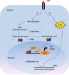
The model for the role of ATM in TNF pathway. Binding of TNF to the TNF receptor on the plasma membrane activates two parallel pathways: (1) IKK complex activation resulting in IκBα phosphorylation and (2) ROS generation leading to DNA double-strand breaks. The latter produces ATM activation and IKKγ-dependent nuclear export. Once in the cytosol, ATM facilitates two important steps of NF-κB activation pathway: rapid IκBα degradation by forming a complex with β-TrCP and RelA Ser 276 phosphorylation by interacting with PKAc.

Similar articles

Cited by

References

    1. Hayden M.S., Ghosh S. Signaling to NF-kappaB. Genes Dev. 2004;18:2195–2224. - PubMed
    1. Ghosh S., May M.J., Kopp E.B. NF-kappa B and Rel proteins: evolutionarily conserved mediators of immune responses. Annu. Rev. Immunol. 1998;16:225–260. - PubMed
    1. Medzhitov R. Toll-like receptors and innate immunity. Nat. Rev. Immunol. 2001;1:135–145. - PubMed
    1. Brasier A.R. The NF-kappaB regulatory network. Cardiovasc. Toxicol. 2006;6:111–130. - PubMed
    1. Jamaluddin M., Wang S., Boldogh I., Tian B., Brasier A.R. TNF-alpha-induced NF-kappaB/RelA Ser(276) phosphorylation and enhanceosome formation is mediated by an ROS-dependent PKAc pathway. Cell. Signal. 2007;19:1419–1433. - PubMed

Publication types

MeSH terms