. 2014 Jul 10;8(1):114-25.
doi: 10.1016/j.celrep.2014.05.038. Epub 2014 Jun 19.

The Mammalian response to virus infection is independent of small RNA silencing

Affiliations

The Mammalian response to virus infection is independent of small RNA silencing

Simone Backes et al. Cell Rep. .

Abstract

A successful cellular response to virus infection is essential for evolutionary survival. In plants, arthropods, and nematodes, cellular antiviral defenses rely on RNAi. Interestingly, the mammalian response to virus is predominantly orchestrated through interferon (IFN)-mediated induction of antiviral proteins. Despite the potency of the IFN system, it remains unclear whether mammals also have the capacity to employ antiviral RNAi. Here, we investigated this by disabling IFN function, small RNA function, or both activities in the context of virus infection. We find that loss of small RNAs in the context of an in vivo RNA virus infection lowers titers due to reduced transcriptional repression of the host antiviral response. In contrast, enabling a virus with the capacity to inhibit the IFN system results in increased titers. Taken together, these results indicate that small RNA silencing is not a physiological contributor to the IFN-mediated cellular response to virus infection.


Figures

Figure 1
Figure 1. Small RNA profiling of virus-infected cells
(A) 18–25 nucleotides (nts) RNA sequencing reads obtained from glial cells persistently infected with Borna disease virus, or fibroblasts infected with Influenza A virus, Sindbis virus and Vesicular stomatitis virus (VSV) (MOI of 1 for 12hrs) were analyzed for size distribution. (B) VSV-derived reads from (A) were aligned for their distribution across the genome. (C) Northern blot analysis of wildtype (MEF) and dicer-deficient cells (Dcr−/−) infected with VSV (MOI of 1 for 9 hrs) for VSV-derived small RNA VSV318, miR-93 and U6. (D) Northern blot analysis of MEF and Dcr−/− infected with VSV (MOI of 1 for 9 hrs) for VSV-derived small RNA VSV11161, miR-93 and U6.
Figure 2
Figure 2. VSV does not encode an inhibitor of RNAi
(A) Schematic of VSV genome engineered to encode 4 perfectly complementary miR-142 target (VSV142T) or 4 scrambled sites (VSVscbl) in the 3' UTR of the mRNA encoding for the L protein. (B) and (C) Multicycle growth curve of VSVctrl, VSV142T and VSVscbl on (B) RAW cells or (C) dicer-deficient cells (Dcr−/−). Supernatants were analyzed at the indicated time points by plaque assay. (D) Northern blot analysis of BHK transfected with miR-124 expressing plasmid (p124) or infected with VSV expressing miR-124 (VSV-124, MOI of 1 for 16 hr) for miR-124 and U6.
Figure 3
Figure 3. Engineering VSV to antagonize small RNAs
(A) Schematic of Vaccinia virus (VACV) VP55 insertion into the VSV genome. Each independent transcript is shown as it would be generated. “An”, polyadenylated tail. (B) Western blot analysis of VACV, VSVctrl or VSV-VP55-infected BHK for VACV VP55 and E3 and for VSV G expression. (C) Northern blot analysis of BHK co-infected with VSV expressing miR-124 (VSV-124) and VACV, VSVctrl or VSV-VP55 (MOI of 1 for 12hrs) for miR-124, VSV-leader RNA and U6. Relative density depicts mature miR-124/U6. (D) Analysis of murine fibroblasts transfected with control (scbl) or VSV-N-specific (VSV-N) siRNAs. Six hours posttransfection, cells were infected with VSVctrl or VSV-VP55 for 10 hrs (MOI of 1). Western blot was probed for VSV proteins and actin. (E) Schematic of premiR-146b-5p processing and corresponding miRNA duplex (middle) and mature miRNAs (bottom). The mature miRNA sequence-specific reads were determined by sequencing of the 20–25 nt fraction of VSV-VP55-infected cells. Adenosines (A) in black depict nontemplated bases and percent representation reflects the portion of the corresponding sequence in the total miRNA-specific tailed fraction.
Figure 4
Figure 4. Small RNAs do not impact VSV replication
(A) and (B) Multicycle growth curve of VSVctrl and VSV-VP55 (MOI of 0.01) on dicer-deficient cells (Dcr−/−). (A) Cells were analyzed at the indicated time points for expression of VSV proteins. (B) Supernatants were analyzed at the indicated time points by plaque assay. (C) and (D) Multicycle growth curve of VSVctrl and VSV-VP55 (MOI of 0.01) on wildtype fibroblasts (MEF). (C) Cells were analyzed at the indicated time points for expression of VSV proteins. (D) Supernatants were analyzed at the indicated time points by plaque assay.
Figure 5
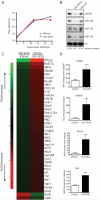
Figure 5. Host miRNAs target interferon-stimulated genes during VSV-infection
(A) to (D) Bone-marrow derived macrophages (BMMs) were infected with VSVctrl or VSV-VP55 (MOI of 5). (A) Viral replication was analyzed by plaque assay at the indicated time points. (B) 24 hrs post infection, BMMs were analyzed by Northern blot for presence of cellular miRNAs (miR-93, miR-146, miR-155, miR142) and U6. Relative density depicts miR-146/U6. (C) 10 hours post infection, BMMs were analyzed by mRNA sequencing. Top 40 of protein-coding transcripts differentially regulated by VSV-VP55 infection are shown. (D) Quantitative reverse transcriptase PCR depicting levels of PTGS2, Ddx60, Rsad2 and Mx1 induction over endogenous tubulin levels from BMMs infected for 10 hrs with VSVctrl or VSV-VP55.
Figure 6
Figure 6. Functional IFN-response impacts VSV replication
(A) 18–25 nucleotides (nts) RNA sequencing reads obtained from the lungs of mice infected intranasally with VSVctrl or VSV-VP55 (1×10⁷ PFU for 24hrs) aligned for their distribution across the VSV-VP55 genome. “goi”, gene of interst. (B) Wildtype mice were infected intranasally with VSVctrl or VSV-VP55 (1×10⁷ PFU; 3 mice per virus). 24hrs post infection lungs were analyzed for miR-146 and U6 expression by Northern blot. “M1, M2, M3”, mouse 1, mouse 2, mouse 3. Relative density depicts mature miR-146/U6. (C) Quantitative reverse transcriptase PCR depicting levels of IFN-β induction over endogenous tubulin levels from wildtype fibroblasts (MEF) infected with VSVctrl, VSV-VP55 or VSVNS1 (MOI of 1 for 16hrs). (D) Wildtype mice were infected intranasally with VSVctrl, VSV-VP55 or VSV-NS1 (1×10⁷ PFU). 24hrs post infection lungs and spleens were analyzed by plaque assay. (E) Interferon I and III receptor knockout mice (Ifnar1−/−/Il28r−/−) were infected intranasally with VSVctrl, VSV-VP55 or VSV-NS1 (1×10⁴ PFU). 48hrs post infection lungs and spleens were analyzed by plaque assay.

Similar articles

Cited by

References

    1. Ameres SL, Horwich MD, Hung JH, Xu J, Ghildiyal M, Weng Z, Zamore PD. Target RNA-directed trimming and tailing of small silencing RNAs. Science. 2010;328:1534–1539. - PMC - PubMed
    1. Aravin AA, Hannon GJ. Small RNA silencing pathways in germ and stem cells. Cold Spring Harb Symp Quant Biol. 2008;73:283–290. - PubMed
    1. Backes S, Shapiro JS, Sabin LR, Pham AM, Reyes I, Moss B, Cherry S, tenOever BR. Degradation of host microRNAs by poxvirus poly(A) polymerase reveals terminal RNA methylation as a protective antiviral mechanism. Cell Host Microbe. 2012;12:200–210. - PMC - PubMed
    1. Barnes D, Kunitomi M, Vignuzzi M, Saksela K, Andino R. Harnessing endogenous miRNAs to control virus tissue tropism as a strategy for developing attenuated virus vaccines. Cell Host Microbe. 2008;4:239–248. - PMC - PubMed
    1. Bartel DP. MicroRNAs: genomics, biogenesis, mechanism, and function. Cell. 2004;116:281–297. - PubMed

Publication types

LinkOut - more resources