Chronic variable stress activates hematopoietic stem cells
- PMID: 24952646
- PMCID: PMC4087061
- DOI: 10.1038/nm.3589
Chronic variable stress activates hematopoietic stem cells
Abstract
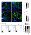
Exposure to psychosocial stress is a risk factor for many diseases, including atherosclerosis. Although incompletely understood, interaction between the psyche and the immune system provides one potential mechanism linking stress and disease inception and progression. Known cross-talk between the brain and immune system includes the hypothalamic-pituitary-adrenal axis, which centrally drives glucocorticoid production in the adrenal cortex, and the sympathetic-adrenal-medullary axis, which controls stress-induced catecholamine release in support of the fight-or-flight reflex. It remains unknown, however, whether chronic stress changes hematopoietic stem cell activity. Here we show that stress increases proliferation of these most primitive hematopoietic progenitors, giving rise to higher levels of disease-promoting inflammatory leukocytes. We found that chronic stress induced monocytosis and neutrophilia in humans. While investigating the source of leukocytosis in mice, we discovered that stress activates upstream hematopoietic stem cells. Under conditions of chronic variable stress in mice, sympathetic nerve fibers released surplus noradrenaline, which signaled bone marrow niche cells to decrease CXCL12 levels through the β3-adrenergic receptor. Consequently, hematopoietic stem cell proliferation was elevated, leading to an increased output of neutrophils and inflammatory monocytes. When atherosclerosis-prone Apoe(-/-) mice were subjected to chronic stress, accelerated hematopoiesis promoted plaque features associated with vulnerable lesions that cause myocardial infarction and stroke in humans.
Figures


Comment in
-
Stressing out stem cells: linking stress and hematopoiesis in cardiovascular disease.Nat Med. 2014 Jul;20(7):707-8. doi: 10.1038/nm.3631. Nat Med. 2014. PMID: 24999940 Free PMC article.
Similar articles
-
Dipeptidyl Peptidase-4 Regulates Hematopoietic Stem Cell Activation in Response to Chronic Stress.J Am Heart Assoc. 2017 Jul 14;6(7):e006394. doi: 10.1161/JAHA.117.006394. J Am Heart Assoc. 2017. PMID: 28710180 Free PMC article.
-
Ischemic stroke activates hematopoietic bone marrow stem cells.Circ Res. 2015 Jan 30;116(3):407-17. doi: 10.1161/CIRCRESAHA.116.305207. Epub 2014 Oct 31. Circ Res. 2015. PMID: 25362208 Free PMC article.
-
Physiologic corticosterone oscillations regulate murine hematopoietic stem/progenitor cell proliferation and CXCL12 expression by bone marrow stromal progenitors.Leukemia. 2013 Oct;27(10):2006-15. doi: 10.1038/leu.2013.154. Epub 2013 May 17. Leukemia. 2013. PMID: 23680895
-
Adrenergic Modulation of Hematopoiesis.J Neuroimmune Pharmacol. 2020 Mar;15(1):82-92. doi: 10.1007/s11481-019-09840-7. Epub 2019 Feb 14. J Neuroimmune Pharmacol. 2020. PMID: 30762159 Review.
-
Regulation of hematopoietic stem cells by bone marrow stromal cells.Trends Immunol. 2014 Jan;35(1):32-7. doi: 10.1016/j.it.2013.10.002. Epub 2013 Nov 5. Trends Immunol. 2014. PMID: 24210164 Free PMC article. Review.
Cited by
-
Association Between Social Isolation With Age-Gap Determined by Artificial Intelligence-Enabled Electrocardiography.JACC Adv. 2024 Mar 20;3(9):100890. doi: 10.1016/j.jacadv.2024.100890. eCollection 2024 Sep. JACC Adv. 2024. PMID: 39372468 Free PMC article.
-
Chronic social defeat stress induces meningeal neutrophilia via type I interferon signaling.bioRxiv [Preprint]. 2024 Aug 31:2024.08.30.610447. doi: 10.1101/2024.08.30.610447. bioRxiv. 2024. PMID: 39257811 Free PMC article. Preprint.
-
The impact of social relationships on the risk of stroke and post-stroke mortality: a systematic review and meta-analysis.BMC Public Health. 2024 Sep 4;24(1):2403. doi: 10.1186/s12889-024-19835-6. BMC Public Health. 2024. PMID: 39232685 Free PMC article.
-
Systemic and local regulation of hematopoietic homeostasis in health and disease.Nat Cardiovasc Res. 2024 Jun;3(6):651-665. doi: 10.1038/s44161-024-00482-4. Epub 2024 Jun 12. Nat Cardiovasc Res. 2024. PMID: 39196230 Review.
-
Social defeat stress impairs systemic iron metabolism by activating the hepcidin-ferroportin axis.FASEB Bioadv. 2024 Jul 2;6(8):263-275. doi: 10.1096/fba.2024-00071. eCollection 2024 Aug. FASEB Bioadv. 2024. PMID: 39114446 Free PMC article.
References
-
- Black PH. The inflammatory response is an integral part of the stress response: Implications for atherosclerosis, insulin resistance, type II diabetes and metabolic syndrome X. Brain Behav Immun. 2003;17:350–364. - PubMed
-
- Rosengren A, et al. Association of psychosocial risk factors with risk of acute myocardial infarction in 11119 cases and 13648 controls from 52 countries (the INTERHEART study): case-control study. Lancet. 2004;364:953–962. - PubMed
-
- Glaser R, Kiecolt-Glaser JK. Stress-induced immune dysfunction: implications for health. Nat Rev Immunol. 2005;5:243–251. - PubMed
-
- Cohen S, Kamarck T, Mermelstein R. A global measure of perceived stress. J Health Soc Behav. 1983;24:385–396. - PubMed
SUPPLEMENTARY REFERENCES
-
- Purton LE, Scadden DT. Limiting factors in murine hematopoietic stem cell assays. Cell Stem Cell. 2007;1:263–270. - PubMed
-
- Hu Y, Smyth GK. ELDA: extreme limiting dilution analysis for comparing depleted and enriched populations in stem cell and other assays. J Immunol Methods. 2009;347:70–78. - PubMed
Publication types
MeSH terms
Substances
Grants and funding
LinkOut - more resources
Full Text Sources
Other Literature Sources
Medical
Molecular Biology Databases
Miscellaneous

