

Characterization of NO-producing neurons in the rat corpus callosum
- PMID: 24944862
- PMCID: PMC4055183
- DOI: 10.1002/brb3.218
Characterization of NO-producing neurons in the rat corpus callosum
Abstract
Introduction: The aim of this study was to determine the presence and distribution of nitric oxide (NO)-producing neurons in the rat corpus callosum (cc).
Material and methods: To investigate this aspect of cc organization we used nicotinamide adenine dinucleotide phosphate diaphorase (NADPH-d) histochemistry and neuronal NO synthase (nNOS) immunocytochemistry.
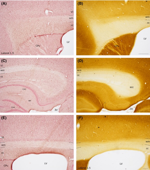
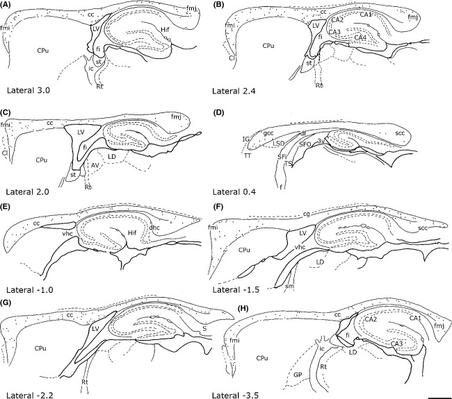
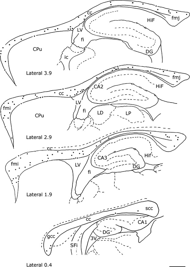
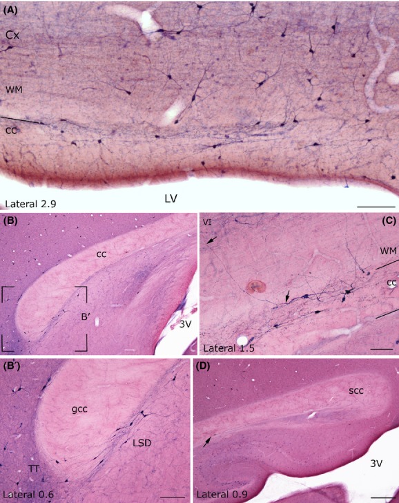
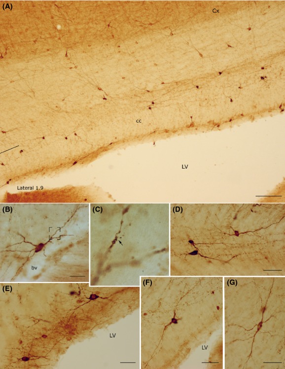
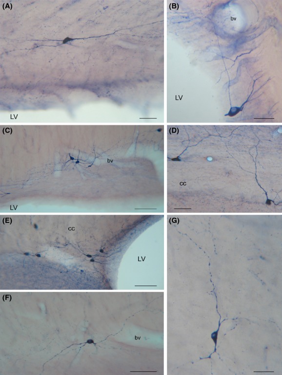
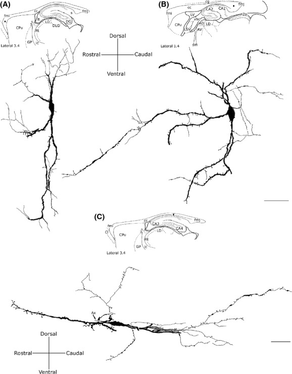
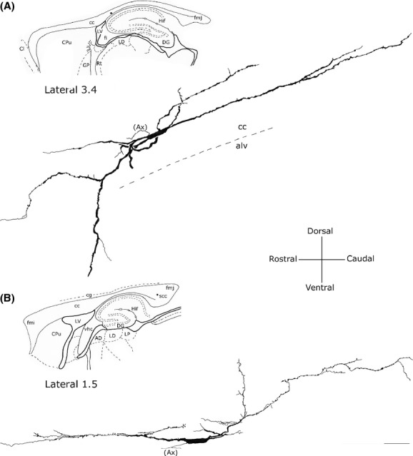
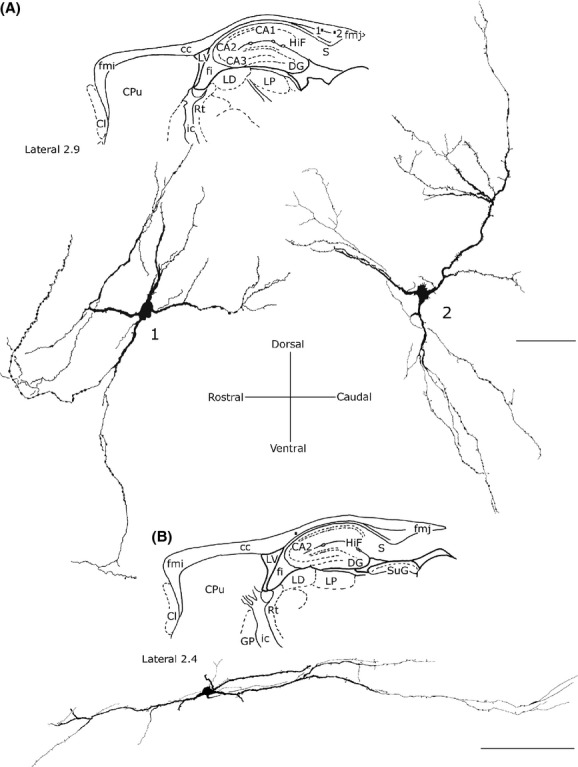
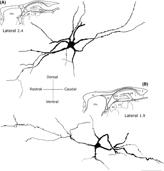
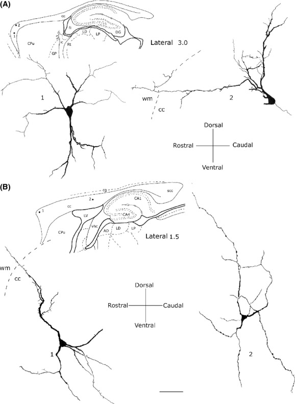
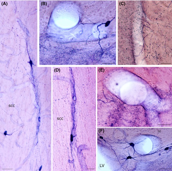
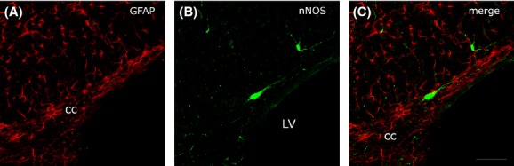
Results: Intense NADPH-d-positive (NADPH-d+) neurons were found along the rostrocaudal extension of the cc (sagittal sections). They were more numerous in the lateral cc and gradually decreased in the more medial regions, where they were very few or absent. The Golgi-like appearance of NADPH-d+ intracallosal neurons allowed dividing them into five morphological types: (1) bipolar; (2) fusiform; (3) round; (4) polygonal; and (5) pyramidal. The number of NADPH-d+ neurons (both hemispheres) was counted in two brains using 50-μm thick sections. In the first brain, counts involved 145 sections and neurons were 2959; in the second, 2227 neurons were counted in 130 sections. The distribution and morphology of nNOS-immunopositive (nNOSIP) neurons was identical to that of NADPH-d+neurons. Some of these neurons were observed in the cc ependymal region, where they might be in contact with cerebrospinal fluid (CSF), monitoring its composition, pH, and osmolality changes, or playing a role in regulating the synthesis and release of several peptides. The somatic, dendritic, and axonal processes of many NADPH-d+/nNOSIP neurons were closely associated with intracallosal blood vessels.
Conclusions: Such close relationship raises the possibility that these neurons are a major source of NO during neural activity. As NO is a potent vasodilator, these findings strongly suggest that NO-positive neurons transduce neuronal signals into vascular responses in selected cc regions, thus giving rise to hemodynamic changes detectable by neuroimaging.
Keywords: Colocalization; GFAP; NADPH-d; immunocytochemistry; nNOS; nitric oxide.
Figures
Similar articles
-
Intracallosal neuronal nitric oxide synthase neurons colocalize with neurokinin 1 substance P receptor in the rat.J Comp Neurol. 2015 Mar 1;523(4):589-607. doi: 10.1002/cne.23695. Epub 2014 Nov 18. J Comp Neurol. 2015. PMID: 25312245
-
Postnatal development of the distribution of nitric oxide-producing neurons in the rat corpus callosum.Neurosci Res. 2020 Feb;151:15-30. doi: 10.1016/j.neures.2019.02.005. Epub 2019 Feb 20. Neurosci Res. 2020. PMID: 30796928
-
Distribution of calbindin-D28k, neuronal nitric oxide synthase, and nicotinamide adenine dinucleotide phosphate diaphorase (NADPH-d) in the lateral nucleus of the sheep amygdaloid complex.Anat Embryol (Berl). 2006 Nov;211(6):707-20. doi: 10.1007/s00429-006-0133-x. Epub 2006 Oct 18. Anat Embryol (Berl). 2006. PMID: 17047987
-
Differential distribution of parvalbumin- and calbindin-D28K-immunoreactive neurons in the rat periaqueductal gray matter and their colocalization with enzymes producing nitric oxide.Brain Res Bull. 2013 Oct;99:48-62. doi: 10.1016/j.brainresbull.2013.09.004. Epub 2013 Oct 6. Brain Res Bull. 2013. PMID: 24107244
-
Traumatic injury of the spinal cord and nitric oxide.Prog Brain Res. 2007;161:171-83. doi: 10.1016/S0079-6123(06)61011-X. Prog Brain Res. 2007. PMID: 17618976 Review.
Cited by
-
Evidence for Functional Networks within the Human Brain's White Matter.J Neurosci. 2017 Jul 5;37(27):6394-6407. doi: 10.1523/JNEUROSCI.3872-16.2017. Epub 2017 May 25. J Neurosci. 2017. PMID: 28546311 Free PMC article.
-
Does functional MRI detect activation in white matter? A review of emerging evidence, issues, and future directions.Front Neurosci. 2014 Aug 8;8:239. doi: 10.3389/fnins.2014.00239. eCollection 2014. Front Neurosci. 2014. PMID: 25152709 Free PMC article. Review.
-
White Matter Neurons in Young Adult and Aged Rhesus Monkey.Front Neuroanat. 2016 Feb 22;10:15. doi: 10.3389/fnana.2016.00015. eCollection 2016. Front Neuroanat. 2016. PMID: 26941613 Free PMC article.
-
Substance P NK1 receptor in the rat corpus callosum during postnatal development.Brain Behav. 2017 May 2;7(6):e00713. doi: 10.1002/brb3.713. eCollection 2017 Jun. Brain Behav. 2017. PMID: 28638718 Free PMC article.
-
Neuronal nitric oxide synthase positive neurons in the human corpus callosum: a possible link with the callosal blood-oxygen-level dependent (BOLD) effect.Brain Struct Funct. 2023 Mar;228(2):511-523. doi: 10.1007/s00429-022-02599-3. Epub 2022 Dec 3. Brain Struct Funct. 2023. PMID: 36460768
References
-
- Arnold PB, Li CX, Waters RS. Thalamocortical arbors extend beyond single cortical barrels: an in vivo intracellular tracing study in rat. Exp. Brain Res. 2001;136:152–168. - PubMed
-
- Barbaresi P, Fabri M, Conti F, Manzoni T. D-[3H]Aspartate retrograde labelling of callosal and association neurones of somatosensory areas I and II of cats. J. Comp. Neurol. 1987;263:159–178. - PubMed
-
- Barbaresi P, Quaranta A, Amoroso S, Mensà E, Fabri M. Immunocytochemical localization of calretinin-containing neurons in the rat periaqueductal gray and colocalization with enzymes producing nitric oxide: a double, double-labeling study. Synapse. 2012;66:291–307. - PubMed
-
- Barrera A, Jiménez L, González GM, Montiel J, Aboitiz F. Dendritic structure of single hippocampal neurons according to sex and hemisphere of origin in middle-aged and elderly human subjects. Brain Res. 2001;906:31–37. - PubMed
MeSH terms
Substances
LinkOut - more resources
Full Text Sources
Other Literature Sources
Research Materials
Miscellaneous
