Exendin-4 pretreated adipose derived stem cells are resistant to oxidative stress and improve cardiac performance via enhanced adhesion in the infarcted heart
- PMID: 24915574
- PMCID: PMC4051823
- DOI: 10.1371/journal.pone.0099756
Exendin-4 pretreated adipose derived stem cells are resistant to oxidative stress and improve cardiac performance via enhanced adhesion in the infarcted heart
Abstract
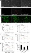
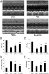
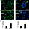
Reactive oxygen species (ROS), which were largely generated after myocardial ischemia, severely impaired the adhesion and survival of transplanted stem cells. In this study, we aimed to determine whether Exendin-4 pretreatment could improve the adhesion and therapeutic efficacy of transplanted adipose derived stem cells (ADSCs) in ischemic myocardium. In vitro, H2O2 was used to provide ROS environments, in which ADSCs pretreated with Exendin-4 were incubated. ADSCs without pretreatment were used as control. Then, cell adhesion and viability were analyzed with time. Compared with control ADSCs, Exendin-4 treatment significantly increased the adhesion of ADSCs in ROS environment, while reduced intracellular ROS and cell injury as determined by dihydroethidium (DHE) staining live/Dead staining, lactate dehydrogenase-release assay and MTT assay. Western Blotting demonstrated that ROS significantly decreased the expression of adhesion-related integrins and integrin-related focal adhesion proteins, which were significantly reversed by Exendin-4 pretreatment and followed by decreases in caspase-3, indicating that Exendin-4 may facilitate cell survival through enhanced adhesion. In vivo, myocardial infarction (MI) was induced by the left anterior descending artery ligation in SD rats. Autologous ADSCs with or without Exendin-4 pretreatment were injected into the border area of infarcted hearts, respectively. Multi-techniques were used to assess the beneficial effects after transplantation. Longitudinal bioluminescence imaging and histological staining revealed that Exendin-4 pretreatment enhanced the survival and differentiation of engrafted ADSCs in ischemic myocardium, accompanied with significant benefits in cardiac function, matrix remodeling, and angiogenesis compared with non-pretreated ADSCs 4 weeks post-transplantation. In conclusion, transplantation of Exendin-4 pretreated ADSCs significantly improved cardiac performance and can be an innovative approach in the clinical perspective.
Conflict of interest statement
Figures


Similar articles
-
The stem cell adjuvant with Exendin-4 repairs the heart after myocardial infarction via STAT3 activation.J Cell Mol Med. 2014 Jul;18(7):1381-91. doi: 10.1111/jcmm.12272. Epub 2014 Apr 30. J Cell Mol Med. 2014. PMID: 24779911 Free PMC article.
-
Stimulation of glucagon-like peptide-1 receptor through exendin-4 preserves myocardial performance and prevents cardiac remodeling in infarcted myocardium.Am J Physiol Endocrinol Metab. 2014 Oct 15;307(8):E630-43. doi: 10.1152/ajpendo.00109.2014. Epub 2014 Aug 12. Am J Physiol Endocrinol Metab. 2014. PMID: 25117407 Free PMC article.
-
Exendin-4 in combination with adipose-derived stem cells promotes angiogenesis and improves diabetic wound healing.J Transl Med. 2017 Feb 15;15(1):35. doi: 10.1186/s12967-017-1145-4. J Transl Med. 2017. PMID: 28202074 Free PMC article.
-
A brief review: adipose-derived stem cells and their therapeutic potential in cardiovascular diseases.Stem Cell Res Ther. 2017 Jun 5;8(1):124. doi: 10.1186/s13287-017-0585-3. Stem Cell Res Ther. 2017. PMID: 28583198 Free PMC article. Review.
-
Cardiac Adipose Tissue Contributes to Cardiac Repair: a Review.Stem Cell Rev Rep. 2021 Aug;17(4):1137-1153. doi: 10.1007/s12015-020-10097-4. Epub 2021 Jan 3. Stem Cell Rev Rep. 2021. PMID: 33389679 Review.
Cited by
-
L-Carnitine Attenuates Cardiac Dysfunction by Ischemic Insults Through Akt Signaling Pathway.Toxicol Sci. 2017 Dec 1;160(2):341-350. doi: 10.1093/toxsci/kfx193. Toxicol Sci. 2017. PMID: 28973678 Free PMC article.
-
Cerium oxide nanoparticles-carrying human umbilical cord mesenchymal stem cells counteract oxidative damage and facilitate tendon regeneration.J Nanobiotechnology. 2023 Oct 3;21(1):359. doi: 10.1186/s12951-023-02125-5. J Nanobiotechnology. 2023. PMID: 37789395 Free PMC article.
-
Exenatide (a GLP-1 agonist) improves the antioxidative potential of in vitro cultured human monocytes/macrophages.Naunyn Schmiedebergs Arch Pharmacol. 2015 Sep;388(9):905-19. doi: 10.1007/s00210-015-1124-3. Epub 2015 May 19. Naunyn Schmiedebergs Arch Pharmacol. 2015. PMID: 25980358 Free PMC article.
-
Cytoglobin Promotes Cardiac Progenitor Cell Survival against Oxidative Stress via the Upregulation of the NFκB/iNOS Signal Pathway and Nitric Oxide Production.Sci Rep. 2017 Sep 7;7(1):10754. doi: 10.1038/s41598-017-11342-6. Sci Rep. 2017. PMID: 28883470 Free PMC article.
-
Pretreatment of Adipose Derived Stem Cells with Curcumin Facilitates Myocardial Recovery via Antiapoptosis and Angiogenesis.Stem Cells Int. 2015;2015:638153. doi: 10.1155/2015/638153. Epub 2015 May 5. Stem Cells Int. 2015. PMID: 26074974 Free PMC article.
References
-
- Dow J, Simkhovich BZ, Kedes L, Kloner RA (2005) Washout of transplanted cells from the heart: a potential new hurdle for cell transplantation therapy. Cardiovasc Res 67: 301–307. - PubMed
-
- Toma C, Pittenger MF, Cahill KS, Byrne BJ, Kessler PD (2002) Human mesenchymal stem cells differentiate to a cardiomyocyte phenotype in the adult murine heart. Circulation 105: 93–98. - PubMed
-
- Ingber DE (2002) Mechanical signaling and the cellular response to extracellular matrix in angiogenesis and cardiovascular physiology. Circ Res 91: 877–887. - PubMed
-
- Giancotti FG, Ruoslahti E (1999) Integrin signaling. Science 285: 1028–1032. - PubMed
Publication types
MeSH terms
Substances
Grants and funding
LinkOut - more resources
Full Text Sources
Other Literature Sources
Medical
Research Materials

