Review
. 2014:83:585-614.
doi: 10.1146/annurev-biochem-060713-035513.

Mechanism and function of oxidative reversal of DNA and RNA methylation

Affiliations
Review

Mechanism and function of oxidative reversal of DNA and RNA methylation

Li Shen et al. Annu Rev Biochem. 2014.

Abstract

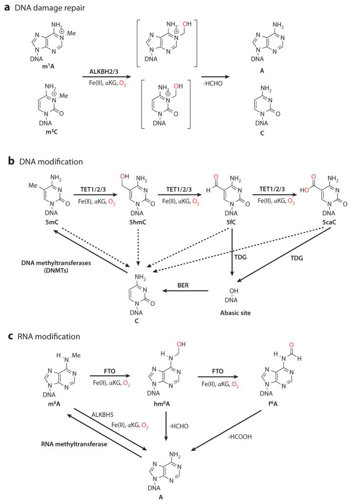
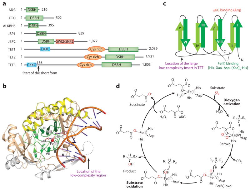
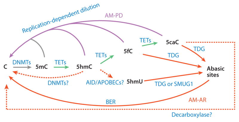
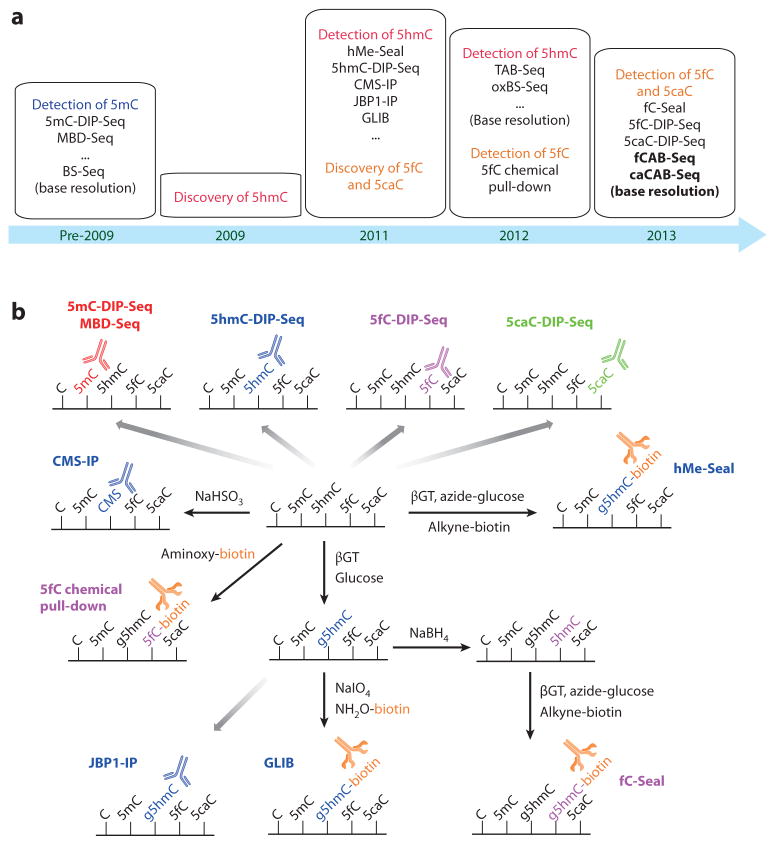
The importance of eukaryotic DNA methylation [5-methylcytosine (5mC)] in transcriptional regulation and development was first suggested almost 40 years ago. However, the molecular mechanism underlying the dynamic nature of this epigenetic mark was not understood until recently, following the discovery that the TET proteins, a family of AlkB-like Fe(II)/α-ketoglutarate-dependent dioxygenases, can oxidize 5mC to generate 5-hydroxymethylcytosine (5hmC), 5-formylcytosine (5fC), and 5-carboxylcytosine (5caC). Since then, several mechanisms that are responsible for processing oxidized 5mC derivatives to achieve DNA demethylation have emerged. Our biochemical understanding of the DNA demethylation process has prompted new investigations into the biological functions of DNA demethylation. Characterization of two additional AlkB family proteins, FTO and ALKBH5, showed that they possess demethylase activity toward N(6)-methyladenosine (m(6)A) in RNA, indicating that members of this subfamily of dioxygenases have a general function in demethylating nucleic acids. In this review, we discuss recent advances in this emerging field, focusing on the mechanism and function of TET-mediated DNA demethylation.

Keywords: 5-methylcytosine oxidation; DNA/RNA demethylation; Fe(II)/α-ketoglutarate-dependent dioxygenases; N6-methyladenosine; Tet.


Figures

Figure 1
Figure 1
Overview of the oxidative reversal of mammalian DNA and RNA methylation. (a) DNA methylation damage, such as N1-methyladenosine (m1A) and N3-methylcytosine (m3C), can be repaired by the AlkB family dioxygenases ALKBH2 and ALKBH3 through an unstable hemiaminal intermediate (brackets). (b) DNA methylation at the 5-position of cytosine is deposited by DNA methyltransferases (DNMTs). The Fe(II)/α-ketoglutarate (αKG)-dependent dioxygenases TET1, TET2, and TET3 can oxidize 5-methylcytosine (5mC) to the chemically stable intermediates 5-hydroxymethylcytosine (5hmC), 5-formylcytosine (5fC), and 5-carboxylcytosine (5caC) in a stepwise manner. 5mC, 5hmC, 5fC, and 5caC can be passively converted back to cysteine during DNA replication, when DNMTs fail to methylate the newly synthesized strand (dashed arrows). In addition, 5fC and 5caC can be excised by thymine-DNA glycosylase (TDG) and repaired through base excision repair (BER). (c) RNA methylation modification in the form of N6-methyladenosine (m6A) is generated from adenine by RNA methyltransferase. The AlkB family dioxygenases FTO (fat mass– and obesity-associated protein) and ALKBH5 remove the methyl group of m6A by oxidation. FTO oxidizes m6A to form metastable products of N6-hydroxymethyladenosine (hm6A) and N6-formyladenosine (f 6A), which decompose back to adenosine.
Figure 2
Figure 2
The structural and chemical basis of methyl oxidation by Fe(II)/α-ketoglutarate (αKG)-dependent dioxygenases. (a) Domain structures of Escherichia coli AlkB, mouse FTO (fat mass– and obesity-associated protein), ALKBH5, JBPs ( J-binding proteins), and TET proteins. TET proteins harbor three conserved domains, including the CXXC zinc finger, the cysteine-rich (Cys-rich) domain, and the double-stranded β-helix (DSBH) fold. (b) Crystal structure of human TET2 catalytic domain with DNA and an αKG analog, N-oxalylglycine (NOG) (Protein Data Bank identifier 4NM6). The conserved β-strands in the DSBH fold are colored green, the α-helixes in the DSBH fold are in yellow, the large insert is in gray, and the Cys-rich domain is in brown. The red and gray spheres represent Fe(II) and Zn(II), respectively. (c) The topology diagram for the conserved DSBH fold. The locations of Fe(II) and the αKG-binding sites are indicated. (d ) The chemical mechanism underlying Fe(II)/αKG-dependent dioxygenase–mediated 5mC oxidation. In the dioxygen activation stage, Fe(II) and αKG activate dioxygen to form a highly active Fe(IV)-oxo species. In the substrate oxidation stage, Fe(IV)-oxo inserts the oxygen atom into the C–H bond of the substrate, and Fe(IV) is reduced back to Fe(II) to complete the catalytic cycle.
Figure 3
Figure 3
Proposed mechanisms of TET-mediated DNA demethylation. TET proteins can oxidize 5-methylcytosine (5mC) to generate 5-hydroxymethylcytosine (5hmC), which is recognized poorly by DNA methyltransferase 1 (DNMT1) and thus can be diluted during DNA replication. 5hmC can also be further oxidized by TET proteins to produce 5-formylcytosine (5fC) and 5-carboxylcytosine (5caC). Alternatively, 5hmC may be deaminated by AID and APOBECs to become 5-hydroxymethyluracil (5hmU). 5fC, 5caC, and 5hmU can be excised from DNA by glycosylases. In addition, DNMT3A and DNMT3B may directly dehydroxymethylate 5hmC to generate cytosine, and a putative decarboxylase may also directly convert 5caC to cytosine. TET-catalyzed reactions are colored green, AM-AR (active modification followed by active restoration) pathways red, and AM-PD (active modification followed by passive dilution) pathways magenta. Solid lines represent processes with strong evidence; dashed lines indicate processes that either are hypothetical or need further verification. Abbreviation: BER, base excision repair.
Figure 4
Figure 4
Sequencing methods for 5-methylcytosine (5mC) derivatives. (a) Timeline of sequencing-method development for cytosine derivatives. (b) Affinity-based profiling methods for oxidized 5mC derivatives. Antibody-based DNA immunoprecipitation (DIP) methods are available for 5mC (5mC-DIP-Seq, MBD-Seq), 5-hydroxymethylcytosine (5hmC) (5hmC-DIP-Seq, CMS-IP, JBP1-IP), 5-formylcytosine (5fC) (5fC-DIP-Seq), and 5-carboxylcytosine (5caC) (5caC-DIP-Seq). 5hmC, 5fC, and 5caC can also be chemically and/or enzymatically labeled with biotin. These methods include 5hmC-selective chemical labeling (hMe-Seal) and GLIB (glucosylation, periodate oxidation, biotinylation) for 5hmC profiling, 5fC chemical pull-down, and 5fC-selective chemical labeling (fC-Seal) for 5fC profiling. Abbreviations: CMS, 5-methylenesulfonate; DIP-Seq, DNA immunoprecipitation sequencing; JBP, J-binding protein; MBD-Seq, methyl-binding protein sequencing.
Figure 5
Figure 5
Bisulfite sequencing (BS-Seq) and modified BS-Seq for base-resolution detection of oxidized 5-methylcytosine (5mC). With bisulfite treatment and polymerase chain reaction (PCR) amplification, BS-Seq reads out the sum of 5mC and 5-hydroxymethylcytosine (5hmC). Oxidative bisulfite sequencing (oxBS-Seq) oxidizes DNA with potassium perruthenate (KRuO4) before bisulfite treatment and PCR amplification. It reads out 5hmC through subtraction from BS-Seq signals. TET-assisted bisulfite sequencing (TAB-Seq) requires the protection of 5hmC with glucosylation before oxidizing DNA with TET proteins. After bisulfite treatment and PCR amplification, TAB-Seq directly reads out 5hmC. 5fC chemical modification–assisted bisulfite sequencing (fCAB-Seq) protects 5fC with EtONH2 before bisulfite treatment and PCR amplification, and after subtracting BS-Seq signals, it reads out 5fC. 5caC chemical modification–assisted bisulfite sequencing (caCAB-Seq) protects 5-carboxylcytosine (5caC) with 1-ethyl-3-(3-dimethylaminopropyl) carbodiimide (EDC) coupling before bisulfite treatment and PCR amplification, and after subtracting BS-Seq signals, it reads out 5caC.
Figure 6
Figure 6
Dynamics of 5-methylcytosine (5mC) and its oxidation derivatives during preimplantation development. After fertilization, 5mC in the paternal genome is quickly converted to 5-hydroxymethylcytosine/5-formylcytosine/5-carboxylcytosine (5hmC/5fC/5caC), which is then diluted through a replication-dependent process, whereas the maternal genome simply goes through replication-dependent passive DNA demethylation. At the blastocyst stage, DNA methylation patterns in the inner cell mass are quickly reestablished. The expression levels of Tet1, Tet2, Tet3, and Tdg are indicated (the dashed line represents cytoplasmic localization).
Figure 7
Figure 7
DNA methylation dynamics during primordial germ cell (PGC) reprogramming. Genome-wide DNA demethylation in developing PGCs takes place in two steps. First, during embryonic day (E)7.25 to E9.5, the bulk of the genome is demethylated in a replication-dependent but TET-independent manner. Second, at E9.5, TET1 and possibly TET2 convert the remaining 5-methylcytosine (5mC) to 5-hydroxymethylcytosine (5hmC), which is then diluted through a replication-dependent process to complete the demethylation in PGC reprogramming. The expression levels of Tet1, Tet2, Tet3, and Tdg are indicated.

Similar articles

Cited by

References

    1. Goll MG, Bestor TH. Eukaryotic cytosine methyltransferases. Annu Rev Biochem. 2005;74:481–514. - PubMed
    1. Jia G, Fu Y, Zhao X, Dai Q, Zheng G, et al. N6-Methyladenosine in nuclear RNA is a major substrate of the obesity-associated FTO. Nat Chem Biol. 2011;7:885–87. - PMC - PubMed
    1. Klose RJ, Bird AP. Genomic DNA methylation: the mark and its mediators. Trends Biochem Sci. 2006;31:89–97. - PubMed
    1. Smith ZD, Meissner A. DNA methylation: roles in mammalian development. Nat Rev Genet. 2013;14:204–20. - PubMed
    1. Fu Y, He C. Nucleic acid modifications with epigenetic significance. Curr Opin Chem Biol. 2012;16:516–24. - PMC - PubMed

Publication types