. 2014 Aug 15;112(4):942-50.
doi: 10.1152/jn.00175.2014. Epub 2014 May 21.

Changes in input strength and number are driven by distinct mechanisms at the retinogeniculate synapse

Affiliations

Changes in input strength and number are driven by distinct mechanisms at the retinogeniculate synapse

David J Lin et al. J Neurophysiol. .

Abstract

Recent studies have demonstrated that vision influences the functional remodeling of the mouse retinogeniculate synapse, the connection between retinal ganglion cells and thalamic relay neurons in the dorsal lateral geniculate nucleus (LGN). Initially, each relay neuron receives a large number of weak retinal inputs. Over a 2- to 3-wk developmental window, the majority of these inputs are eliminated, and the remaining inputs are strengthened. This period of refinement is followed by a critical period when visual experience changes the strength and connectivity of the retinogeniculate synapse. Visual deprivation of mice by dark rearing from postnatal day (P)20 results in a dramatic weakening of synaptic strength and recruitment of additional inputs. In the present study we asked whether experience-dependent plasticity at the retinogeniculate synapse represents a homeostatic response to changing visual environment. We found that visual experience starting at P20 following visual deprivation from birth results in weakening of existing retinal inputs onto relay neurons without significant changes in input number, consistent with homeostatic synaptic scaling of retinal inputs. On the other hand, the recruitment of new inputs to the retinogeniculate synapse requires previous visual experience prior to the critical period. Taken together, these findings suggest that diverse forms of homeostatic plasticity drive experience-dependent remodeling at the retinogeniculate synapse.

Keywords: critical period; synapse development; synaptic plasticity; thalamus; vision.


Figures

Fig. 1.
Fig. 1.
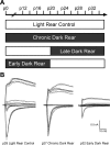
A: experimental paradigm of early dark rear (early DR) experiment. Filled blocks show time periods in development when mice were dark-reared. In early DR, mice were dark-reared from birth until postnatal day (P)20, at which time they were exposed to normal 12:12-h light-dark cycles for the rest of development. The early DR experiment is the inverse of the late DR experiment, previously published and shown in schematic form (Hooks and Chen 2006). Chronic DR mice were reared in the dark for all of development. Light rear (LR) control mice were exposed to normal 12:12-h light-dark cycles for all of development. All recordings were conducted at mature ages (P27–34). B: representative whole cell patch-clamp recordings from different experimental groups. Left, superimposed traces of evoked excitatory postsynaptic currents (EPSCs) recorded while increasing stimulus intensity from a dorsal lateral geniculate nucleus (LGN) relay neuron of a P28 LR control mouse. Middle, representative LGN relay neuron from a P27 chronic DR mouse. Right, representative LGN relay neuron from a P32 early DR mouse. Stimulus intensity ranged from 10 μA to 1 mA in all experiments. Note the reduction in currents elicited by all levels of stimulation in early DR mice compared with other experimental groups.
Fig. 2.
Fig. 2.
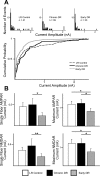
Early visual deprivation decreases input strength at the retinogeniculate synapse. A, top: analysis of single-fiber AMPAR currents for LR control, chronic DR, and early DR mice. Single-fiber AMPAR currents were measured at a holding potential of −70 mV. Histograms are divided into bins of 50 pA. Bottom, cumulative probability plots for the 3 conditions. B: synaptic currents from mice subjected to different visual manipulations measured at −70 mV (AMPAR) and +40 mV (NMDAR, slow component). The peak current amplitude in response to activation of a single retinal afferent was measured as single-fiber current, and maximal current was measured in response to maximal excitation of the optic tract. Average single-fiber (left) and maximal current amplitude (right; note changes in scale) for AMPAR (top) and NMDAR current (bottom) was assessed in LR control, chronic DR, and early DR. Data are means ± SE. All recordings occurred at mature ages (P27–34). LR control, 26 cells from 14 animals; chronic DR, 28 cells from 15 animals; early DR, 25 cells from 12 animals. *P < 0.05; **P < 0.01.
Fig. 3.
Fig. 3.
Early visual deprivation does not significantly alter retinal input number. Estimates of number of retinal ganglion cell (RGC) inputs were calculated by 2 different methods. A: fibers remaining for experimental groups, computed as ratio of average maximal current to average single-fiber current (Avg Max/SF), calculated independently for AMPAR and NMDAR currents. B: fiber fraction for chronic and early DR manipulations. No significant changes in connectivity were observed in early DR mice compared with LR control and chronic DR mice. Data are means ± SE; n = 82, 78, and 66 for LR control, chronic DR, and early DR mice, respectively.
Fig. 4.
Fig. 4.
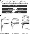
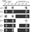
A: experimental paradigm and example traces of repeat dark rear (RDR) experiments. Filled blocks show time periods in development when mice were dark-reared. All recordings occurred at mature ages (P27–34) at least 7 days after the last manipulation of visual experience. B: representative electrophysiological recordings for different experimental groups in A. Left, superimposed traces of evoked EPSCs recorded while increasing stimulus intensity from an LGN relay neuron of a P27 chronic DR mouse. Middle, representative relay neuron from a P31 RDR(20,24) mouse. Right, representative relay neuron from a P32 RDR(18,22) mouse. Stimulus intensity ranged from ∼10 μA to 1 mA in all experiments. Note the dramatic increase in distinct steps of elicited current for RDR(18,22) mice with a relative preservation of maximal current elicited compared with the other experimental groups.
Fig. 5.
Fig. 5.
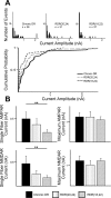
The timing of visual experience is critical for rewiring the retinogeniculate synapse. A, top: analysis of single-fiber AMPAR currents for chronic DR, RDR(20,24), and RDR(18,22) mice. Single-fiber AMPAR currents were measured at a holding potential of −70 mV. Histograms are divided into bins of 50 pA. Bottom, cumulative probability plots for the same experimental groups. B: synaptic currents after manipulations of visual experience measured at −70 mV (AMPAR) and +40 mV (NMDAR, slow component). Average single-fiber (left) and maximal current amplitude (right; note changes in scale) for AMPAR (top) and NMDAR current (bottom) assessed in chronic DR, RDR(20,24), and RDR(18,22) mice. All recordings occurred at mature ages (P27–34) at least 7 days after final manipulation of visual experience. Data are means ± SE. Chronic DR, 28 cells from 15 animals (data also shown in Fig. 2); RDR(20,24), 20 cells from 11 animals; RDR(18,22), 24 cells from 11 animals. *P < 0.05; **P < 0.01.
Fig. 6.
Fig. 6.
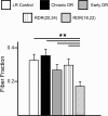
Visual experience in the precritical period is necessary for triggering recruitment of new inputs to the retinogeniculate synapse. Fiber fraction ratio is shown for all experimental groups. Each single-fiber current divided by the maximal current for the same relay neuron estimates the fraction of the cell's total current contributed by the single-fiber input. The population average of all fiber fraction ratios for a given experimental condition gives an estimate of retinogeniculate connectivity: a decrease in fiber fraction ratio signifies a relative increase in the number of connected afferents. Note that the RDR(18,22) group exhibits a significantly decreased fiber fraction ratio compared with each and all of the other experimental groups tested. Data are means ± SE; n = 82, 78, 66, 62, and 74 for LR control, chronic DR, early DR, RDR(20,24) and RDR(18,22) mice, respectively. Data for LR control, chronic DR, and early DR are also represented in Fig. 3. **P < 0.01 for all comparisons.
Fig. 7.
Fig. 7.
Role of visual experience in wiring and rewiring the retinogeniculate synapse. Summary schematic shows different experimental paradigms and how they each affect retinal afferent maturation and pruning. In early stages of development (P8–16), regardless of visual experience, LGN relay neurons undergo pruning of initially weak and redundant inputs. At older ages (P20+), late visual deprivation, but not chronic dark rearing, results in the weakening of existing inputs and the recruitment of additional afferents. Early visual deprivation (early DR experiment) results in the weakening of retinal afferents but no recruitment of additional inputs. Repeat dark rear experiments [RDR(20,24) and RDR(18,22)] reveal that visual experience before P20 is critical for triggering the later recruitment of additional inputs in response to visual deprivation. Thus the timing of early visual experience is critical for the expression of plasticity at the retinogeniculate synapse.

Similar articles

Cited by

References

    1. Blackman MP, Djukic B, Nelson SB, Turrigiano GG. A critical and cell-autonomous role for MeCP2 in synaptic scaling up. J Neurosci 32: 13529–13536, 2012 - PMC - PubMed
    1. Butts DA, Kanold PO, Shatz CJ. A burst-based “Hebbian” learning rule at retinogeniculate synapses links retinal waves to activity-dependent refinement. PLoS Biol 5: e61, 2007 - PMC - PubMed
    1. Campbell G, Shatz CJ. Synapses formed by identified retinogeniculate axons during the segregation of eye input. J Neurosci 12: 1847–1858, 1992 - PMC - PubMed
    1. Carrasco MM, Razak KA, Pallas SL. Visual experience is necessary for maintenance but not development of receptive fields in superior colliculus. J Neurophysiol 94: 1962–1970, 2005 - PubMed
    1. Chandrasekaran AR, Plas DT, Gonzalez E, Crair MC. Evidence for an instructive role of retinal activity in retinotopic map refinement in the superior colliculus of the mouse. J Neurosci 25: 6929–6938, 2005 - PMC - PubMed

Publication types

LinkOut - more resources