

A membrane reservoir at the cell surface: unfolding the plasma membrane to fuel cell shape change
- PMID: 24844289
- PMCID: PMC4199810
- DOI: 10.4161/bioa.29069
A membrane reservoir at the cell surface: unfolding the plasma membrane to fuel cell shape change
Abstract
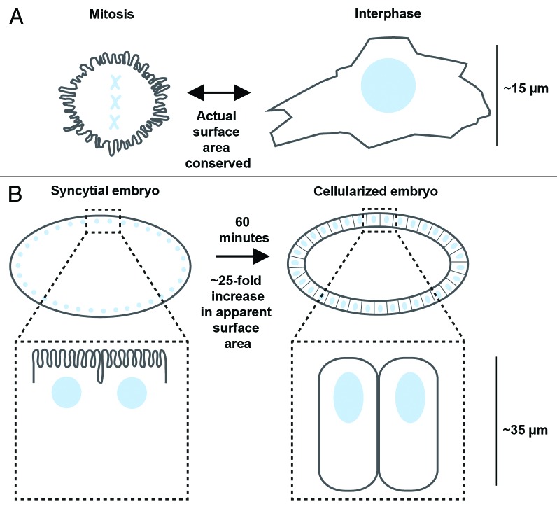
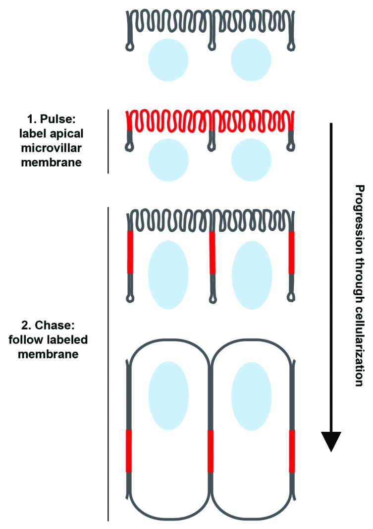
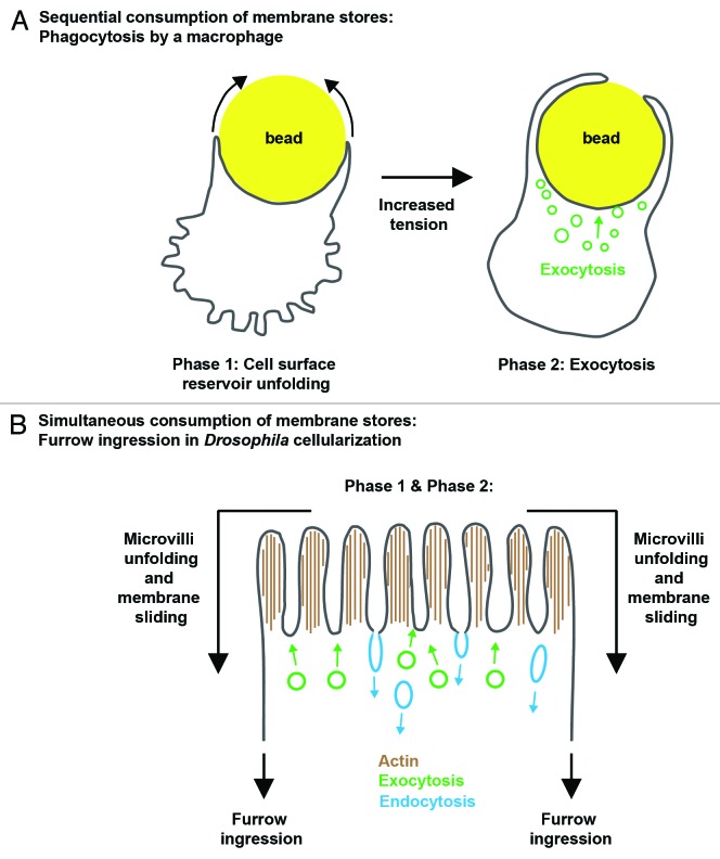
Cell surface expansion is a necessary part of cell shape change. One long-standing hypothesis proposes that membrane for this expansion comes from the flattening out of cell surface projections such as microvilli and membrane folds. Correlative EM data of cells undergoing phagocytosis, cytokinesis, and morphogenesis has hinted at the existence of such an unfolding mechanism for decades; but unfolding has only recently been confirmed using live-cell imaging and biophysical approaches. Considering the wide range of cells in which plasma membrane unfolding has now been reported, it likely represents a fundamental mechanism of cell shape change.
Keywords: actin; cell shape change; cell surface area regulation; cellularization; exocytosis; microvilli; morphogenesis; plasma membrane tension.
Figures
Similar articles
-
Membrane Supply and Demand Regulates F-Actin in a Cell Surface Reservoir.Dev Cell. 2016 May 9;37(3):267-78. doi: 10.1016/j.devcel.2016.04.010. Dev Cell. 2016. PMID: 27165556 Free PMC article.
-
The plasma membrane flattens out to fuel cell-surface growth during Drosophila cellularization.Dev Cell. 2013 Dec 23;27(6):648-55. doi: 10.1016/j.devcel.2013.11.006. Epub 2013 Dec 5. Dev Cell. 2013. PMID: 24316147 Free PMC article.
-
Mechanics of neutrophil phagocytosis: behavior of the cortical tension.J Cell Sci. 2005 May 1;118(Pt 9):1789-97. doi: 10.1242/jcs.02275. Epub 2005 Apr 12. J Cell Sci. 2005. PMID: 15827090
-
Membrane Tension and the Role of Ezrin During Phagocytosis.Adv Exp Med Biol. 2020;1246:83-102. doi: 10.1007/978-3-030-40406-2_6. Adv Exp Med Biol. 2020. PMID: 32399827 Review.
-
Cell surface mechanics and the control of cell shape, tissue patterns and morphogenesis.Nat Rev Mol Cell Biol. 2007 Aug;8(8):633-44. doi: 10.1038/nrm2222. Nat Rev Mol Cell Biol. 2007. PMID: 17643125 Review.
Cited by
-
Rab5ab-Mediated Yolk Cell Membrane Endocytosis Is Essential for Zebrafish Epiboly and Mechanical Equilibrium During Gastrulation.Front Cell Dev Biol. 2021 Oct 29;9:697097. doi: 10.3389/fcell.2021.697097. eCollection 2021. Front Cell Dev Biol. 2021. PMID: 34778246 Free PMC article.
-
Genetic impairment of parasite myosin motors uncovers the contribution of host cell membrane dynamics to Toxoplasma invasion forces.BMC Biol. 2016 Nov 9;14(1):97. doi: 10.1186/s12915-016-0316-8. BMC Biol. 2016. PMID: 27829452 Free PMC article.
-
Membrane-actin interactions in morphogenesis: Lessons learned from Drosophila cellularization.Semin Cell Dev Biol. 2023 Jan 15;133:107-122. doi: 10.1016/j.semcdb.2022.03.028. Epub 2022 Apr 5. Semin Cell Dev Biol. 2023. PMID: 35396167 Free PMC article. Review.
-
Combining patch-clamping and fluorescence microscopy for quantitative reconstitution of cellular membrane processes with Giant Suspended Bilayers.Sci Rep. 2019 May 10;9(1):7255. doi: 10.1038/s41598-019-43561-4. Sci Rep. 2019. PMID: 31076583 Free PMC article.
-
The Giardia ventrolateral flange is a lamellar membrane protrusion that supports attachment.PLoS Pathog. 2022 Apr 28;18(4):e1010496. doi: 10.1371/journal.ppat.1010496. eCollection 2022 Apr. PLoS Pathog. 2022. PMID: 35482847 Free PMC article.
References
Publication types
MeSH terms
LinkOut - more resources
Full Text Sources
Other Literature Sources