. 2014 May 15;10(5):e1004117.
doi: 10.1371/journal.ppat.1004117. eCollection 2014 May.

Papillomavirus genomes associate with BRD4 to replicate at fragile sites in the host genome

Affiliations

Papillomavirus genomes associate with BRD4 to replicate at fragile sites in the host genome

Moon Kyoo Jang et al. PLoS Pathog. .

Abstract

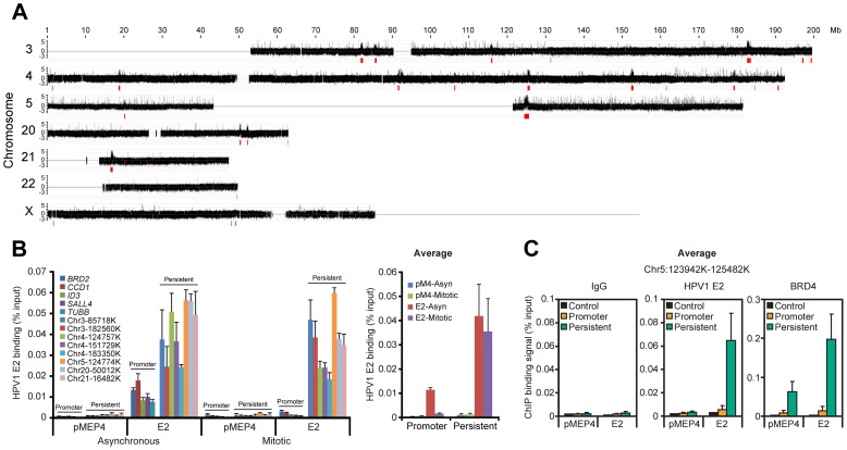
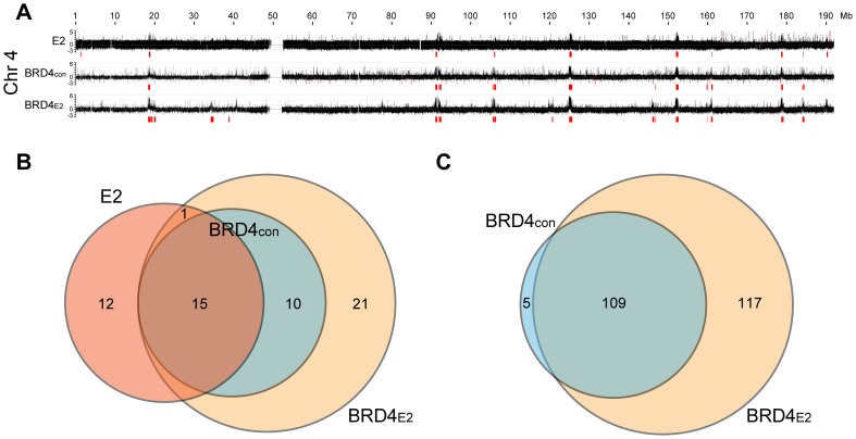
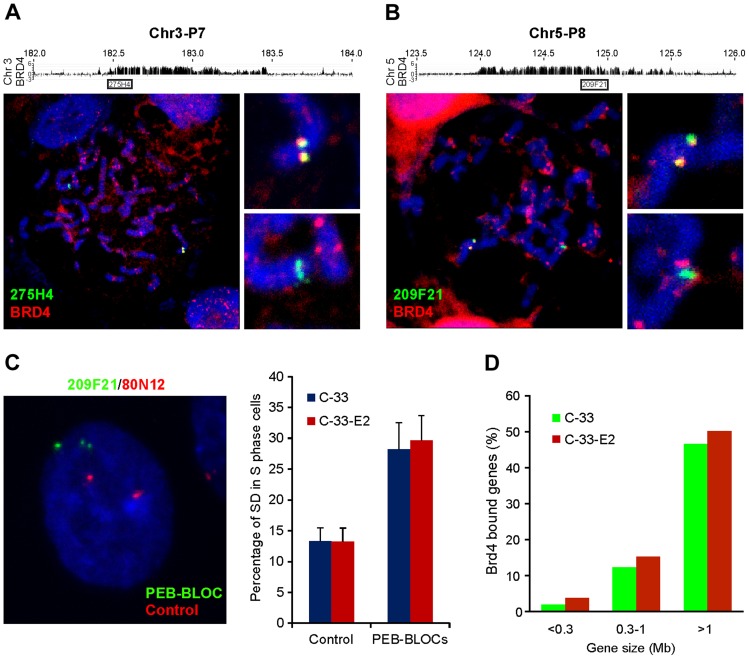
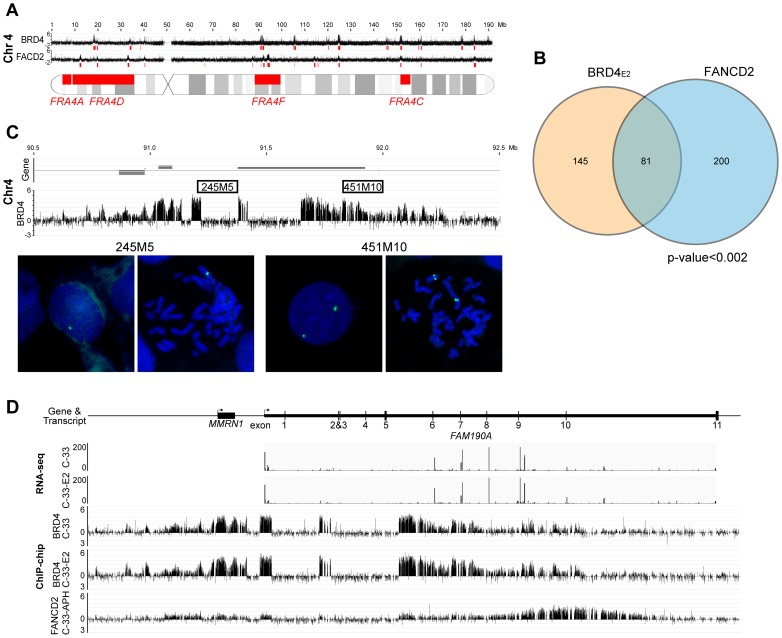
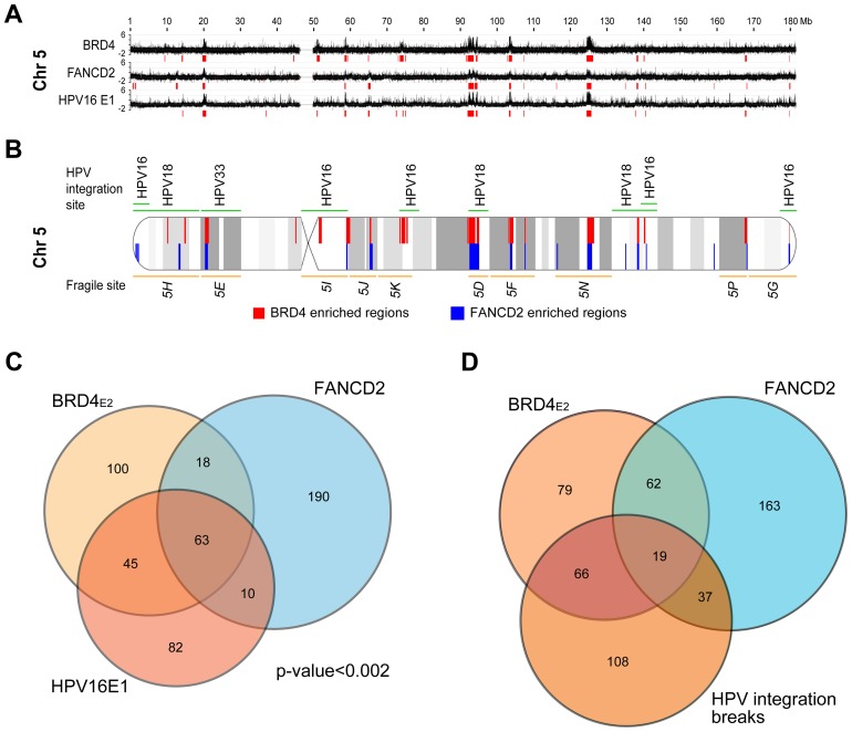
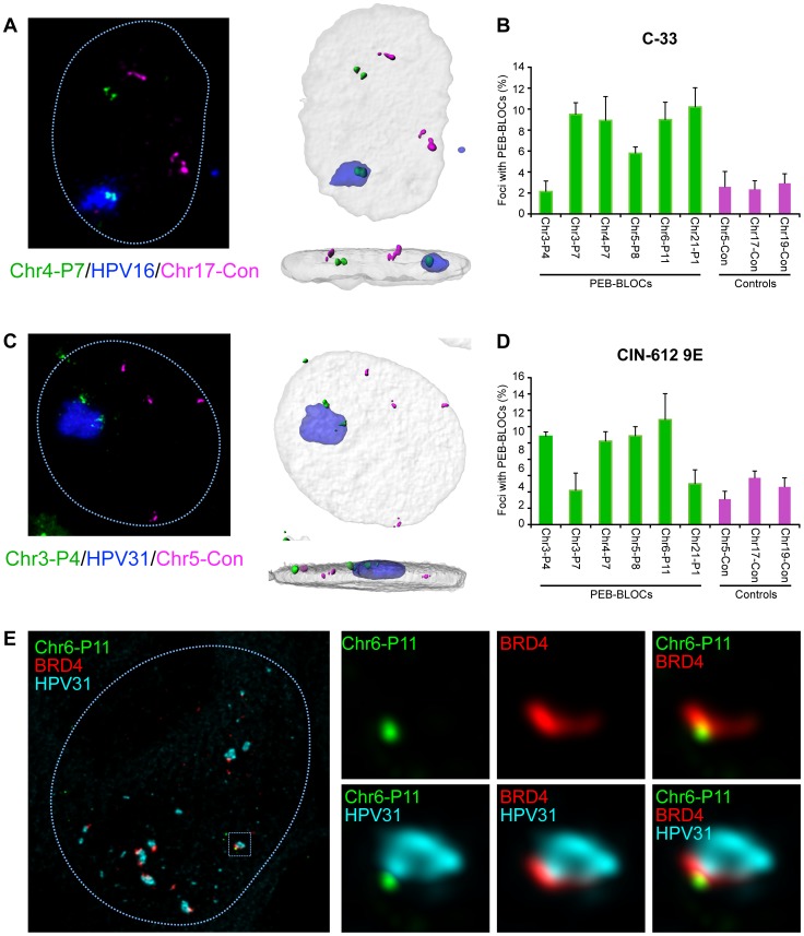
It has long been recognized that oncogenic viruses often integrate close to common fragile sites. The papillomavirus E2 protein, in complex with BRD4, tethers the viral genome to host chromatin to ensure persistent replication. Here, we map these targets to a number of large regions of the human genome and name them Persistent E2 and BRD4-Broad Localized Enrichments of Chromatin or PEB-BLOCs. PEB-BLOCs frequently contain deletions, have increased rates of asynchronous DNA replication, and are associated with many known common fragile sites. Cell specific fragile sites were mapped in human C-33 cervical cells by FANCD2 ChIP-chip, confirming the association with PEB-BLOCs. HPV-infected cells amplify viral DNA in nuclear replication foci and we show that these form adjacent to PEB-BLOCs. We propose that HPV replication, which hijacks host DNA damage responses, occurs adjacent to highly susceptible fragile sites, greatly increasing the chances of integration here, as is found in HPV-associated cancers.


Conflict of interest statement

The authors have declared that no competing interests exist.

Figures

Figure 1
Figure 1. HPV1 E2 binds to broad regions of mitotic chromatin.
A. ChIP-chip binding profile of E2 on a subset of human chromosomes. E2-bound mitotic chromatin was hybridized to microarray chips containing the chromosomes shown. The Y-axis is a scaled log2-ratio of bound to input signal. Large chromatin regions enriched for E2 binding were identified as described in Methods. They are indicated in red underneath the signal map and in Table S2. Chromosomes 18 and 19 were also analyzed but showed no binding peaks and are not shown. B. E2 binding to the large peaks persists throughout the cell cycle. Chromatin was isolated from asynchronous and mitotic fractions from C-33 cells containing empty vector (pMEP4 or pM4) or C-33-1E2 cells (E2) and analyzed by ChIP and Q-PCR using primers (Table S9) for broad E2 binding regions (persistent: throughout the cell cycle) or promoter regions previously shown to bind E2/BRD4 (only in interphase) . Average E2 binding levels were calculated from two independent experiments for five promoter regions and eight broad E2 binding regions. C. Average BRD4 and E2 binding levels on mitotic chromatin to five sites within a broad E2 binding region, chr5:123,942,100–125,482,100 in the absence (pMEP4 empty vector) or presence (E2) of E2 expression. E2 and BRD4 binding was analyzed by ChIP using Q-PCR with the primers described in Table S9 and Figure S1C. Average E2 and BRD4 binding levels and STDEV are presented as calculated from four promoter regions (BRD2, CCD1, SALL4, TUBB) and five binding sites within a broad E2 binding peak in Chr5 (See Figure S1 for complete data).
Figure 2
Figure 2. Profile of BRD4 binding to human chromosomes in C-33-1E2 cells.
E2 expression was induced in C-33-1E2 cells (and E2 negative control cells) and chromatin was isolated with an anti-BRD4 antibody (C-term) and hybridized to 2.1M human whole genome arrays (NimbleGen). BRD4CON represents negative control C-33 cells and BRD4E2 represents C-33 cells expressing HPV1 E2. A. The BRD4 binding profiles are shown for chromosome 4 (the complete data set is in Figure S2). The data is aligned with the E2 binding profile for chromosome 4 from Figure 1A. Broad regions of enriched binding were defined computationally and are shown in red (the coordinates are listed in Table S2). B. Regions enriched for E2 binding (E2), and BRD4 binding in absence (BRD4CON) and presence of E2 (BRD4E2), were analyzed and computationally identified in a subset of human chromosomes (chromosome 3, 4, 5, 21, 22, and X) as described in Methods. The overlap between the enriched regions was further calculated as described in Methods and is represented in the Venn diagram. C. Broad enriched regions of BRD4 binding in the absence (BRD4CON) and presence of E2 (BRD4E2) were identified computationally for the entire human genome (Figure S2). The overlap between the enriched regions was calculated as described in Methods and is represented in the Venn diagram.
Figure 3
Figure 3. BRD4 is essential for persistent HPV1 E2 binding to host mitotic chromatin.
A. E2 expression was induced in asynchronous C-33 cells expressing either wild-type or R37A/I73A E2 proteins. Chromatin was isolated with FLAG antibodies (against E2) or with BRD4 antiserum. ChIP DNA was quantitated by Q-PCR using primers specific for the PEB-BLOCs listed. Average values and STDEV were calculated from two independent experiments. B. The location of E2 wild-type or E2 R37A/I73A (green) and BRD4 (red) as detected by immunofluorescence. Cellular DNA is counterstained with DAPI in blue. Approximately 50 mitotic cells were analyzed for E2 and BRD4 chromosomal speckles. Average values and STDEV were calculated for three independent experiments. C. C-33-1E2 cells were treated with BRD4 siRNA for 3 days and stained for E2 (green), BRD4 (red) and cellular DNA (blue). Approximately 50 mitotic cells were analyzed for E2 and BRD4 chromosomal speckles. Average values and STDEV were calculated for three independent experiments. D. C-33 cells expressing HPV1 E2 were treated with DMSO, GSK525762+ (GSK+), or GSK525762 (GSK−), for 24 h and E2 expression was induced for 4 h before fixation. Chromatin was isolated with FLAG M2 or with BRD4 immune serum. ChIP DNA was quantitated by Q-PCR using primers specific for the PEB-BLOCs shown. Average values and STDEV were calculated from two independent experiments. E. C-33-1E2 cells were treated with DMSO, GSK525762+ (GSK+), or GSK525762 (GSK−), for 24 h and E2 expression was induced before fixation. Cells were stained for E2 (green), BRD4 (red) and cellular DNA (blue). Greater than 50 interphase cells were analyzed for E2 and BRD4 colocalization. Average values and STDEV were calculated for three independent experiments.
Figure 4
Figure 4. Persistent E2 binding correlates with histone acetylation through CREBBP/EP300 HAT activity.
A. Mitotic chromatin was isolated from C-33-1E2 cells and immunoprecipitated with control serum or specific antibodies to E2, BRD4, H3K56ac, H4K8ac, H3K4me1, H3K4me2, H3K4me3, and histone H3. ChIP DNA was analyzed by Q-PCR for specific PEB-BLOC regions (listed in Table S9). Average values and STDEV are shown for three independent experiments on four non-E2 binding regions, four active promoters, and six PEB-BLOCs. B. ChIP-chip analysis of E2, BRD4, H4K8ac, and H3K4me1 binding in C-33-1E2 cells. Chromatin was prepared from asynchronous cells and isolated using antibodies against E2, BRD4, acH4K8, and H3Kme14. ChIP DNA was hybridized to one HD microarray chip (Nimblegen). The binding profile for E2, BRD4, and histones on chromosome 3 and 4 is shown. Broad regions of enriched binding were defined computationally and are shown in red and are listed in Table S4. C. Venn diagram showing the overlap among the enriched binding regions defined in B. D. C-33-1E2 cells were treated with CREBBP, EP300 or KAT5 siRNA for 3 days. Cells were stained by immunofluorescence for anti-BRD4 (green), anti-H4K8ac (red), cellular DNA (blue), and anti-CREBBP, -EP300, or -KAT5 antibodies (cyan). The bar chart to the right shows quantification of BRD4 speckle formation in these Interphase cells were analyzed for. Average values and STDEV were calculated for three independent experiments (75–150 cells counted per experiment).
Figure 5
Figure 5. PEB-BLOCs share properties with fragile sites and are targets for chromosomal deletion.
A, B. Chromosomes were spread from mitotic C-33-1E2 cells induced for E2 expression. BRD4 speckles were identified by immunofluorescence, fixed, and hybridized with PEB-BLOC specific FISH probes (107E21, 209F21, 274H4, or 365P10). A representative example is shown for probes 275H4 (A, Chr3-P7) and 209F21 (B, Chr5-P8). The BRD4 signal is red and the FISH signal is green. C. C-33-1E2 cells, induced for E2 expression were analyzed with specific FISH probes. PEB-BLOCs (209F21, 90I21, 274H4, 365P10, 1062A20; green) and control regions (80N12 and 379K17; red). Approximately 50 S-phase cells were analyzed for the SD pattern (one single, one double dot) FISH signals for each PEB-BLOCs or control region. This SD pattern represents asynchronous replication at the allele being examined. Average values and STDEV were calculated for two independent experiments on two control regions (80N12 and 379K17), and 4 PEB-BLOCs (Chr3-P7, Chr5-P8, Chr10-P4, Chr12-P3). D. Correlation between gene size and overlapping PEB-BLOC. The overlap was calculated for a region encompassing the gene body plus 5 kb upstream.
Figure 6
Figure 6. PEB-BLOCs are closely associated with common fragile sites.
A. An alignment of BRD4 enriched regions (PEB-BLOCs), FANCD2 enriched regions and known common fragile sites on chromosome 4. The red blocks under the signalmap represent computationally defined enriched regions. B. Venn diagram showing the overlap in PEB-BLOCs and FANCD2 enriched regions in the entire human genome. The significance of overlap was calculated by the permutation method. C. Binding profile of BRD4 in C-33-1E2 cells on PEB-BLOC Chr4-P4, which overlaps the known common fragile site FRA4F. FISH analysis with 245M5 or 451M10 probes on interphase and mitotic cells reveal a deletion one allele overlapping the 245M5 probe. D. RNAseq analysis of the long FAM190A gene in Chr4-P4 PEB-BLOC. Reads of transcripts were aligned with BRD4 ChIP-chip binding data from C-33 and C-33-1E2 cells and FANCD2 binding data from aphidicolin treated C-33 cells.
Figure 7
Figure 7. HPV16 E1/E2 binding sites are closely associated with PEB-BLOCs and common fragile sites.
A. HPV16 E1 and E2 were transiently expressed in C-33 cells. ChIP-chip was performed using an anti-EE (E1) antibody. The Y-axis corresponds to a scaled log2-ratio of E1 signal to input signal. Previously obtained BRD4 (C-33-1E2 cells), and FANCD2 (aphidicolin-treated cells) binding profiles are aligned. Enriched regions were computationally defined and are shown in red under the signal map profile. Chromosome 5 is shown here and the entire data is shown in Figure S7. B. Diagram of chromosome 5 showing the overlap of BRD4 enriched regions (red), FANCD2 enriched regions (blue), common fragile sites (orange), and HPV integration sites (green) with cytogenetic bands in grey. C. A Venn diagram showing the overlap among the enriched BRD4, E1 and FANCD2 binding region regions, as defined by the permutation method. D. A Venn diagram showing the overlap among the enriched BRD4 and FANCD2 binding region regions and HPV integration breakpoints as defined by the permutation method. See Table 3 for statistics.
Figure 8
Figure 8. Papillomavirus replication factories are associated with PEB-BLOCs.
A. C-33 cells containing replicating HPV16 genomes were analyzed by FISH. The representative image shown was probed with HPV16 DNA (blue), Chr4-P7 PEB-BLOC (107E21 BAC clone; green) and a control region (304M13; magenta). 3D image stacks were deconvolved using Huygens Essential software. High resolution surface renderings of all objects were generated by Bitplane Imaris software. B. PEB-BLOCs were probed in the cells described in A, using a combination of labeled BAC clones (124A13, 1079D6, 107E21, 126M10, 1062A20 corresponding to PEB-BLOCs Chr2-P6, Chr3-P7, Chr4-P7, Chr5-P8, Chr6-P11), control BAC clones (182E4, 304M13, 568L16 representing negative regions) and HPV16 DNA. Approximately 100 cells were analyzed for colocalization of HPV16, PEB-BLOCs or control regions. Average values and STDEV were calculated for three independent experiments. C. Differentiated CIN-612 9E cells were analyzed by FISH. The representative image shown was probed with HPV31 DNA (blue), Chr3-P4 PEB-BLOC (452E16 BAC clone; green) and control region (182E4; magenta). 3D image stacks were deconvolved using Huygens Essential software. High resolution surface renderings of all objects were generated by Bitplane Imaris software. D. PEB-BLOCs, HPV31, and negative regions were detected using BAC clones (452E16, 1079D6, 107E21, 126M10, 1062A20, 812E1 corresponding to PEB-BLOCs Chr3-P4, Chr3-P7, Chr4-P7, Chr5-P8, Chr6-P11, Chr21-P1;green), HPV31 DNA (blue), and BAC clones (182E4, 304M13, 568L16 corresponding to negative regions; magenta). >50 HPV31 foci were analyzed for colocalization with PEB-BLOCs or control regions. Average values and STDEV were calculated for three independent experiments. E. Differentiated CIN-612 9E cells were analyzed by IF-FISH. BRD4 speckles were identified by immunofluorescence, fixed, and hybridized with a PEB-BLOC specific FISH probe and HPV31 FISH probe. The representative image shown was probed with Chr6-P11 PEB-BLOC (1062A20 BAC clone; green), BRD4 (red), and HPV31 DNA (cyan).

Similar articles

Cited by

References

    1. Skiadopoulos MH, McBride AA (1998) Bovine papillomavirus type 1 genomes and the E2 transactivator protein are closely associated with mitotic chromatin. J Virol 72: 2079–2088. - PMC - PubMed
    1. Ilves I, Kivi S, Ustav M (1999) Long-term episomal maintenance of bovine papillomavirus type 1 plasmids is determined by attachment to host chromosomes, which is mediated by the viral E2 protein and its binding sites. Journal of Virology 73: 4404–4412. - PMC - PubMed
    1. Lehman CW, Botchan MR (1998) Segregation of viral plasmids depends on tethering to chromosomes and is regulated by phosphorylation. ProcNatlAcadSci USA 95: 4338–4343. - PMC - PubMed
    1. You J, Croyle JL, Nishimura A, Ozato K, Howley PM (2004) Interaction of the bovine papillomavirus E2 protein with Brd4 tethers the viral DNA to host mitotic chromosomes. Cell 117: 349–360. - PubMed
    1. Baxter MK, McPhillips MG, Ozato K, McBride AA (2005) The mitotic chromosome binding activity of the papillomavirus E2 protein correlates with interaction with the cellular chromosomal protein, Brd4. J Virol 79: 4806–4818. - PMC - PubMed

Publication types

Substances