. 2014 Jun 27;289(26):18347-59.
doi: 10.1074/jbc.M113.504894. Epub 2014 May 12.

The Cdc42 guanine nucleotide exchange factor FGD6 coordinates cell polarity and endosomal membrane recycling in osteoclasts

Affiliations

The Cdc42 guanine nucleotide exchange factor FGD6 coordinates cell polarity and endosomal membrane recycling in osteoclasts

Charlotte Steenblock et al. J Biol Chem. .

Abstract

The initial step of bone digestion is the adhesion of osteoclasts onto bone surfaces and the assembly of podosomal belts that segregate the bone-facing ruffled membrane from other membrane domains. During bone digestion, membrane components of the ruffled border also need to be recycled after macropinocytosis of digested bone materials. How osteoclast polarity and membrane recycling are coordinated remains unknown. Here, we show that the Cdc42-guanine nucleotide exchange factor FGD6 coordinates these events through its Src-dependent interaction with different actin-based protein networks. At the plasma membrane, FGD6 couples cell adhesion and actin dynamics by regulating podosome formation through the assembly of complexes comprising the Cdc42-interactor IQGAP1, the Rho GTPase-activating protein ARHGAP10, and the integrin interactors Talin-1/2 or Filamin A. On endosomes and transcytotic vesicles, FGD6 regulates retromer-dependent membrane recycling through its interaction with the actin nucleation-promoting factor WASH. These results provide a mechanism by which a single Cdc42-exchange factor controlling different actin-based processes coordinates cell adhesion, cell polarity, and membrane recycling during bone degradation.

Keywords: Actin; CDC42; FGD6; Membrane Recycling; Osteoclast; Podosome; Src.


Figures

FIGURE 1.
FIGURE 1.
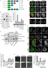
FGD6 localizes to sealing zones and transcytotic vesicles of osteoclasts and regulates their polarity. A, intracellular distribution of Myc- or GFP-tagged FGD6 (green) expressed in osteoclasts grown on glass or ODs. Cells were stained with phalloidin (red) and DAPI (blue) and analyzed by confocal microscopy. Bars, 20 μm. B, intracellular distribution of GFP-Rab38 (green) and digested calcium (red) in osteoclasts grown on ODs. See also supplemental Videos S1 and S2. C, dynamics of GFP-FGD6 and RFP-Ezrin actin-binding domain at podosomes of osteoclasts grown on glass analyzed by time lapse video microscopy (one frame per min). Fluorescence intensities associated with GFP or RFP on single podosomes were quantified. See also supplemental Video S3. D, dynamics of GFP-FGD6 in osteoclasts grown on ODs analyzed by time lapse microscopy (one frame per min). See also supplemental Video S4. E, osteoclasts grown on ODs were either mock-transfected, or transfected with scrambled siRNAs or with siRNAs targeting FGD6. Cells were stained with phalloidin (red) and DAPI (blue) and analyzed by confocal microscopy. Bars, 20 μm. F, FGD6 expression levels were quantified by RT-PCR. G, the number of osteoclasts with sealing zones was quantified. Values are mean ± S.D. from 3 experiments (n = 300 osteoclasts per experiment). All groups were compared with control by applying a Dunnett one-way ANOVA test. *, p < 0.05. **, p < 0.01. H, distribution of integrin β3 and Lamp1 in control and FGD6-depleted osteoclasts grown on ODs. Bars, 20 μm. Representative images are shown.
FIGURE 2.
FIGURE 2.
The PH and FYVE domains of FGD6 are required for sealing zone formation and surface resorption. A, the FGD6 mutants used in this study. B and C, binding of His-Myc-tagged FGD6 lacking its N-terminal domain (aa 843–1398) to the indicated lipids present on nitrocellulose strips (B) or liposomes (C). D, osteoclasts grown on ODs expressing GFP-FGD6 full-length or truncated from its N-terminal domain (green) were left untreated or treated with either 500 nm wortmannin or 10 μm PP2 for 1 h. Cells were stained with phalloidin (red) and DAPI (blue) and analyzed by confocal microscopy. Bars, 20 μm. E, cell lysates from similarly treated osteoclasts were immunoprecipitated with anti-GFP. After Western blotting immunoprecipitates (IP) and cell lysates were probed for GFP-FGD6 or phosphotyrosine. F-H, in osteoclasts depleted from endogenous FGD6 different GFP-tagged FGD6 proteins (wild type and mutants) were expressed as indicated (green). F, cells were stained with phalloidin (red) and DAPI (blue), and analyzed by confocal microscopy. Bars, 20 μm. G, expression levels of the FGD6 full-length and mutants were quantified by RT-PCR. Values are mean ± S.D. from 3 experiments. H, the corresponding digested areas on ODs (appearing as black spots on a gray background) were quantified. Values are mean ± S.D. from 3 experiments. All groups were compared with control by applying a Dunnett one-way ANOVA test. *, p < 0.05. **, p < 0.01. Representative images are shown. The abbreviations are: PI, phosphatidylinositol; PE, phosphatidylethanolamine; LPA, lysophosphatidic acid; LPC, lysophosphatidylcholine; S1P, sphingosin-1-phosphate; WB, Western blot.
FIGURE 3.
FIGURE 3.
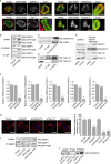
FGD6 is a Cdc42 GEF. A, osteoclasts were mock-transfected or transfected with siRNAs targeting FGD6. Cell lysates were prepared and GTP-Cdc42 was immunoprecipitated with anti-GTP-Cdc42. After Western blotting (WB) immunoprecipitates (IP) and cell lysates were probed for Cdc42 and GAPDH. B, similarly treated osteoclasts were grown on ODs and stained with anti-GTP-Cdc42 (green), phalloidin (red), and DAPI (blue) and analyzed by confocal microscopy. C–E, osteoclasts were either mock-transfected, or transfected with scrambled siRNAs or with siRNAs targeting Cdc42. C, Cdc42 expression levels were quantified by RT-PCR. Values are mean ± S.D. from 3 experiments. D, similarly treated osteoclasts were grown on ODs and stained with phalloidin (red) and DAPI (blue) and analyzed by confocal microscopy. Bars, 20 μm. E, the number of osteoclasts with sealing zones was quantified. Values are mean ± S.D. from 3 experiments (n = 300 osteoclasts/experiment). All groups were compared with control by applying a Dunnett one-way ANOVA test. *, p < 0.05. **, p < 0.01. Representative images are shown.
FIGURE 4.
FIGURE 4.
FGD6 interacts with IQGAP1, ARHGAP10, Talin-1, and Filamin A at the plasma membrane. A, osteoclasts expressing GFP-FGD6 (green) were grown on ODs and stained with DAPI (blue) and anti-IQGAP1, anti-ARHGAP10, anti-Talin-1, or anti-Filamin A (red). Bars, 50 μm. B and C, cell lysates from control osteoclasts or osteoclasts expressing GFP-FGD6 were used for immunoprecipitation of (B) IQGAP1 or GFP-FGD6 or (C) ARHGAP10 with anti-IQGAP1, anti-GFP, or anti-ARHGAP10. After Western blotting (WB) immunoprecipitates and cell lysates were probed for IQGAP1, GFP-FGD6, or ARHGAP10. D, endogenous FGD6 was immunoprecipitated from osteoclasts lysates with anti-FGD6 and after Western blotting the immunoprecipitate (IP) was probed for Filamin A or Talin-1/2. E–H, osteoclasts were either mock-transfected, or transfected with scrambled siRNAs or with siRNAs targeting IQGAP1, ARHGAP10, Talin-1, Talin-2, or Filamin A. E, osteoclasts were collected and lysed. Lysates were analyzed by SDS-PAGE followed by Western blotting using antibodies directed against the indicated gene products. F, the expression levels were quantified by RT-PCR. Values are mean ± S.D. from 3 experiments. G, cells grown on ODs were stained with phalloidin (red) and DAPI (blue), and analyzed by confocal microscopy. Bars, 50 μm. H, the number of osteoclasts with sealing zones was quantified. Values are mean ± S.D. from 3 experiments (n = 200 osteoclasts/experiment). All groups were compared with control by applying a Dunnett one-way ANOVA test. *, p < 0.05. **, p < 0.01. See also supplemental Videos S5–S8. I and J, control and GFP-FGD6-expressing osteoclasts were left untreated or treated with 10 μm PP2 for 1 h. I, IQGAP1, or J, FGD6 was immunoprecipitated from cell lysates using the corresponding antibodies, and after Western blotting the immunoprecipitates and lysates were probed for IQGAP1, GFP-FGD6, or ARHGAP10. Each experiment was repeated at least 3 times. Representative images are shown.
FIGURE 5.
FIGURE 5.
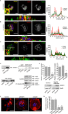
FGD6 interacts with WASH1 on endosomes. Osteoclasts expressing GFP-FGD6 (green) were grown on ODs and stained with DAPI (blue) and anti-EEA1 (A), anti-WASH1 (B), or anti-Vps35 (C) (red), and analyzed by confocal microscopy. Bars, 20 μm. Arrows show early endosomes around transcytotic vesicles. The corresponding fluorescence intensity profiles along the white line are shown. D, osteoclast lysates were incubated with GST, GST-FGD6(1–1039), or GST-Cdc42 loaded with GDP or GTPγS, or with anti-FGD6 antibodies. After pull down, the interacting material was analyzed by Western blotting (WB) using anti-WASH1. GTP-Cdc42 was immunoprecipitated from lysates of osteoclasts depleted or not from FGD6, and after Western blotting the immunoprecipitates (IP) were probed for Cdc42 and WASH1. E–G, osteoclasts grown on ODs were mock-transfected, or transfected with scrambled siRNAs or with siRNAs targeting WASH1 or Vps35. E, the expression levels were determined by quantitative RT-PCR. Osteoclasts were also collected and lysed. Lysates were analyzed by SDS-PAGE followed by Western blotting using antibodies directed against the indicated gene products. F, osteoclasts were also stained with phalloidin (red) and DAPI (blue) and analyzed by confocal microscopy. Bars, 50 μm. G, the number of osteoclasts with sealing zones was quantified. Values are mean ± S.D. from 3 experiments (n = 200 osteoclasts per experiment). All groups were compared with control by applying a Dunnett one-way ANOVA test. *, p < 0.05. **, p < 0.01. Representative images are shown. See also supplemental Videos S9 and S10.
FIGURE 6.
FIGURE 6.
Model of FGD6 function in podosome dynamics and retromer-dependent recycling (1). At the plasma membrane, integrins bind to RGD motifs of extracellular bone matrix proteins. This triggers the activation of the PI 3-kinase, which produces PI(3,4,5)P3 in the vicinity of the engaged integrins (2). Upon Src-dependent phosphorylation, FGD6 binds to PI(3,4,5)P3 and potentially PI(4,5)P2, and exchanges GDP for GTP on Cdc42 (3). FGD6 interacts with IQGAP1, which binds Cdc42 and PI(3,4,5)P3. IQGAP1 can then trigger F-actin polymerization. FGD6 also interacts with Talin-1/2 or Filamin A (FLNa) and can coordinate Cdc42-dependent cell adhesion and F-actin polymerization required for podosome formation. These complexes may contain ARHGAP10 whose GAP activity is inhibited by Pyk2-dependent phosphorylation (4). On early endosomes and transcytotic vesicles, phosphorylated FGD6 binds to PI(3)P and to the WASH complex. FGD6 also exchanges GDP for GTP on Cdc42, which activates WASH. WASH triggers F-actin polymerization to sustain retromer (Vps35)-dependent recycling of membrane components.

Similar articles

Cited by

References

    1. Crockett J. C., Rogers M. J., Coxon F. P., Hocking L. J., Helfrich M. H. (2011) Bone remodelling at a glance. J. Cell Sci. 124, 991–998 - PubMed
    1. Teitelbaum S. L. (2011) The osteoclast and its unique cytoskeleton. Ann. N.Y. Acad. Sci. 1240, 14–17 - PubMed
    1. Stenbeck G., Horton M. A. (2004) Endocytic trafficking in actively resorbing osteoclasts. J. Cell Sci. 117, 827–836 - PubMed
    1. Lowell C. A., Soriano P. (1996) Knockouts of Src-family kinases: stiff bones, wimpy T cells, and bad memories. Genes Dev. 10, 1845–1857 - PubMed
    1. Czupalla C., Mansukoski H., Pursche T., Krause E., Hoflack B. (2005) Comparative study of protein and mRNA expression during osteoclastogenesis. Proteomics 5, 3868–3875 - PubMed

Publication types

Substances

LinkOut - more resources