. 2014 Jun 12;510(7504):273-7.
doi: 10.1038/nature13233. Epub 2014 Apr 30.

Human embryonic-stem-cell-derived cardiomyocytes regenerate non-human primate hearts

Affiliations

Human embryonic-stem-cell-derived cardiomyocytes regenerate non-human primate hearts

James J H Chong et al. Nature. .

Abstract

Pluripotent stem cells provide a potential solution to current epidemic rates of heart failure by providing human cardiomyocytes to support heart regeneration. Studies of human embryonic-stem-cell-derived cardiomyocytes (hESC-CMs) in small-animal models have shown favourable effects of this treatment. However, it remains unknown whether clinical-scale hESC-CM transplantation is feasible, safe or can provide sufficient myocardial regeneration. Here we show that hESC-CMs can be produced at a clinical scale (more than one billion cells per batch) and cryopreserved with good viability. Using a non-human primate model of myocardial ischaemia followed by reperfusion, we show that cryopreservation and intra-myocardial delivery of one billion hESC-CMs generates extensive remuscularization of the infarcted heart. The hESC-CMs showed progressive but incomplete maturation over a 3-month period. Grafts were perfused by host vasculature, and electromechanical junctions between graft and host myocytes were present within 2 weeks of engraftment. Importantly, grafts showed regular calcium transients that were synchronized to the host electrocardiogram, indicating electromechanical coupling. In contrast to small-animal models, non-fatal ventricular arrhythmias were observed in hESC-CM-engrafted primates. Thus, hESC-CMs can remuscularize substantial amounts of the infarcted monkey heart. Comparable remuscularization of a human heart should be possible, but potential arrhythmic complications need to be overcome.


Figures

Extended Data Figure 1
Extended Data Figure 1. Cryopreservation does not affect hESC-CM engraftment
(a) Schematic representation of experimental design for cryopreservation testing experiments. (b) Human genomes detected after injection of cryopreserved or non-cryopreserved hESC-CM were not significantly different (p>0.05, t-test). Mean ± standard error is shown (n=9 biological replicates).
Extended Data Figure 2
Extended Data Figure 2. Creation and validation of the GCaMP3-expressing hESC lines
(a) Targeting construct for zinc finger nuclease engineering of GCaMP3 into the AAVS1 locus. The endogenous genomic probe and neomycin resistance gene probe binding sites used for Southern blotting are shown. (b) Southern blot analysis demonstrates a single integration event by hybridization for neomycin resistance cassette (left) and heterozygous AAVS1 integration by genomic probe labelling (right).
Extended Data Figure 3
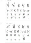
Extended Data Figure 3. Chromosomal analysis of hESCs modified to encode GCaMP3
(a) H7-GCaMP3 ESC line demonstrate an isochrome of the chromosome 20 long arm (arrow). (b) RUES2-GCaMP3 ESC line show normal karyotype.
Extended Data Figure 4
Extended Data Figure 4. Flow cytometry for cardiomyocyte differentiation of hESCs
Representative histogram of hESC-CM after differentiation shows 73% cTnT-expressing cells. cTnT = cardiac troponin T.
Extended Data Figure 5
Extended Data Figure 5. Schematic representation of experimental design
Myocardial infarction (MI) was created by advancing a balloon catheter into the distal left anterior descending artery and inflating it to create ischemia (90 minutes) followed by reperfusion. The infarct was induced 14 days prior to hESC-CM delivery via left thoracotomy. Immunosuppression using cyclosporine A, methylprednisolone and abatacept (CTLA4-Ig) was delivered 5 days prior to cell delivery continuing until euthanasia of animals. Primary endpoints were 1) histologically based morphometric calculations of infarct and graft size with analysis of graft composition, 2) ex vivo analysis of graft-host electromechanical coupling enabled by GCaMP3 fluorescence detection. Secondary endpoints were 1) detection of arrhythmias by telemetric electrocardiogram analysis and 2) analysis of left ventricular functional change by trans-esophageal echocardiography.
Extended Data Figure 6
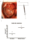
Extended Data Figure 6. Technique for hESC-CM injection to infarct region and border zones using “mattress” suture strategy
(a) The macaque infarcted ventricular apex is seen by blanched region (dotted line) during left thoracotomy. A total of 15 aliquots, each containing 100 μL of hESC-CM in pro-survival cocktail, were delivered via 5 epicardial puncture sites (arrows, note one further puncture site not seen is on posterior aspect). (b) hESC-CM retention after injection was increased by use of a “matress suture”. X = insertion points of suture with dotted lines representing path of suture (exaggerated size for diagrammatic representation). Needle tip was inserted into the resulting rectangular area and suture tightened after series of three injections (altering trajectory of needle) but before withdrawal of needle tip. (c) Quantitation of India ink retention after injection into left ventricular myocardium of anaesthetised macaques with or without use of the mattress suture technique (n=3 each group). A trend favouring greater retention with the mattress suture is seen.
Extended Data Figure 7
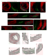
Extended Data Figure 7. Remuscularization of the infarcted macaque heart
(a–f) Single channels of confocal immunofluorescence shown in Fig. 1 (a–f). Macaque heart shown was subjected to myocardial infarction and transplantation of hESC-CM 14 days prior to sacrifice. GFP=green fluorescence protein. (a–f′) Picrosirius Red staining of sections in close proximity to confocal immunofluorescence in (a–f) shows lack of fibrosis within hESC-CM grafts.
Extended Data Figure 8
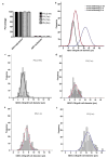
Extended Data Figure 8. Remuscularized infarct region is composed of engrafted cardiomyocytes that increase in size with time
(a) Quantification of the sarcomeric protein α-Actinin expression in green fluoresnce protein (GFP) expressing grafts. The vast majority (>98%) of GFP-expressing cells co-expressed α-Actinin. P3-P6 represent individual animals (n=1) sacrificed at 2 weeks (2 wk) 1 month (m) or 3 months after hESC-CM delivery. 500–700 cells were counted from 3 different graft regions of each heart. Percentage of cells GFP/α-Actinin double positive and GFP positive/α-Actinin negative are shown as mean +/− SD. (b) Normal curve from histograms showing the distribution of hESC derived cardiomyocyte diameters (graft) in monkey hearts 2 weeks, 1 or 3 months after cell delivery. (e–f) Individual histograms with superimposed normal curve of each animal P3-P6 (as above).
Extended Data Figure 9
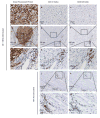
Extended Data Figure 9. No evidence of human graft rejection
Representative low (d–f) and high (a–c and g–i) power magnification of hESC-CM graft 28 days after cell delivery to infarcted macaque heart. Representative low (j–k) and high (l–m) power magnification of infarct region from control macaque 28 days after sham treatment. The hESC-CM graft is detected by anti-green fluorescent protein primary antibody with 3,3′-Diaminobenzidine (DAB) detection of secondary antibody (brown). Few CD3+ T-lymphocytes or CD20+ B-lymphocytes are seen surrounding the hESC-CM grafts. Comparable numbers of T and B cells are seen in control infarcts receiving no hESC-CM treatment. Boxed inset regions show areas of higher magnification.
Extended Data Figure 10
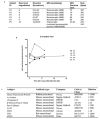
Extended Data Figure 10. Summary of ventricular tachycardia and echocardiographic assessment of left ventricular function
(a) Table characterizing episodes of ventricular tachycardia after engraftment of hESC-CM (detailed in Fig. 5). Note that P5 demonstrated no discernible sinus rhythm on telemetric recording of electrocardiogram 14 days after hESC-CM delivery. Although QRS morphology varied the tachyarrhythmia comprised sustained periods of stable monomorphic QRS morphology. LBBB=left bundle branch block. RBBB=Right bundle branch block. ms=milliseconds. bpm=beats per minute. (b) Left ventricular function was assessed by transesophageal echocardiography at the following time points: prior to myocardial infarct (MI) creation, prior to hESC-CM delivery (2 weeks after MI) and prior to euthanasia (2, 4 or 12 weeks after MI). P7 received no-cells/vehicle only. All other animals received hESC-CM. Results shown are for left ventricular ejection fraction calculated by two blinded cardiologists from the 2-chamber view of the left ventricle. Note that this view best captures the infarcted antero-apical wall. The vehicle-treated control monkey showed a modest diminution in ejection fraction post-infarction. The cell-treated animals showed variable responses, with some having increased function and some having decreased function. Because of small group size, no statistical effects of hESC-CM therapy can be discerned. (c) Table of antibodies used. Abbreviations: DSHB = Developmental Studies Hybridoma Bank.
Figure 1
Figure 1. Remuscularization of the infarcted macaque heart with human cardiomyocytes
Confocal immunofluorescence of macaque hearts subjected to myocardial infarction and transplantation of hESC-CM. Grafts studied at 14 days (a–g) and 84 days post-engraftment (h–i). (a) Remuscularization of a substantial portion of the infarct region (dashed line) with hESC-CM co-expressing green fluorescent protein (GFP). The contractile protein α-Actinin (red) is expressed by both monkey and human cardiomyocytes. Scale bar, 1000 μm. (b–f) Images from the peri-infarct region of the same heart shown in (a), demonstrating significant hESC-CM engraftment. Scale bar, 1000 μm. (g) Graft-host interface at 14 days with interconnected α-Actinin (red) expressing cardiomyocytes (arrows). Note host sarcomeric cross-striations (asterisk) show greater alignment than hESC-CM graft. Scale bar 25 μm (h–i) Day-84 hESC-CM grafts contain host-derived blood vessels lined by CD31+ endothelial cells. Scale bar 20 μm. Inset scale bar 10 μm.
Figure 2
Figure 2. Human cardiomyocyte grafts exhibit maturation with time from engraftment
(a) Cardiomyocyte diameter of hESC-CM shows significant increase from 14 (n=1) to 28 (n=2) days and from 28 to 84 (n=1) days after engraftment. Adult monkey (n=2). From each animal (n), 200–400 cells were counted from 3 histological sections at varied left ventricular levels. Mean ± standard error is shown. (b–q) Confocal immunofluorescence of macaque hearts subjected to myocardial infarction and transplantation of hESC-CM 14 days (b,d, f–h, l–n) or 84 days (c,e, i–k, o–q) after engraftment. Increased sarcomere alignment, cardiomyocyte size in hESC-CM (GFP+) is seen in longer term grafts (b–c). Cx43 expression is not evident in hESC-CM grafts at 14 days but is seen at 84 days (d–e). Cardiomyocytes at the edges of grafts (g,j,m,p) display greater maturation compared to those at central core (h,k,n,q) evidenced by increased size, α-actinin staining intensity, sarcomere alignment (g–k) and N-cadherin expression (m–q). Scale bar for panels f,i,l and o = 200μm. All other scale bars 25 μm. GFP = green fluorescent protein. Cx43 = connexin 43. Yellow and white boxes correspond to higher power fields of graft edge and core respectively.
Figure 3
Figure 3. Blood vessels extend from the host coronary network into the graft
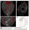
(a–c) 3-dimensional rendered microcomputed tomography of heart perfused with Microfil at 3 months after hESC-CM injection. (b) shows higher power view of boxed area from (a). (c) shows cross-sectional cut plane through the heart at the location of the dotted line in (a). Arteries perfusing the graft are red, other vessels are gray in the uninjured cardiac tissue, or white within the graft. The vessels within the graft are better visualized in Supplementary Video 6. (d) Histological section of heart shown in (a–c) immunostained with an anti-green fluorescence protein antibody to mark the hESC-CM graft (brown). This section corresponds to the same location of the cross-sectional cut plane in panel (c). Black dots are Microfil within coronary vessels.
Figure 4
Figure 4. Human cardiomyocytes are electrically coupled 1:1 to the infarcted host macaque heart after transplantation
(a) Diagram showing regions of the infarcted macaque heart visualized in (b–d). Analysis shown is from ex vivo imaging 14 days after hESC-CM delivery. (b) Still image from low power fluorescence video showing regions of hESC-CM engraftment (red and blue rectangles). (c–d) Still images of calcium indicator GCaMP3-positive hESC-CM grafts (bottom left of panel b) during diastole and systole. Note the gain of fluorescence during systole. (e–h) GCaMP3 fluorescence intensity (AU = arbitrary units) and electrocardiogram (ECG) versus time for the grafted regions of interest shown in (b). Each graft region shows 1:1 coupling synchronous with host ventricular contraction (ECG QRS complex) during (e) spontaneous rhythm or (f–h) atrial pacing. All hESC-CM grafts identified in every transplanted animal showed 1:1 coupling.
Figure 5
Figure 5. Ventricular arrhythmias after hESC-CM transplantation
(a–d) Representative traces from macaque telemetric electrocardiogram recordings showing (a) Normal sinus rhythm (SR), (b) Accelerated idioventricular rhythm (AIVR) (c) Ventricular tachycardia (VT) and (d) Non-sustained VT (NSVT). Scale bar 1 sec. (e–h) Frequency of arrhythmias is highest within the first 2 weeks after hESC-CM transplantation. P2-7 designations are animal identifiers. Animals receiving vehicle only (no cells, P2 and P7) remained in SR throughout. Interrupted Y-axis in (e–f) denotes reduced number of episodes but increased total duration of arrhythmias (VT or AIVR more than 18 hr per 24 hr period).

Comment in

Similar articles

Cited by

References

    1. Lozano R, et al. Global and regional mortality from 235 causes of death for 20 age groups in 1990 and 2010: a systematic analysis for the Global Burden of Disease Study 2010. Lancet. 2012;380:2095–2128. doi: 10.1016/S0140-6736(12)61728-0. S0140-6736(12)61728-0 [pii] - DOI - PMC - PubMed
    1. Laflamme MA, Murry CE. Heart regeneration. Nature. 2011;473:326–335. doi: 10.1038/nature10147. nature10147 [pii] - DOI - PMC - PubMed
    1. Caspi O, et al. Transplantation of human embryonic stem cell-derived cardiomyocytes improves myocardial performance in infarcted rat hearts. J Am Coll Cardiol. 2007;50:1884–1893. doi: 10.1016/j.jacc.2007.07.054. S0735-1097(07)02635-6 [pii] - DOI - PubMed
    1. van Laake LW, et al. Human embryonic stem cell-derived cardiomyocytes survive and mature in the mouse heart and transiently improve function after myocardial infarction. Stem Cell Res. 2007;1:9–24. doi: 10.1016/j.scr.2007.06.001. - DOI - PubMed
    1. Laflamme MA, et al. Formation of human myocardium in the rat heart from human embryonic stem cells. Am J Pathol. 2005;167:663–671. doi: 10.1016/S0002-9440(10)62041-X. S0002-9440(10)62041-X [pii] - DOI - PMC - PubMed

Publication types