. 2014 Jun 6;289(23):16551-64.
doi: 10.1074/jbc.M114.557959. Epub 2014 Apr 22.

Domain contributions to signaling specificity differences between Ras-guanine nucleotide releasing factor (Ras-GRF) 1 and Ras-GRF2

Affiliations

Domain contributions to signaling specificity differences between Ras-guanine nucleotide releasing factor (Ras-GRF) 1 and Ras-GRF2

Shan-Xue Jin et al. J Biol Chem. .

Abstract

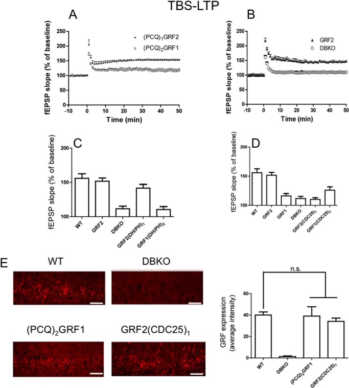
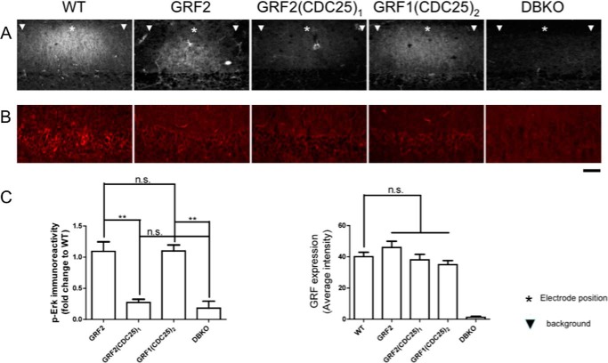
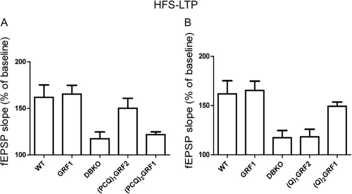
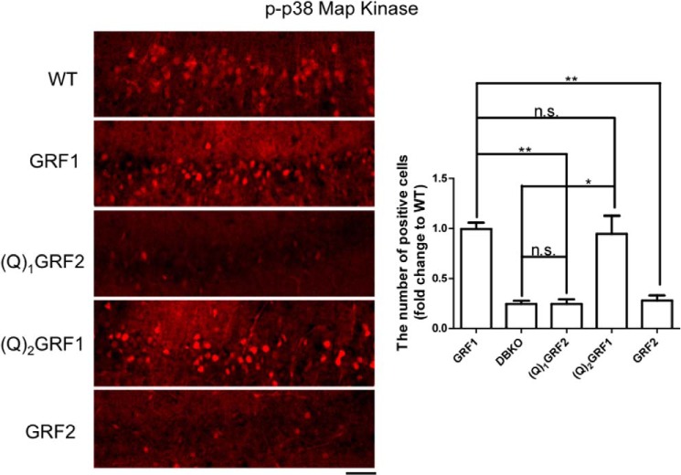
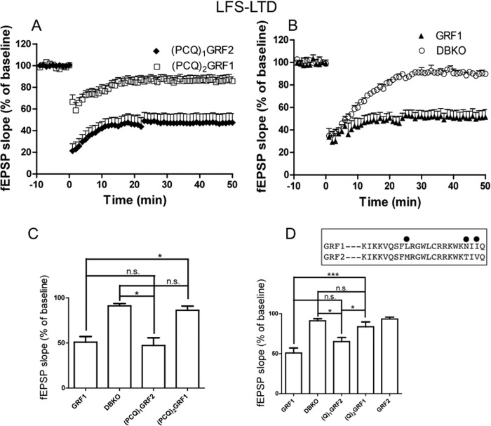
Ras-GRF1 (GRF1) and Ras-GRF2 (GRF2) constitute a family of similar calcium sensors that regulate synaptic plasticity. They are both guanine exchange factors that contain a very similar set of functional domains, including N-terminal pleckstrin homology, coiled-coil, and calmodulin-binding IQ domains and C-terminal Dbl homology Rac-activating domains, Ras-exchange motifs, and CDC25 Ras-activating domains. Nevertheless, they regulate different forms of synaptic plasticity. Although both GRF proteins transduce calcium signals emanating from NMDA-type glutamate receptors in the CA1 region of the hippocampus, GRF1 promotes LTD, whereas GRF2 promotes θ-burst stimulation-induced LTP (TBS-LTP). GRF1 can also mediate high frequency stimulation-induced LTP (HFS-LTP) in mice over 2-months of age, which involves calcium-permeable AMPA-type glutamate receptors. To add to our understanding of how proteins with similar domains can have different functions, WT and various chimeras between GRF1 and GRF2 proteins were tested for their abilities to reconstitute defective LTP and/or LTD in the CA1 hippocampus of Grf1/Grf2 double knock-out mice. These studies revealed a critical role for the GRF2 CDC25 domain in the induction of TBS-LTP by GRF proteins. In contrast, the N-terminal pleckstrin homology and/or coiled-coil domains of GRF1 are key to the induction of HFS-LTP by GRF proteins. Finally, the IQ motif of GRF1 determines whether a GRF protein can induce LTD. Overall, these findings show that for the three forms of synaptic plasticity that are regulated by GRF proteins in the CA1 hippocampus, specificity is encoded in only one or two domains, and a different set of domains for each form of synaptic plasticity.

Keywords: Calcium; Calmodulin (CaM); IQ Motif; LTD; LTP; Mitogen-activated Protein Kinase (MAPK); Ras Protein; Ras-GRF1; Ras-GRF2; Synaptic Plasticity.


Figures

FIGURE 1.
FIGURE 1.
The CDC25 domain of GRF2 is critical for GRF proteins to induce TBS-LTP. TBS-LTP was generated in hippocampal brain slices from WT mice or Grf1/Grf2 knock-out (DBKO) mice re-expressing: A, PCQ chimeras, (PCQ)1GRF2 or (PCQ)2GRF1; B, WT GRF2 and control DBKO mice; C, DH/PH chimeras GRF2(DH/PH)1 and GRF1(DH/PH)2; D, CDC25 chimeras GRF2(CDC25)1 and GRF1(CDC25)2. Panels A and B are fEPSP slopes representative of results from at least 6 slices from 3 mice each. Panels C and D quantify average fEPSP slopes for WT mice expressing endogenous GRF2 and various GRF proteins and chimeras. Experiments are from at least 6 slices from 5 mice and data show mean ± S.E. E, functionally inactive GRF1/GRF2 chimeras expressed at similar levels as endogenous WT GRF2 in the CA1. Scale bar, 50 μm. Bar graph represents the average ± S.E. of three tissue sections. n.s., not statistically different.
FIGURE 2.
FIGURE 2.
Activation of ERK MAPK is associated with the presence of the CDC25 domain of GRF2 and is necessary but not sufficient to promote TBS-LTP by GRF proteins. A, hippocampal brain slices from WT mice (WT), DBKO mice expressing GRF2, or the chimeric GRF proteins indicated and DBKO mice (DBKO), were exposed to TBS (140 μA) and then 5 min later samples were frozen and immunostained with anti-p-ERK MAPK antibodies. Representative p-ERK MAPK signals from slices are shown (top) (*, shows approximate stimulating electrode position; ▾, shows background area chosen at least 200 μm from electrode where GRF proteins are expressed, but stimulated cells are not observed). B, representative images of stimulated brain slices stained with GRF antibodies. C, quantification of fluorescence intensity in A and B. The left bar graph represents the signals obtained from samples described in A. The signal represents that from the stimulated area minus the signal from the neighboring unstimulated area both of which express exogenous GRF proteins. This value was then compared with that obtained with brain slices from WT mice expressing endogenous GRF2. The final results are expressed as fold-change compared with WT. The right bar graph represents the signals obtained from the GRF immunostaining. All data are the average ± S.E. of at least 3 independent experiments; **, p ≤ .01; n.s., not statistically different. Scale bar, 50 μm.
FIGURE 3.
FIGURE 3.
The PH and/or coiled-coil domains of GRF1 determine whether GRF proteins induce HFS-LTP. A and B, HFS-LTP was generated in hippocampal brain slices from WT mice and DBKO mice re-expressing indicated GRF proteins or various GRF1/GRF2 chimeras. Data show quantified average fEPSP slopes from at least 6 slices from 4 mice and data show mean ± S.E.
FIGURE 4.
FIGURE 4.
Activation of p38 MAPK mediated by the PH and/or coiled-coil domains of GRF1 is necessary and sufficient to induce HFS-LTP by GRF proteins. Hippocampal brain slices from WT mice or DBKO mice expressing the indicated WT and chimeric GRF proteins were exposed to HFS stimulation (140 μA) and then 10 min later samples were frozen and immunostained with anti-p-p38 MAPK antibodies. Representative signals from slices are shown (left) and then quantified for phospho-p38 MAPK-stained cells (right). Data represent the mean ± S.E. for at least three independent experiments; *, p ≤ 0.05; **, p ≤ 0.01; n.s., not statistically different; scale bar, 50 μm.
FIGURE 5.
FIGURE 5.
The IQ domain of GRF1 is necessary and sufficient for GRF proteins to induce LFS-LTD. LFS-LTD was generated in hippocampal brain slices from DBKO mice re-expressing various GRF proteins and chimeras. Panels A and B show fEPSP slopes representative of results from at least 6 slices from 3 mice each. Panels C and D show quantified average fEPSP slopes from at least 5 slices from 3 mice and data show mean ± S.E. *, p ≤ 0.05; ***, p ≤ 0.001; n.s., not statistically different; inset, a comparison of the IQ motif sequences between GRF1 and GRF2 with differences marked.
FIGURE 6.
FIGURE 6.
Fluorescence measurement of GRF peptide binding to CaM. Steady-state fluorescence was accomplished in a PTI fluorimeter as described under “Experimental Procedures.” Excitation was at 375 nm and emission scans were collected from 380 to 580, respectively. For panels A and B, each experiment began by adding CaM-ACR to 150 nm final concentration (black squares) to buffer containing 100 μm EGTA, scans were taken, and then Ca2+ was added to 400 μm final concentration (black circles). Then either the GRF1 (GRF1p; panel A) or GRF2 (GRF2p; panel B) peptide was added and traces were again collected (black triangles). Finally, EDTA to a final concentration of 10 mm was added (decreasing free Ca2+ to <5 nm) and traces were taken. Note the blue shift in the emission spectra both when Ca2+ is added and again with peptide along with increased fluorescence intensities. The addition of EDTA largely abrogated the fluorescence intensity but there was a persistent blue shift in the emission spectra indicating the peptides were interacting with apo-CaM. To more directly investigate this possibility, the impact of peptide binding to apo-CaM was addressed. Fluorescence scans of 150 nm CaM-ACR (black squares) in the presence of 1 mm EDTA were followed by the addition of 1 μm GRF1p (panel C) or 1 μm GRF2p (panel D) and scans again taken (black circles). Note the blue shift in the spectra indicating binding of the peptides to apo-CaM, but only a modest intensity increase was evident. Ca2+ was then added to 1.6 mm (to give 400 μm final; black triangles) and scans were again taken. Notice that there is no further blue shift in the spectra but a significant increase in intensity is evident with Ca2+ (black triangles). Following addition of EDTA, to 10 mm final concentration (inverted black triangles), the fluorescence intensity returned to that slightly below those before the addition of Ca2+ (black circles) but note that there remains a blue shift in the spectra consistent with binding to apo-CaM.
FIGURE 7.
FIGURE 7.
The IQ domain of GRF1 binds to apo-calmodulin with higher affinity than the IQ domain of GRF2. ITC was accomplished on a VP-ITC unit at 25 °C as described under “Experimental Procedures.” Reactions were accomplished in 50 mm MOPS, 100 mm KCl with either 2 mm Ca2+ (panels A and B) or 1 mm EDTA (panels C and D). The reaction cell contained 5 μm CaM and the injection pipette was filled with GRF1 peptide (GRF1p; panels A and C) or GRF2 peptide (GRF2p; panels B and D). For Ca2+/CaM reactions (panels A and B) the peptide concentration was 75 μm in the pipette and for the apo-CaM conditions (panels C and D) the peptide was at 150 μm. The recorded heat signatures (all were exothermic) were normalized to the mole of injectant and the resulting data were fit with a single site-binding model using Microcal software. E, table summarizing the values from fits to the data. Kd was calculated as 1/Ka.

Similar articles

Cited by

References

    1. Feig L. A. (2011) Regulation of neuronal function by Ras-GRF exchange factors. Genes Cancer 2, 306–319 - PMC - PubMed
    1. Li S., Tian X., Hartley D. M., Feig L. A. (2006) Distinct roles for Ras-guanine nucleotide-releasing factor 1 (Ras-GRF1) and Ras-GRF2 in the induction of long-term potentiation and long-term depression. J. Neurosci. 26, 1721–1729 - PMC - PubMed
    1. Jin S. X., Arai J., Tian X., Kumar-Singh R., Feig L. A. (2013) Acquisition of contextual discrimination involves the appearance of a Ras-GRF1/p38 MAP kinase-mediated signaling pathway that promotes LTP. J. Biol. Chem. 288, 21703–21713 - PMC - PubMed
    1. Arai J. A., Li S., Hartley D. M., Feig L. A. (2009) Transgenerational rescue of a genetic defect in long-term potentiation and memory formation by juvenile enrichment. J. Neurosci. 29, 1496–1502 - PMC - PubMed
    1. d'Isa R., Clapcote S. J., Voikar V., Wolfer D. P., Giese K. P., Brambilla R., Fasano S. (2011) Mice lacking Ras-GRF1 show contextual fear conditioning but not spatial memory impairments: convergent evidence from two independently generated mouse mutant lines. Front. Behav. Neurosci. 5, 78. - PMC - PubMed

Publication types

Substances