. 2014 Aug;62(8):1241-53.
doi: 10.1002/glia.22677. Epub 2014 Apr 21.

Sumoylation of the astroglial glutamate transporter EAAT2 governs its intracellular compartmentalization

Affiliations

Sumoylation of the astroglial glutamate transporter EAAT2 governs its intracellular compartmentalization

E Foran et al. Glia. 2014 Aug.

Abstract

EAAT2 is a predominantly astroglial glutamate transporter responsible for the majority of synaptic glutamate clearance in the mammalian central nervous system (CNS). Its dysfunction has been linked with many neurological disorders, including amyotrophic lateral sclerosis (ALS). Decreases in EAAT2 expression and function have been implicated in causing motor neuron excitotoxic death in ALS. Nevertheless, increasing EAAT2 expression does not significantly improve ALS phenotype in mouse models or in clinical trials. In the SOD1-G93A mouse model of inherited ALS, the cytosolic carboxy-terminal domain is cleaved from EAAT2, conjugated to SUMO1, and accumulated in astrocytes where it triggers astrocyte-mediated neurotoxic effects as disease progresses. However, it is not known whether this fragment is sumoylated after cleavage or if full-length EAAT2 is already sumoylated prior to cleavage as part of physiological regulation. In this study, we show that a fraction of full-length EAAT2 is constitutively sumoylated in primary cultures of astrocytes in vitro and in the CNS in vivo. Furthermore, the extent of sumoylation of EAAT2 does not change during the course of ALS in the SOD1-G93A mouse and is not affected by the expression of ALS-causative mutant SOD1 proteins in astrocytes in vitro, indicating that EAAT2 sumoylation is not driven by pathogenic mechanisms. Most interestingly, sumoylated EAAT2 localizes to intracellular compartments, whereas non-sumoylated EAAT2 resides on the plasma membrane. In agreement, promoting desumoylation in primary astrocytes causes increased EAAT2-mediated glutamate uptake. These findings could have implications for optimizing therapeutic approaches aimed at increasing EAAT2 activity in the dysfunctional or diseased CNS.

Keywords: GLT-1; amyotrophic lateral sclerosis; excitotoxicity; protein trafficking.


Conflict of interest statement

CONFLICT OF INTEREST

The Authors declare that they have no conflict of interest

Figures
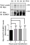
Fig 1
Fig 1. EAAT2 is sumoylated in primary astrocytes
Primary astrocytes were treated with AdV-Myc-EAAT2 (MOI 3) and collected at 8, 16, 24 or 32 hours. Virus treated astrocytes were analyzed for transduction by western blotting for the Myc tag (anti-Myc, Clontech, Cat #631206, 1:1000). Exogenous Myc-EAAT2 was immunoprecipitated using an antibody to the myc tag (anti-Myc, Clontech, Cat #631206, 10 μg) and was immunoblotted for SUMO1 (anti-SUMO1, SantaCruz, cat.# sc-5308, 1:500), showing de novo sumoylation of the exogenous Myc-EAAT2. The immunoprecipitated Myc was also positive for Myc indicating a successful immunoprecipitation (not shown). All immunoprecipitations were normalized to their own immunoprecipitation control. There was no increase in the sumoylation of the exogenous EAAT2 over time, indicating that the SUMO1 conjugation occurs quickly and reaches equilibrium within 8 hours of protein expression (n=3 per group ANOVA ((F3,11)=0.15, p=.92)).
Fig 2
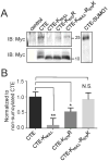
Fig 2. Lysine 570 is the primary target of sumoylation in the c-terminal tail of EAAT2
(A) Cos7 cells were transfected with Myc-CTE, Myc-CTEKnull, Myc-CTEK570R or Myc-CTEKnullR570K in conjunction with SUMO1 and UBC9. The immunoblot of Myc (anti-Myc, Clontech, Cat #631206, 10 μg) showed a 25 kDa band in the CTE lane indicating sumoylation of CTE (Gibb et al. 2007). There was no 25 kDa band in the CTEKnull lane indicating that no sumoylation occurs. The band in the CTEK570R lane was reduced indicating that there is less sumoylation of CTE when the consensus site was destroyed. Sumoylation was nearly restored when the only lysine available was the lysine 570. All transfected cells had a 10 kDa band, indicating mostly unsumoylated exogenous CTE. (B) Sumoylation of CTE was quantified with all sumoylated CTE bands normalized to their non-sumoylated CTE bands (n=3, ANOVA ((F3,11)=12.47, p=.002) with post hoc Dunnet test, Myc-CTEKnull p<0.01, Myc-CTEK570R, p<0.05, Myc-CTEKnullR570K, p>0.05).
Fig. 3
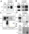
Fig. 3. EAAT2 is sumoylated in human and rat CNS
(A) Rat cortices were immunoprecipitated for EAAT2 (using custom antibody against residues 518-536) and analyzed by western blotting with anti-SUMO1 antibody, showing sumoylated EAAT2. IgGs (Santa Cruz, cat.#sc-2027, 10 μg) were used as control for immunoprecipitation. The immunoprecipitated EAAT2 (ABR518-536, 10 μg) was also positive for another epitope of EAAT2 (a.a. 1-85, Santa Cruz, cat.# sc-15317, 1:1000) indicating a successful immunoprecipitation (not shown). (B) Rat cortices were also immunoprecipitated for SUMO1 (anti-SUMO1, Santa Cruz, cat.# sc-5308, 10 μg) and analyzed by western blotting with anti-EAAT2 antibody (ABR518-536, 1:10,000), confirming sumoylated EAAT2. The immunoprecipitated SUMO1 was also positive for SUMO1 indicating a successful immunoprecipitation (not shown). (C) Rat cortex homogenate was treated without (-) or with (+) activate Caspase-3 (Sigma, cat.#C5482, 200 ng, 5hr, 37°C) and the products were analyzed by western blotting with anti-EAAT2 antibody (ABR518-536; 1:10,000). Samples treated with activated caspase-3 showed both a 10 kDa and a 25 kDa band, the latter of which was also positive for SUMO1 (anti-SUMO1, Santa Cruz, cat.# sc-5308, 10 μg), indicating that the EAAT2 had been sumoylated prior to caspase-3 treatment. The lysate incubated without active caspase-3 showed no cleavage of EAAT2, indicating that the caspase-3 cleavage was specific. (D) EAAT2 was immunoprecipitated (ABR518-536, 10 μg) from human post-mortem spinal cord homogenates and stained for SUMO1 (anti-SUMO1, Santa Cruz, cat.# sc-5308, 1:500), indicating some sumoylated EAAT2 in normal human spinal cord. (E) SUMO1 (anti-SUMO1, Santa Cruz, cat.# sc-5308, 10 μg) was also immunoprecipitated from human post-mortem spinal cord homogenates and stained for EAAT2 (ABR518-536, 1:10,000), confirming sumoylated EAAT2 in normal human spinal cord.
Fig 4
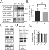
Fig 4. Sumoylation of EAAT2 is not altered by disease progression in the SOD1-G93A mouse
Spinal cord homogenate from both presymptomatic (65 days old) and symptomatic (120 days old) G93A-SOD1 mice were analyzed by western blotting for key proteins involved in sumoylation. (A) Homogenates were immunoprecipitated with anti-SUMO1 antibody (anti-SUMO1, Santa Cruz, cat.# sc-5308, 10 μg) and immunoblotted for EAAT2 (ABR518-536, dil. 1:10,000) or immunoprecipitated with anti-EAAT2 antibody (ABR518-536, 10 μg) and immunoblotted for SUMO1 (anti-SUMO1, Santa Cruz, cat.# sc-5308, dil. 1:500), demonstrating similar levels of sumoylation of EAAT2. (B) Three spinal cords from each age group were analyzed for quantification of EAAT2-SUMO1. (n=3, Two-tailed T-test (P>0.05)). (C) Homogenates were stained for proteins involved in sumoylation; UBC9 (Cell Signaling, cat.# 4786, dil.1:1000) and Senp1 (Abcam, cat.# 58417, dil. 1:1000). (D.) Homogenates were stained with anti-SUMO1 antibody (Santa Cruz, cat. # sc-5308, dil. 1:500). (E) Three spinal cords from each age group were used for analysis. All blots were normalized to the expression levels of GAPDH (Fitzgerald, cat.# 10R-G109A, dil. 1:25,000) and quantified. No change was seen between the two groups (n=3, two tailed T-test, p>0.05).
Fig 5
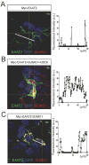
Fig 5. Sumoylation of EAAT2 alters EAAT2 localization in a heterologous system
HEK293T cells were transfected with myc-EAAT2 alone (A), myc-EAAT2 with UBC9 and SUMO1 (B), or myc-EAAT2-SUMO1 (C). (A) Confocal analysis of HEK293T cells expressing Myc-EAAT2 and stained with anti-EAAT2 (ABR518-536, dil. 1:100) and anti-SUMO1 (Santa Cruz, cat.# sc-5308, dil. 1:50) showed that Myc-EAAT2 was primarily localized to the plasma membrane. The intensity of the fluorescence along the central axis of a representative astrocyte was graphed to show the subcellular localization of EAAT2 (ImageJ). (B) HEK293T cells transfected with myc-EAAT2 as well as SUMO1 and UBC9 showed increased cytoplasmic localization of EAAT2 (anti-EAAT2, ABR518-536, dil. 1:100, anti-SUMO1, Santa Cruz, cat# sc-5308, 1:50). (C.) Myc-EAAT2-SUMO1 had a propensity to form cytoplasmic aggregates (anti-EAAT2, ABR518-536, dil. 1:100, anti-SUMO1, Santa Cruz, cat# sc-5308, 1:50).
Fig 6
Fig 6. Sumolyation alters EAAT2 localization in primary astrocytes
Astrocytes in culture were transfected (0.8 μg plasmid DNA/condition) with: (A) WT-UBC9, (B) dominant negative-UBC (HA-dn-UBC9), or (C) Myc-trEAAT2 (EAAT2 missing the last 67 amino acids of the c-terminus). (A) Intracellular localization of EAAT2 is increased by overexpression of wild type UBC9. Astrocytes were stained with anti-EAAT2 antibody (ABR518-536, dil. 1:100) and anti-UBC9 (Cell Signaling, cat.# 4786,1:100). (B) Expression of dominant negative UBC9 (HA-dnUBC9) causes dispersal of EAAT2 relative to wild type UBC9 overexpression. Astrocytes were stained with anti-EAAT2 antibody (ABR518-536, 1:100) and anti-HA (Clontech, cat #631207, dil.1:100). (C) The truncated form of EAAT2 (myc-trEAAT2), which lacks the c-terminus domain, is robustly expressed at the plasma membrane, away from the intracellular pools of the endogenous EAAT2. Staining for trEAAT2 was performed with anti-Myc (Clontech, cat.# 631206, dil. 1:1000), while staining for endogenous EAAT2 was completed with the anti-EAAT2 antibody (ABR518-536, dil. 1:100).
Fig 7
Fig 7. Increasing desumoylation disperses intracellular pools of EAAT2
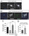
Primary astrocytes were transfected with control plasmid (pcDNA3.1+) or Flag-Senp1 (0.8 μg of plasmid DNA/condition). (A) EAAT2 formed intracellular pools in control transfected astrocytes transfected. Similar pattern of staining is observed in non-transfected astrocytes (not shown), suggesting that transfection of pcDNA3.1 per se does not affect the physiological localization pattern of EAAT2 (anti-EAAT2, ABR518-536, dil. 1:100). (B) These intracellular pools were not present when astrocytes were transfected with Flag-SENP1 (anti-EAAT2, ABR518-536, 1:100, anti-Flag, Sigma, cat.# F3165, dil. 1:200). (C, D) Uptake of 3H-L-glutamate was measured at room temperature for 10 minutes without (-) or with (+) DHK (1mM, Tocris, cat.# 0111). Overexpression of Senp1 increased the total uptake in astrocytes, but did not alter the DHK-insensitive uptake. (D) The DHK-sensitive, or EAAT2-dependent uptake, was increased in the astrocytes overexpressing SENP1 (n=3, two tailed T-test, ***p<0.05).
Fig 8
Fig 8. Sumoylated EAAT2 partitions prevalently to intracellular compartments in vivo
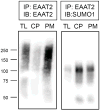
EAAT2 was immunoprecipitated from 1 mg each of normal mouse brain homogenate (total lysate = TL), cytoplasm-enriched fraction (CP), or plasma membrane-enriched fraction (PM). (A) Half of the immunoprecipated protein from each fraction was resolved on 7.5% SDS-PAGE gel and stained for EAAT2 (ABR518-536, dil. 1:500), or (B) stained for SUMO1 (Santa Cruz cat# 5308, dil. 1:100) demonstrating unsumoylated EAAT2 is highly enriched in the plasma membrane fraction and SUMO1-bound EAAT2 is enriched in the cytoplasm fraction of astrocytes in vivo.
Fig 9
Fig 9. SUMO1 attachment to EAAT2 dramatically reduces its plasma membrane and glutamate uptake in a heterologous system
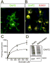
HEK293T cells were plated on 3mm plates and transfected with either myc-EAAT2, (A) or myc-EAAT2-SUMO1, (B) (1.5 μg of plasmid DNA/condition). (C) Uptake of 3H-L-glutamate was measured for 10 minutes at room temperature and compared between the groups. The HEK293T transfected with myc-EAAT2 had nearly double the maximum uptake compared to HEK293T transfected with myc-EAAT2-SUMO1. The uptake rate, however, was similar between the two groups. (D) Transfections showed similar levels of EAAT2 expression by western blot analysis (anti-Myc, Clontech, cat.# 631206, dil. 1:1000), eliminating the possibility that different translation levels accounted for the difference in uptake capacity.

Similar articles

Cited by

References

    1. Benediktsson AM, Marrs GS, Tu JC, Worley PF, Rothstein JD, Bergles DE, Dailey ME. Neuronal activity regulates glutamate transporter dynamics in developing astrocytes. Glia. 2012;60:175–88. - PMC - PubMed
    1. Berry JD, Shefner JM, Conwit R, Schoenfeld D, Keroack M, Felsenstein D, Krivickas L, David WS, Vriesendorp F, Pestronk A, et al. Design and initial results of a multi-phase randomized trial of ceftriaxone in amyotrophic lateral sclerosis. PLoS One. 2013;8:e61177. - PMC - PubMed
    1. Boston-Howes W, Gibb SL, Williams EO, Pasinelli P, Brown RH, Jr, Trotti D. Caspase-3 cleaves and inactivates the glutamate transporter EAAT2. J Biol Chem. 2006;281:14076–84. - PubMed
    1. Bristol LA, Rothstein JD. Glutamate transporter gene expression in amyotrophic lateral sclerosis motor cortex. Ann Neurol. 1996;39:676–9. - PubMed
    1. Butchbach ME, Tian G, Guo H, Lin CL. Association of excitatory amino acid transporters, especially EAAT2, with cholesterol-rich lipid raft microdomains: importance for excitatory amino acid transporter localization and function. J Biol Chem. 2004;279:34388–96. - PubMed

Publication types

MeSH terms