. 2014 Jun;15(6):546-53.
doi: 10.1038/ni.2876. Epub 2014 Apr 20.

The transcription factor Zbtb32 controls the proliferative burst of virus-specific natural killer cells responding to infection

Affiliations

The transcription factor Zbtb32 controls the proliferative burst of virus-specific natural killer cells responding to infection

Aimee M Beaulieu et al. Nat Immunol. 2014 Jun.

Abstract

Natural killer (NK) cells are innate lymphocytes that exhibit many features of adaptive immunity, including clonal proliferation and long-lived memory. Here we demonstrate that the BTB-ZF transcription factor Zbtb32 (also known as ROG, FAZF, TZFP and PLZP) was essential for the proliferative burst and protective capacity of virus-specific NK cells. Signals from proinflammatory cytokines were both necessary and sufficient to induce high expression of Zbtb32 in NK cells. Zbtb32 facilitated NK cell proliferation during infection by antagonizing the anti-proliferative factor Blimp-1 (Prdm1). Our data support a model in which Zbtb32 acts as a cellular 'hub' through which proinflammatory signals instruct a 'proliferation-permissive' state in NK cells, thereby allowing their prolific expansion in response to viral infection.


Figures
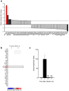
Figure 1
Figure 1. Zbtb32 is highly upregulated in NK cells during viral infection
(a) Expression of 47 BTB-ZF genes in splenic Ly49H+ NK cells sorted from uninfected and MCMV-infected animals on day 1.5 p.i., as assessed by microarray (data provided by the Immunological Genome Consortium). Shown as fold microarray signal intensity for the infected versus uninfected samples (n = 3 biological replicates per group). Solid black bars denote significant upregulation or downregulation. (b) Top 30 most highly induced genes in splenic Ly49H+ NK cells from MCMV-infected versus uninfected control animals. Heat map shows mean microarray signal intensity (n = 3 biological replicates per time point). (c) Zbtb32 mRNA abundance measured by qRT-PCR in splenic Ly49H+ NK cells sorted from MCMV-infected animals on day 2 (n = 6 mice), 4 (n = 3 mice), and 7 (n = 3 mice) shown as fold expression relative to day 0 (n = 5 mice). Data are representative of 3 independent experiments.
Figure 2
Figure 2. Zbtb32 is required for protective anti-viral NK cell responses
(a) Kaplan-Meier survival curves for neonate Ly49H-deficient hosts receiving splenic Ly49H+ NK cells from WT (n = 12 mice) or Zbtb32−/− (n = 12 mice) donors, or receiving PBS only (n = 14 mice), one day prior to infection with MCMV. Data are pooled from 4 independent experiments. (b) Kaplan-Meier survival curves for adult Ly49H-deficient hosts receiving splenic Ly49H+ NK cells from WT (n = 9 mice) or Zbtb32−/− (n = 8 mice) donors, or receiving PBS only (n = 9 mice), one day prior to infection with VSV-m157. Data are pooled from 3 independent experiments.
Figure 3
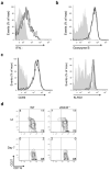
Figure 3. Zbtb32 is dispensable for NK cell activation, maturation, and expression of effector molecules
(a) Intracellular IFN-γ and (b) granzyme B in splenic WT or Zbtb32−/− Ly49H+ NK cells from mixed bone marrow chimeric animals on day 2 p.i. with MCMV. Representative of n = 5 (infected) and n = 2 (uninfected) animals from 2 independent experiments. (c) WT and Zbtb32−/− Ly49H+ NK cells were co-transferred into Ly49H-deficient mice and CD69 and KLRG1 expression was assessed on transferred cells in the spleen on day 2 (for CD69) or day 7 (for KLRG1) p.i. with MCMV. Representative of n = 5 animals per time point from 2 independent experiments. (d) CD27 and CD11b expression were used to assess the relative percentage of immature (CD27hiCD11blo and CD27hiCD11bhi) and mature (CD27loCD11bhi) Ly49H+ NK cells from uninfected (UI) or MCMV-infected (day 7 p.i.) mixed bone marrow chimeric animals. Representative of n = 7 (infected) and n = 2 (uninfected) animals from 2 independent experiments.
Figure 4
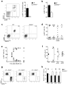
Figure 4. Zbtb32 is essential for NK cell expansion during viral infection
(a – b) and (d – f) Equal numbers of WT (CD45.1+) and Zbtb32−/− (CD45.2+) Ly49H+ NK cells were co-transferred into Ly49H-deficient hosts prior to MCMV infection. (a) The percentage of transferred Ly49H+ NK cells (within the total NK cell gate) in the spleen on day 7 p.i. (n = 3 mice). Representative of 4 independent experiments. P < 0.05 in a ratio paired two-tailed t-test. (b) As in (a), except showing absolute number. Representative of 2 independent experiments. (c) As in (a), except WT or Zbtb32−/− NK cells (CD45.2+) were separately transferred into independent Ly49H-deficient hosts (CD45.1+). Representative of n = 4 mice per group from 2 independent experiments. (d) Relative percentages of co-transferred WT and Zbtb32−/− Ly49H+ NK cells in various organs on day 7 p.i. Symbols represent individual mice from 3 independent experiments. (e) Relative percentages of co-transferred WT and Zbtb32−/− Ly49H+ NK cells in the spleen at various time points. Symbols represent individual mice. (f) Relative percentages of co-transferred Ly49H+ WT and Zbtb32−/− NK cells in the spleen on day 8 p.i. with the indicated viruses. Symbols represent individual mice. Representative of at least 2 independent experiments. (g) Ly49H+ NK cells from Zbtb32−/−, Zbtb32+/−, or Zbtb32+/+ littermates (CD45.2+) were co-transferred with equal numbers of Ly49H+ NK cells from WT animals (CD45.1+). Plots show the relative percentages of transferred Ly49H+ WT and Zbtb32 littermate NK cells in the spleen on day 7 p.i. with MCMV (n = 3 animals per group). Representative of 2 independent experiments. (h) Relative percentage of WT or Zbtb32−/− tetramer+ CD8+ T cells in WT:Zbtb32−/− mixed bone marrow chimeric mice (n = 8 animals) on day 7 p.i. (for M45+) or day 14 p.i. (for m139+ or M38+) with MCMV. Representative of 3 independent experiments.
Figure 5
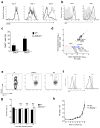
Figure 5. Zbtb32 regulates NK cell proliferation following MCMV infection
(a–f) Equal numbers of WT (CD45.1+; black lines in histograms) and Zbtb32−/− (CD45.2+; red lines in histograms) Ly49H+ NK cells were co-transferred into Ly49H-deficient hosts one day prior to MCMV infection. (a) CFSE dilution of transferred Ly49H+ NK cell populations in the spleen at indicated time points p.i. Representative of n = 6 mice per time point from 3 independent experiments. (b) Intracellular Ki67 expression in transferred Ly49H+ NK cells on day 4 and 7 p.i. (shaded histograms show uninfected controls). Representative of n = 4 mice per time point from 2 independent experiments. (c) Percentage of BrdU incorporation in transferred Ly49H+ NK cells in the spleen at indicated time points p.i. with MCMV (n = 2 mice per time point). Representative of 2 independent experiments. (d) Expression of 84 genes involved in cell cycle regulation on day 4 p.i. as measured by Cell Cycle PCR Array™ (Qiagen). Genes up- (red) or down-regulated (blue) >4-fold in Zbtb32−/− versus WT Ly49H+ splenic NK cells are annotated. (e) Pan-caspase activation in transferred Ly49H+ NK cell populations in the spleens day 4 p.i. or ininfected (UI) mice. Representative of n = 2 animals per group from 2 independent experiments. (f) Intracellular Bcl-2 expression in transferred Ly49H+ NK cells in the spleen on day 4 and 7 p.i. (shaded histogram shows uninfected controls). Representative of n = 2 – 3 animals per group from 3 independent experiments. (g) WT and Zbtb32−/− Ly49H+ NK cells were co-transferred into Rag2−/− × Il2rg−/− hosts and the relative percentage of each population in the peripheral blood (0, 1, and 2 weeks) or spleen (10 weeks) was determined (n = 3 mice per time point). Representative of 2 independent experiments. (h) Sorted splenic NK cells from WT and Zbtb32−/− mice were incubated with IL-2 and IL-15 and the absolute number of NK cells was determined daily. Data are representative of n = 3 biological replicates per group from 2 independent experiments.
Figure 6
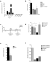
Figure 6. Inflammatory cytokines are necessary and sufficient for maximal Zbtb32 expression in NK cells
(a) Expression of Zbtb32 mRNA in stimulated NK cells, expressed as fold expression after treatment with medium only, as assessed by qRT-PCR (n = 3 biological replicates per condition). Data are representative of 3 independent experiments. (b) WT or mutant Ly49H+ NK cells were sorted from the spleens of mixed bone marrow chimeric mice on day 2 p.i. with MCMV. Zbtb32 mRNA abundance, as a percentage of expression in WT cells, was assessed by qRT-PCR (n = 2 – 8 animals) from 3 pooled experiments. (c) Vista browser image of conserved noncoding sequences (CNS; gray shaded regions) and 2 predicted STAT4 binding sites within the mouse Zbtb32 promoter. (d) Enrichment of acetylated (lysine 27) histone 3 at the Zbtb32 promoter, as assessed by ChIP followed by qPCR on sorted WT NK cells (n = 3 biological replicates). Enrichment of target (Zbtb32 promoter) or control DNA (Gapdh promoter or ‘gene desert’ ∼50 kb upstream of Foxp3 gene) is expressed as percentage of input. Dotted line denotes “background-level” enrichment at the gene desert locus. Representative of 3 independent experiments. (e) As in (b), except using WT:Stat4−/− mixed bone marrow chimeric mice (n = 4 mice from 2 independent experiments). (f) Purified WT NK cells were stimulated for 24 h with IL-12 and IL-18, and STAT4 binding at the Zbtb32 promoter was assessed by ChIP followed by qPCR. Target (Zbtb32) or control (Actb and Gapdh) promoter DNA levels are expressed as a percentage of input. Dotted line denotes “background-level” enrichment, set as the mean enrichment of the pGapdh and pActb control loci. Representative of 2 independent experiments.
Figure 7
Figure 7. Zbtb32 promotes NK cell proliferation by antagonizing Blimp-1
(a) WT and Zbtb32−/− Ly49H+ NK cells were co-transferred into Ly49H-deficient hosts and then recovered from the spleen by sorting on day 4 p.i. with MCMV. Prdm1 mRNA abundance, as a percentage of expression in WT cells, was quantified by qRT-PCR in each population (n = 4 mice). Data are representative of 2 independent experiments. (b,c) Ly49H+ NK cells from Prdm1fl/flNkp46-Cre, Prdm1fl/flNkp46-Cre+, Zbtb32−/−Prdm1fl/flNkp46-Cre, or Zbtb32−/−Prdm1fl/flNkp46-Cre+ littermate animals (CD45.2+) were co-transferred with equal numbers of Ly49H+ NK cells from WT animals (CD45.1+) into Ly49H-deficient hosts one day prior to infection with MCMV. The relative percentages of transferred Ly49H+ WT and CD45.2+ littermate NK cells in the spleen on day 7 p.i. are shown. Depicted are (b) representative responses in individual animals (except the Prdm1fl/flNkp46-Cre+ cohort) and (c) summary data (symbols represent individual mice). Data are representative of 3 independent experiments.

Comment in

Similar articles

Cited by

References

    1. Sun JC, Lanier LL. NK cell development, homeostasis and function: parallels with CD8(+) T cells. Nat Rev Immunol. 2011;11:645–657. - PMC - PubMed
    1. Smith HR, et al. Recognition of a virus-encoded ligand by a natural killer cell activation receptor. Proc Natl Acad Sci U S A. 2002;99:8826–8831. - PMC - PubMed
    1. Arase H, Mocarski ES, Campbell AE, Hill AB, Lanier LL. Direct recognition of cytomegalovirus by activating and inhibitory NK cell receptors. Science. 2002;296:1323–1326. - PubMed
    1. Daniels KA, et al. Murine cytomegalovirus is regulated by a discrete subset of natural killer cells reactive with monoclonal antibody to Ly49H. J Exp Med. 2001;194:29–44. - PMC - PubMed
    1. Brown MG, et al. Vital involvement of a natural killer cell activation receptor in resistance to viral infection. Science. 2001;292:934–937. - PubMed

Publication types

MeSH terms

Associated data