. 2014 Jul 1;544(1):8-18.
doi: 10.1016/j.gene.2014.04.026. Epub 2014 Apr 16.

A conserved RNA polymerase III promoter required for gammaherpesvirus TMER transcription and microRNA processing

Affiliations

A conserved RNA polymerase III promoter required for gammaherpesvirus TMER transcription and microRNA processing

Kevin W Diebel et al. Gene. .

Abstract

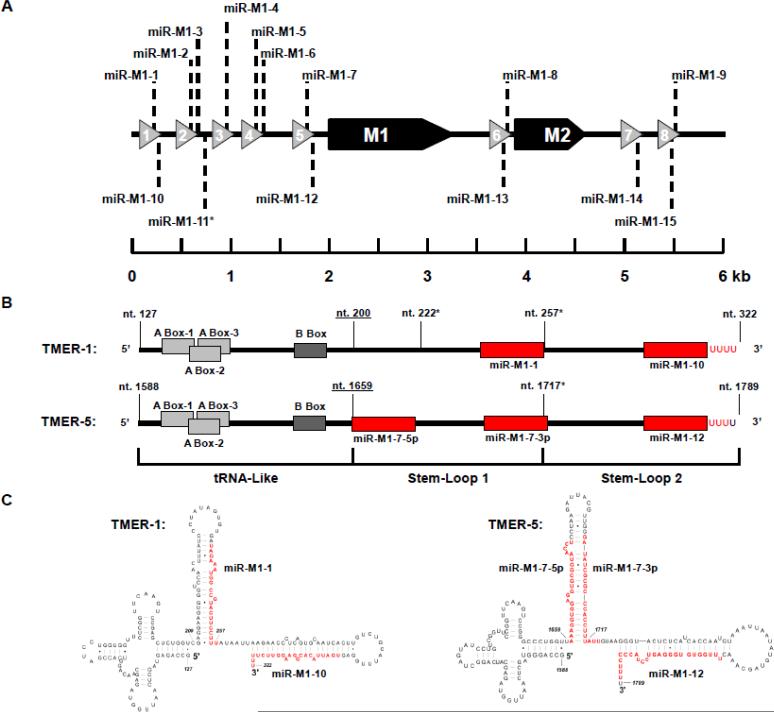
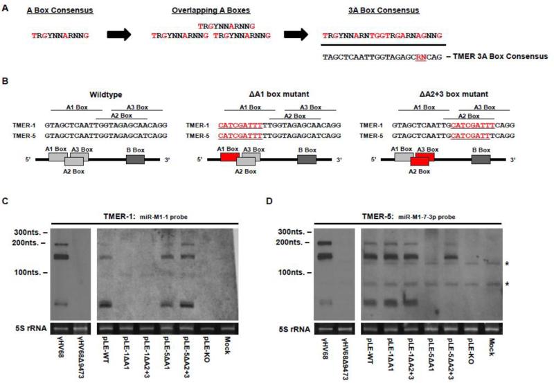
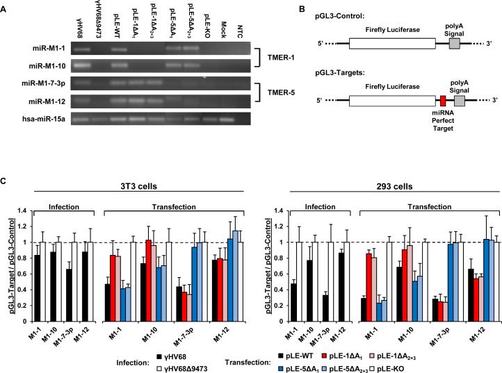
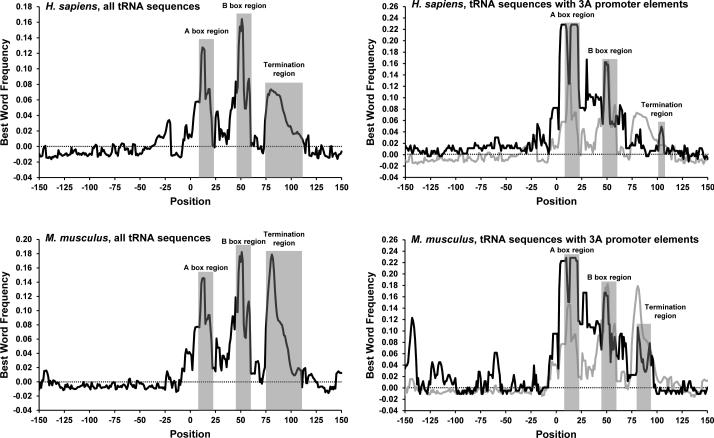
Canonical RNA polymerase III (pol III) type 2 promoters contain a single A and B box and are well documented for their role in tRNA and SINE transcription in eukaryotic cells. The genome of Murid herpesvirus 4 (MuHV-4) contains eight polycistronic tRNA-microRNA encoded RNA (TMER) genes that are transcribed from a RNA pol III type 2-like promoter containing triplicated A box elements. Here, we demonstrate that the triplicated A box sequences are required in their entirety to produce functional MuHV-4 miRNAs. We also identify that these RNA pol III type 2-like promoters are conserved in eukaryotic genomes. Human and mouse predicted tRNA genes containing these promoters also show enrichment of alternative RNA pol III transcription termination sequences and are predicted to give rise to longer tRNA primary transcripts.

Keywords: A box; Gammaherpesvirus 68; Type 2 promoter; tRNA.


Figures

Figure 1
Figure 1. Genomic organization and predicted secondary structures of the γHV68 TMER genes
(A) Schematic of the left end of the γHV68 genome containing all eight of the RNA pol III transcribed TMER genes. The TMER genes are represented as grey arrows labeled 1 through 8. The protein coding transcripts M1 and M2 are shown as black arrows. Locations of the γHV68 encoded miRNA genes are shown as dashed lines and labeled miR-M1-(1-15). *miR-M1-11 is the only γHV68 miRNA that is not directly embedded within a predicted TMER primary transcript (Reese et al., 2010; Zhu et al., 2010). The miRNA genes are labeled here as found on the miRBase website (Kozomara and Griffiths-Jones, 2011). (B) Detailed schematics of the TMER-1 and TMER-5 transcripts. Shown are the predicted start and stop sites of transcription along with the predicted RNaseZL cut site (underlined) and alternative transcriptional stop sites (marked with an asterisk). Positions of the A box promoter elements are shown in light gray and the B box promoter elements in dark gray. miRNA locations associated with these TMER transcripts are shown by red boxes and nucleotides. The boundaries of the three structural domains of each transcript are detailed below the schematics. (C) Predicted secondary structures of the TMER-1 and the TMER-5 transcripts. The positions of the TMER associated miRNAs are highlighted in red.
Figure 2
Figure 2. RNA pol III type 2(3A) A box promoter element requirements in the transcription of the γHV68 TMER-1 and TMER-5 genes
(A) The A box promoter element consensus sequence, the overlapping A box configuration, and the 3A box consensus sequence and its comparison to the γHV68 TMER gene A box consensus sequence is shown. Bases within the A box promoter elements that are highlighted red are invariant in highly expressed human tRNAs (Canella et al., 2010). The underlined R and N bases in the TMER 3A box consensus sequence represent differences between this consensus sequence and the constructed 3A box consensus sequence above it at positions where the bases are predicted to be invariant. (B) Schematic detailing the wildtype and mutant sequences of the RNA pol III type 2(3A) promoter sequences found within the TMER-1 and the TMER-5 genes. Red underlined bases represent replacement mutations created within each TMER gene. Below each sequence is a schematic of the resulting potential RNA pol III promoter. Light grey boxes represent wildtype A box promoter elements while red boxes represent mutated A box promoter elements. Dark grey boxes represent wildtype B box promoter elements. (C, D) Northern blot analysis of γHV68 TMER-1 and TMER-5. (C) Blot probed using a 5’-biotinylated probe designed to hybridize antisense to the miR-M1-1 sequence. (D) Blot probed using a 5’-biotinylated probe designed to hybridize antisense to the miR-M1-7-3p sequence. Two non-specific bands in the miR-M1-7-3p blot are starred to the right of the blot. Below each blot is the ethidium bromide stained 5S rRNA band from each gel photographed to gauge both RNA quality and loading prior to transfer. Labeled below each gel is the infection or transfection condition from which the total RNA was isolated.
Figure 3
Figure 3. Mutation of the 3A box promoter elements inhibits the production of mature miRNAs
(A) RLM-RT-PCR of the viral miRNAs of TMER-1 and TMER-5. 293 cells were either infected with γHV68 or γHV68Δ9473 or transfected with the various Left End plasmid constructs followed by total RNA isolation and RLM-RT-PCR analysis. The human miRNA hsa-miR-15a is an endogenous cellular control for the RLM-RT-PCR assay. NTC – non-template control. (B) Schematics of the pGL3 firefly luciferase reporter system. A single copy of the miR-M1-1, miR-M1-10, miR-M1-7-3p, and miR-M1-12 were each individually cloned into a XbaI cut site located between the firefly luciferase coding region and the polyA signal in the pGL3-Control plasmid. (C) Dual luciferase reporter analysis with the A box mutant plasmids in 3T3 and 293 cells. The pGL3 luciferase reporter target is labeled below each graph. In infected samples, results are shown as a ratio of firefly luciferase expression in γHV68 infected cells divided by firefly luciferase expression in γHV68Δ9473 infected cells. In transfected samples, results are shown as a ratio of firefly luciferase expression in pLE-WT or A box mutant plasmids divided by the firefly luciferase expression in the pLE-KO transfected cells. All firefly luciferase values were normalized to the renilla luciferase transfection control readings prior to the comparative analysis shown on the graphs. Readings below the dashed line represent translational repression by the corresponding viral miRNA. Data represents the average of two experiments with each experiment conducted in triplicate. Error bars represent one standard of deviation.
Figure 4
Figure 4. Sequence context comparison of human and mouse tRNAs with and without the 3A box promoter element
Histograms represent the most common 6 base word within a 9 base sliding window (Best Word Frequency) starting at 150 bases upstream from the predicted transcriptional start site and ending at 150 bases downstream from the predicted transcriptional start site. Any point above the dotted line at zero is considered to have sequence enrichment at that position. The A box and B box promoter regions and the termination regions are highlighted in light grey. The left histograms represent the best word frequency of all human and mouse tRNAs. The right histograms represent the best word frequency of human and mouse tRNAs containing the type 2(3A) A box promoter element (black line) overlaid on top of the best word frequency of all human or mouse tRNAs (grey line).
Figure 5
Figure 5. The impact of canonical RNA pol III termination signals near the predicted tRNA domain of TMER-1 on transcription and miRNA production from the TMER-1 gene
(A) Schematic of TMER-1. Shown is the location and sequence of the hard stop mutation. The alternative transcriptional stop location in wildtype TMER-1 is underlined in the wildtype sequence. The hard stop sequences and locations generated in the hard stop mutant plasmids are underlined and in red below the wildtype sequence. The location of the hard stop probe use for northern analysis of TMER-1 in this figure is shown as a blue line below the TMER-1 diagram. The sequence of the TMER-1 hard stop probe in shown in blue from 3’ to 5’. (B) Northern blot for TMER-1 using a probe immediately downstream from the tRNA region of the TMER-1 transcript. Non-specific bands are indicated with asterisks. Below each blot is the ethidum bromide stained 5S rRNA band from each gel photographed to gauge RNA quality and loading prior to transfer. Labeled below each gel is the transfection condition from which the total RNA was isolated. (C) RLM-RT-PCR for viral miRNAs associated with the transcription of γHV68 TMER-1. The human miRNA, hsa-miR-15a is included as a cellular control for the RLM-RT-PCR assay. NTC – non-template control.

Similar articles

Cited by

References

    1. Andersson MG, Haasnoot PCJ, Xu N, Berenjian S, Berkhout B, Akusjärvi G. Suppression of RNA Interference by Adenovirus Virus-Associated RNA. J. Virol. 2005;79:9556–9565. - PMC - PubMed
    1. Aparicio O, Razquin N, Zaratiegui M, Narvaiza I, Fortes P. Adenovirus Virus-Associated RNA Is Processed to Functional Interfering RNAs Involved in Virus Production. J. Virol. 2006;80:1376–1384. - PMC - PubMed
    1. Barton E, Mandal P, Speck S. Pathogenesis and host control of gammaherpesviruses: lessons from the mouse. Annu. Rev. Immunol. 2011;29:351–397. - PubMed
    1. Blaskovic D, Stanceková M, Svobodová J, Mistríková J. Isolation of five strains of herpesviruses from two species of free living small rodents. Acta Virol. 1980;24:468. - PubMed
    1. Bogenhagen DF, Brown DD. Nucleotide sequences in Xenopus 5S DNA required for transcription termination. Cell. 1981;24:261–270. - PubMed

Publication types

MeSH terms

LinkOut - more resources